Immune System Worksheet 1 Bio 221 Lecture
1/64
There's no tags or description
Looks like no tags are added yet.
Name | Mastery | Learn | Test | Matching | Spaced | Call with Kai |
|---|
No analytics yet
Send a link to your students to track their progress
65 Terms
What is the function of the immune system?
A group of organs and cells that defends the body against pathogens, removes damaged cells, absorbs nutrients, and identifies and destroys abnormal or cancerous cells.
What are the components of the immune system?
The immune system includes white blood cells (leukocytes), lymphatic vessels, lymph nodes, the spleen, thymus gland, bone marrow, and MALT (mucosa-associated lymphoid tissue). (non-encapsulated wbc)
Name the 5 types of white blood cells and their basic functions.
Neutrophils (Innate) —-Phagocytose bacteria and debris.
Lymphocytes (Adaptive except for NK cells)—Include B cells, T cells, and NK cells; responsible for adaptive and innate immunity.
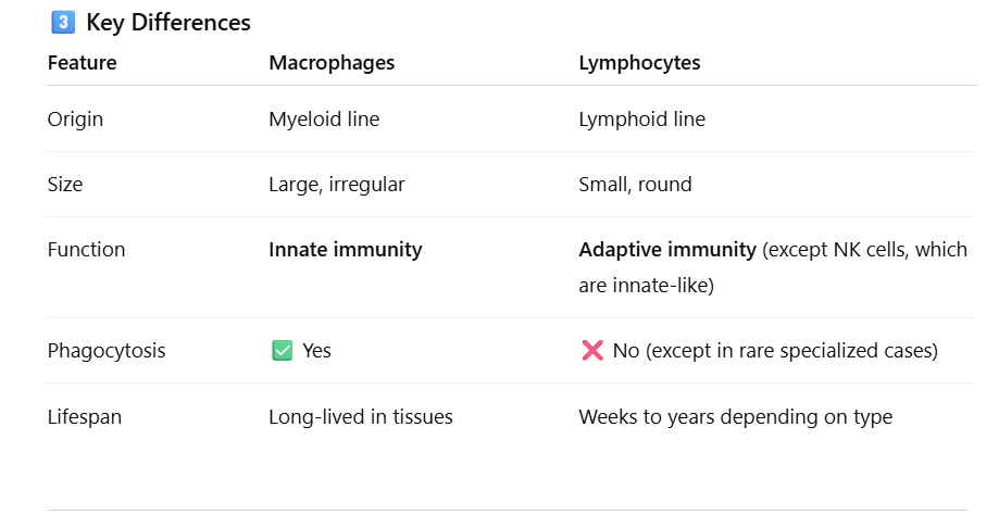
Monocytes (Innate)— Become macrophages; engulf pathogens and dead cells.
Eosinophils (Innate)—Attack parasites and modulate allergic responses.
Basophils (Innate)—Release histamine and heparin during inflammation and allergies.
What are the functions of the lymph vessels?
They transport lymph, return excess interstitial fluid to the bloodstream, and help move immune cells throughout the body.
What are characteristics of lymph vessels?
Lymph vessels have thin walls, valves to prevent backflow, and resemble veins in structure.
What are lymph nodes?
Lymph nodes are small, bean-shaped organs that filter lymph and house lymphocytes to initiate immune responses.
Approximately how many lymph nodes are in the body?
There are about 600 to 700 lymph nodes in the human body.
What fibers and cells are present in lymph nodes?
Lymph nodes contain reticular fibers that trap particles and white blood cells, including:
Lymphocytes (All adaptive T and B cells except for NK cells, innate) – Recognise and respond to specific antigens.
Macrophages (Innate)—Engulf and destroy pathogens and debris.
Dendritic cells (Innate) – Capture antigens and present them to lymphocytes to activate immune responses.
What is MALT?
MALT (mucosa-associated lymphoid tissue) is lymphoid tissue that protects mucosal surfaces—such as those in the digestive, respiratory, urinary, and reproductive tracts—from pathogens.
What are specific types of MALT?
Specific types of MALT include tonsils, Peyer’s patches in the small intestine, and the appendix.
What are the functions of the spleen?
The spleen filters blood, removes old red blood cells, stores platelets, and activates lymphocytes to initiate immune responses.
Distinguish between red pulp and white pulp.
The red pulp filters blood and removes old red blood cells, while the white pulp contains lymphocytes and initiates immune responses.
What is the function of the thymus gland?
The thymus gland is where T lymphocytes (T-cell) mature and differentiate, allowing them to function properly in immune responses.
What is the role of thymosin?
Thymosin is a hormone that promotes the development and maturation of T cells.
Distinguish between nonspecific (innate) and specific immunity.
Innate immunity provides general, immediate defense with no memory, while specific (adaptive) immunity targets particular antigens and has memory for faster future responses.
→ “memory” refers to the immune system’s ability to remember a pathogen it has encountered before.
Name some components of the first line of defense.
The first line of defense consists of physical and chemical barriers such as skin, mucous membranes, tears, saliva, stomach acid, and normal microbiota that prevent pathogens from entering the body.
What are PAMPs and DAMPs?
: PAMPs (pathogen-associated molecular patterns) are molecules present on pathogens that signal infection, while DAMPs (damage-associated molecular patterns) are molecules released by injured or stressed cells to signal tissue damage.
What are PRRs (TLRs)?
PRRs, like Toll-like receptors (TLRs), are on immune cells and detect signs of infection or cell damage:
PAMPs: Pathogen-associated molecular patterns (from microbes)
DAMPs: Damage-associated molecular patterns (from injured cells)
When PRRs detect these signals, they activate the immune system to fight infection or repair damage.
Memory Tip: 🧠 PRRs = Immune “sensors” that detect danger
What cells have PRRs?
Macrophages, dendritic cells, and neutrophils have PRRs, allowing them to detect PAMPs and DAMPs and initiate immune responses.
What happens when PRRs are activated?
When PRRs (Pattern Recognition Receptors) are activated, they trigger immune responses, including inflammation, cytokine release, and activation of other immune cells to fight pathogens or respond to tissue damage.
What are the symptoms of inflammation and name two vasodilators.
The symptoms of inflammation are redness, heat, swelling, pain, and loss of function. Two vasodilators that contribute to these symptoms are histamine and bradykinin.
What is margination and diapedesis?
Margination is when white blood cells stick to the walls of blood vessels at the site of infection.
Diapedesis is when these white blood cells squeeze through the vessel walls to reach the infected or damaged tissue.
What are the steps of phagocytosis?
Chemotaxis – The phagocyte moves toward the pathogen.
Adherence – The phagocyte binds to the pathogen, sometimes aided by opsonins.
Ingestion – The pathogen is engulfed into a phagosome.
Digestion – The phagosome joins with a lysosome to form a phagolysosome, where the pathogen is broken down.
Exocytosis – Waste materials are expelled from the cell.
What is an opsonin?
An opsonin is a molecule, such as an antibody or complement protein, that coats a pathogen, making it easier for immune cells to recognise and engulf, thereby enhancing phagocytosis.
What is complement?
Complement is a group of plasma proteins that enhance immune responses by destroying pathogens (lysis), coating them for easier engulfment (opsonization), and promoting inflammation.
Name the 2 ways complement is activated.
Classical Pathway: Triggered by antibodies bound to pathogens → starts a cascade → leads to opsonization, inflammation, MAC formation.
Alternative Pathway: Triggered directly by pathogen surfaces, no antibodies needed → same cascade and outcomes.
Key Difference: 🧠 Classical = antibody-dependent, Alternative = innate/direct.
Memory Tip: 🧠 “Classical needs a class (antibodies), Alternative acts automatically.”
What is the action of interferon, and what secretes it?
Interferons:
Interferons are proteins released by virus-infected cells that alert and protect nearby healthy cells.
Warn nearby cells to make antiviral proteins that block virus replication.
Activate macrophages and natural killer (NK) cells to destroy infected cells.
Help the immune system spot infected cells by increasing how well they display antigens. (Increases antigen presentation)
Slow the spread of viruses by creating an antiviral shield around neighboring cells.
Summary:
Interferons act as the body’s early warning system against viruses, helping to stop infection and activate other immune defenses.
What is the hypoferremic response?
The hypoferremic response is when the body reduces the amount of iron in the blood during infection to limit bacterial growth, since many bacteria need iron to survive. (The body binds up iron and iron-binding proteins; therefore, it’s less available for pathogens.)
What is a pyrogen, and how does its secretion improve immune response?
A pyrogen is a substance that causes fever, either from pathogens or immune cells. Fever speeds up immune cell activity and slows the growth of pathogens, enhancing the body’s defense.
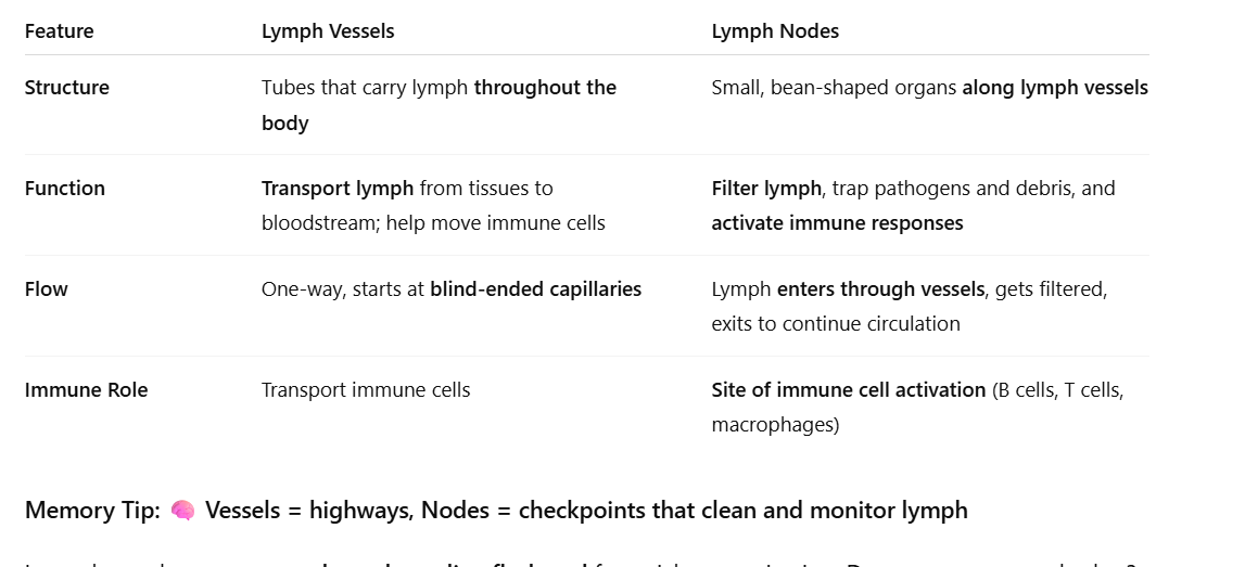
What are lymph vessels and what do they contain?
Lymph vessels are blind-ended tubes filled with lymph, a clear fluid derived from interstitial fluid.
How does lymph move through lymph vessels?
Lymph flows one way through vessels aided by valves, skeletal muscle contraction, and pressure changes during breathing.
What is the main function of lymph vessels?
They collect excess interstitial fluid from capillary beds and return it to the bloodstream.
Example: About 30 L of fluid is filtered from capillaries daily, 27 L reabsorbed, and 3 L recovered by lymph vessels as lymph.
What are the functions of lymph vessels?
Lymph vessels pick up excess filtered fluid from capillary beds, absorb fats from the intestines and transport them to the bloodstream, and expose lymph to white blood cells for immune monitoring.
Blood plasma leaks out of capillaries → becomes interstitial fluid → drains into lymphatic vessels → becomes lymph → returns to bloodstream near the heart. (The excess filtered fluid was blood plasma at first).
What are granulocytes and what are their types and functions?
Granulocytes are a type of white blood cell that contain granules in the cytoplasm.
Types:
Neutrophils – Most abundant; phagocytise pathogens.
Eosinophils – Red granules; involved in allergic reactions and parasitic infections. (Parasite killers + allergy responders).
Basophils – Least abundant; release histamine and mediate allergic reactions.
What are agranulocytes, and what are their types and functions?
Agranulocytes are white blood cells without visible cytoplasmic granules.
Types:
Monocytes – Become phagocytic macrophages and give rise to dendritic cells.
Lymphocytes—Carry out specific immune responses, including B cells, T cells, and NK (natural-killer) cells.
Where do granulocytes and agranulocytes come from?
All WBCs come from hematopoietic stem cells in the bone marrow.
Granulocytes (neutrophils, eosinophils, basophils) develop from myeloid progenitors.
Monocytes (agranulocytes) also come from myeloid progenitors.
Lymphocytes (B cells, T cells, NK cells) come from lymphoid progenitors.
Dendrtic cells from myeloid stem cells.
Memory Tip: 🧠 Myeloid → granulocytes, monocytes& dendritic cells; Lymphoid → lymphocytes
How much of the spleen is made up of red pulp?
Red pulp makes up about three-quarters (¾) of the spleen and is responsible for filtering blood and removing old red blood cells.
What are key features of the thymus gland?
The thymus gland shrinks with age (involutes) and secretes thymosin.
What is the difference between innate (non-specific) and adaptive (specific) immunity?
Innate (non-specific) immunity: Present at birth, works immediately, does not require prior exposure, responds the same way to all pathogens, and does not improve with repeated exposure.
Adaptive (specific) immunity: Develops after first exposure, is not effective at birth, targets specific pathogens, and improves with repeated exposure due to immune memory.
Which white blood cells perform phagocytosis?
The white blood cells that perform phagocytosis are:
Neutrophils – the first responders that rapidly engulf and destroy pathogens.
Monocytes/Macrophages – monocytes circulate in the blood and become macrophages in tissues, where they continue phagocytosis and present antigens to T cells.
Dendritic cells – specialized phagocytes that capture antigens and serve as antigen-presenting cells (APCs) to activate T cells.
What is the activation of complement?
When complement proteins in the blood are triggered by antigens or antibodies, they start a chain reaction that helps fight infections. This cascade:
Opsonizes pathogens: Tags microbes so immune cells can eat them more easily. (Tags pathogens — marks them so immune cells can eat them easily)
Triggers inflammation: Calls immune cells to the infection site.
Causes cell lysis: Forms the membrane attack complex (MAC) that pokes holes in microbes, killing them.
Memory Tip: 🧠 OIL = Opsonization, Inflammation, Lysis
What are the steps of second-line defense?
🛡 Second Line of Defense — Internal Battle
Goal: Destroy pathogens that slip past the first line (skin + mucous membranes).
⚔ Step-by-Step (Easier to Memorize)
1⃣ Pathogen Break-In
→ Germs enter tissues after breaking through skin or mucous membranes.
2⃣ Alarm Sounds (Inflammation Begins)
→ Damaged cells release histamine and other chemicals to signal danger.
3⃣ Vasodilation = Redness & Swelling
→ Blood vessels widen → more blood, fluid, and WBCs rush to the area.
4⃣ Phagocyte Migration (Chemotaxis)
→ Neutrophils and macrophages follow the chemical trail to infection.
5⃣ Phagocytosis = Eat & Destroy
→ Phagocytes engulf and digest pathogens using enzymes.
6⃣ Complement Activation
→ Complement proteins:
Opsonize (coat pathogens for easy eating)
Punch holes (membrane attack complexes)
7⃣ Fever & Antimicrobial Substances
→ Body temp rises to slow microbe growth.
→ Interferons & complement proteins block pathogen spread.
8⃣ Tissue Repair
→ Once pathogens are gone, inflammation decreases and healing begins.
What is the goal of the first line of defense?
To prevent pathogens from entering the body.
What are antigens, antibodies, and pathogens?
🦠 Pathogen
A pathogen is the whole disease-causing organism, like a bacterium, virus, fungus, or parasite.
It’s the invader that causes illness.
Example: The influenza virus (the flu) is a pathogen.
🎯 Antigen
An antigen is a specific molecule or structure on the pathogen that your immune system recognizes as “foreign.”
Usually a protein, sugar, or toxin on the pathogen’s surface.
It’s the target the immune system identifies.
Example: The spike protein on the coronavirus is an antigen.
🛡 Antibody
An antibody is a Y-shaped protein made by B cells (a type of white blood cell) in response to a specific antigen.
It binds exactly to that antigen like a lock and key, neutralizing the pathogen or marking it for destruction by other immune cells.
Example: Anti-spike antibodies bind to the coronavirus spike protein to block infection.
🧩 In short:
The pathogen is the invader,
the antigen is the recognizable part of that invader,
and the antibody is the weapon your immune system makes to fight it.
🦠 The pathogen is the burglar,
🎯 the antigen is the burglar’s fingerprint,
🛡 the antibody is the detective’s handcuff that fits that fingerprint perfectly.
Compare innate and adaptive immunity by giving an example of how each responds to a pathogen. Include whether the response has memory or not.
Innate Immunity (general, immediate, no memory)
Cut on your skin → neutrophils rush to the site to attack any bacteria.
Inhalation of dust containing microbes → macrophages in the lungs engulf and destroy invaders immediately.
Fever response to infection → body raises temperature to slow pathogen growth, regardless of the specific microbe.
Key: Works right away but doesn’t “remember” the pathogen.
Adaptive (Specific) Immunity (targets specific antigens, has memory)
Chickenpox infection in childhood → body produces antibodies and memory B/T cells. If exposed again, the immune system responds faster and stronger, preventing disease.
Flu vaccination → introduces antigens so memory cells are ready for future infections.
Allergy testing → adaptive immune cells react to a specific antigen that the body has seen before.
Key: Slower at first exposure but faster and stronger upon re-exposure.
What is lymph and what does it do?
Lymph is a clear fluid derived from interstitial fluid that circulates in the lymphatic system. It:
Transports immune cells to fight infections
Returns excess tissue fluid to the bloodstream
Carries fats from the digestive system
Memory Tip: 🧠 Lymph = tissue fluid + immune cells on the move
Are PAMPs and DAMPs PRRs? Explain the difference between them.
❌ No — PAMPs and DAMPs are not PRRs.
PAMPs (Pathogen-Associated Molecular Patterns): Molecules found on pathogens (e.g. LPS, flagellin, viral RNA).
DAMPs (Damage-Associated Molecular Patterns): Molecules released by damaged or dying host cells (e.g. ATP, HMGB1, uric acid).
PRRs (Pattern Recognition Receptors): Receptors on immune cells that detect PAMPs and DAMPs, triggering an immune response (e.g. Toll-like receptors, NOD-like receptors, RIG-I).
🧠 Summary:
PAMPs & DAMPs = signals
PRRs = sensors that recognize those signals.
What are the three types of barriers in the first line of defense?
Physical, Chemical, and Biological barriers.
What are examples of physical barriers in the first line of defense?
Skin: tough, waterproof shield
Mucous membranes: trap microbes
Cilia: sweep mucus and germs out of airways
What are examples of chemical barriers in the first line of defense?
Acidic pH (stomach, sweat, vaginal secretions) kills microbes
Lysozyme (in tears, saliva, mucus) breaks bacterial cell walls
Sebum and sweat have antimicrobial substances
What is an example of a biological barrier?
Normal flora (beneficial microbes) that compete with harmful bacteria for space and nutrients.
What’s an easy way to remember the first line of defense?
Think “Physical, Chemical, Biological” = Skin, Secretions, Symbiotic bacteria — your body’s first wall of defense.
What does lymph collect as it drains from tissues?
It picks up bits of bacteria, viruses, toxins, damaged cells, and sometimes cancer cells — giving a “snapshot” of what’s happening in tissues.
Where does lymph flow for immune monitoring?
Through lymph nodes, which act as immune checkpoints containing lymphocytes (B & T cells) and macrophages that inspect the lymph.
How do immune cells monitor lymph inside lymph nodes?
Macrophages filter and destroy debris/pathogens.
Dendritic cells present antigens to T cells.
B cells can become activated to make antibodies.
What happens if lymph contains harmful substances?
Immune cells multiply and launch an immune response, causing lymph nodes to swell as they fight infection.
Why do lymph nodes swell during infection?
Because immune cells inside are rapidly dividing and fighting pathogens — the nodes are “working overtime.
Where do all white blood cells start out?
All immune cells begin in the bone marrow and then circulate through the bloodstream to patrol for threats.
What triggers immune cells to leave the bloodstream and enter tissues?
Infection or tissue damage releases chemical signals (like histamine and cytokines) that attract immune cells to the site.
What is the process of white blood cells moving from the bloodstream into tissues called?
Diapedesis (or extravasation)
What do immune cells do once they enter the tissues?
They fight infection — neutrophils and macrophages phagocytose invaders, dendritic cells collect antigens, and lymphocytes launch adaptive responses.
Next — Adaptive immunity (T & B cells)
T cells → activated by antigens presented by dendritic cells in lymph nodes; then migrate to tissues to kill infected cells or help B cells.
B cells → activated by helper T cells in lymph nodes; then become plasma cells that secrete antibodies targeting the pathogen.
What’s an easy way to remember where immune cells go?
Born in bone, patrol in blood, fight in tissues.”
Do antibodies engulf antigens? How do antibodies, phagocytes, and antigens interact in the immune response?
Antibodies (Immunoglobulins):
Proteins produced by B cells (a type of lymphocyte).
Bind specifically to antigens on pathogens.
Neutralize toxins or viruses by blocking their activity.
Tag pathogens for destruction (opsonization), making it easier for phagocytes to act.
Phagocytes (e.g., macrophages, neutrophils):
White blood cells that engulf and digest pathogens.
Key distinction:
Antibodies do NOT engulf antigens. They act as markers that help phagocytes recognize and destroy invaders.
✅ Analogy: Antibodies = flags marking the enemy; phagocytes = cleanup crew that swallows and digests the marked pathogens.
How do T cells help B cells make antibodies?
B cell finds an antigen and shows it on its surface (MHC II).
Helper T cell sees the antigen and sends signals (cytokines) to the B cell.
B cell turns into:
Plasma cell → makes antibodies
Memory B cell → remembers the antigen for next time
✅ Pathway: Pathogen → Dendritic cell → Helper T cell → B cell → Antibodies
How do B cells find antigens, and what role do T cells play in antibody production?
B cells recognize antigens directly using B cell receptors (BCRs) on their surface.
After binding, the B cell processes the antigen and displays it on MHC II.
Helper T cells, activated by dendritic cells presenting the same antigen, provide cytokines and co-stimulatory signals to fully activate the B cell.
The B cell then differentiates into:
Plasma cells → produce antibodies
Memory B cells → long-term immunity
✅ Analogy:
B cells = detectives who find the suspect (antigen).
T cells = backup officers who give the detectives the green light and extra tools to catch the suspect efficiently.