Axial Skeleton--Pictures
1/84
There's no tags or description
Looks like no tags are added yet.
Name | Mastery | Learn | Test | Matching | Spaced |
|---|
No study sessions yet.
85 Terms
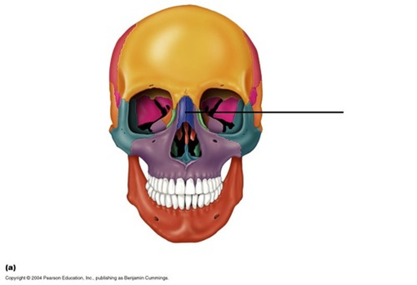
Ethmoid bone
separates nasal & cranial cavities

part of axial. 8
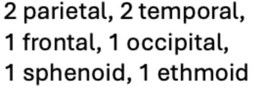
cranial bones
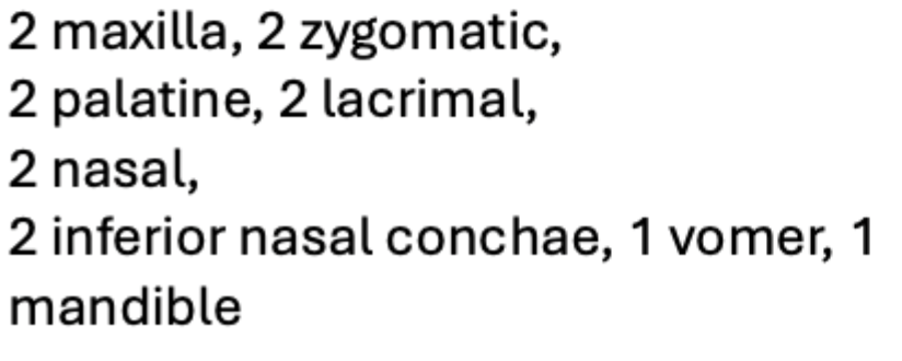
part of axial. 14
facial bones
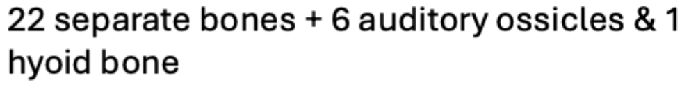
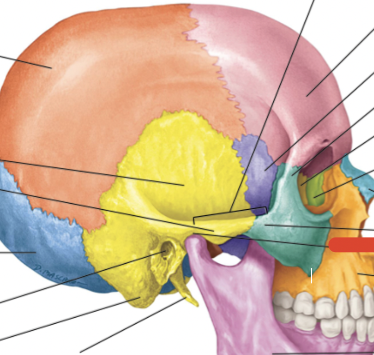
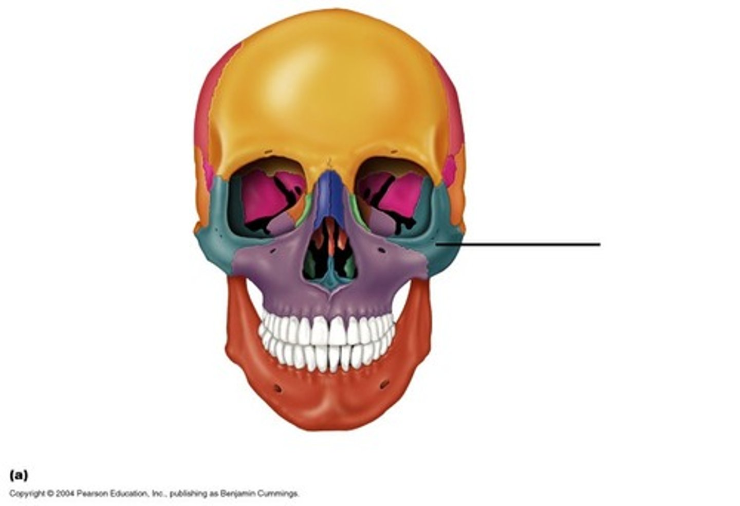
the skull bones
Hyoid Bone
Found below chin under the mandible
attachment point for tongue muscles
Attachment point for neck muscles that elevate larynx during speech & swallowing

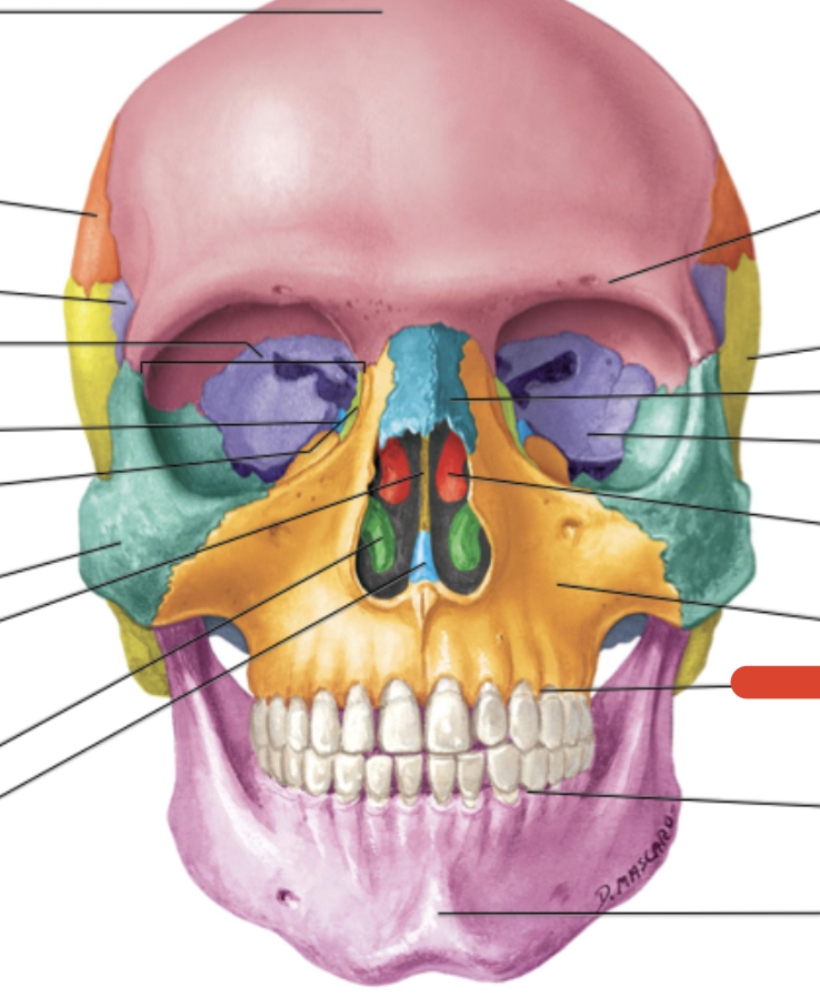
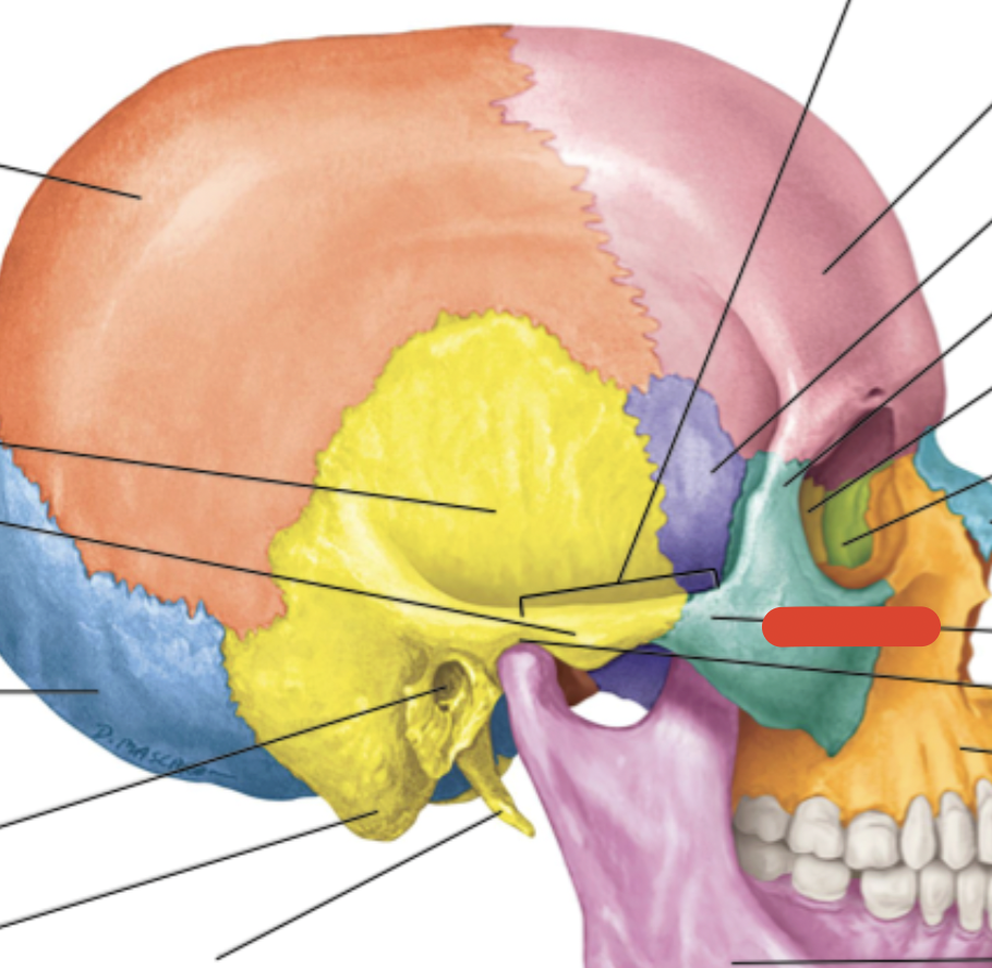
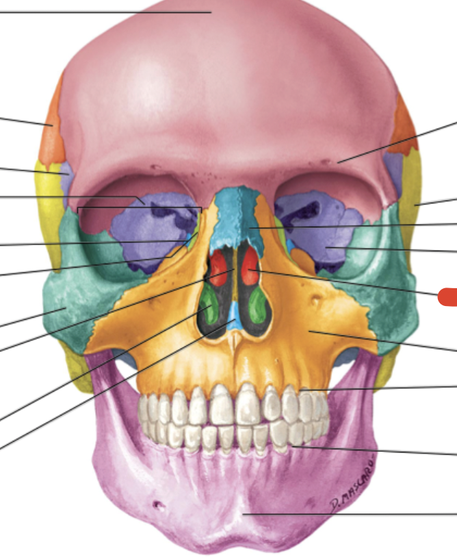
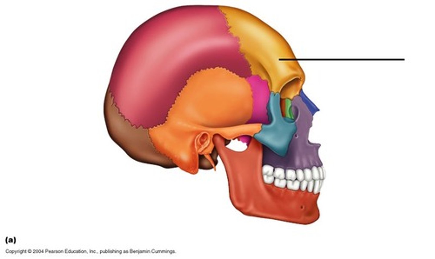
Frontal bone
Sphenoid bone
Zygomatic bone
Maxilla bone
Palatine bone
Lacrimal bone
Ethmoid bone
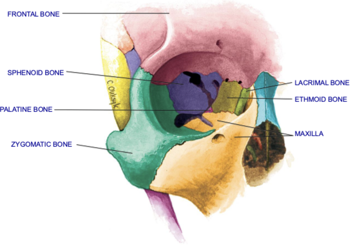
Bones of the Orbit - 7

Roof of orbit: frontal, sphenoid
Lateral wall of orbit: sphenoid, zygomatic
Floor of orbit: zygomatic, maxilla, palatine
Medial wall of orbit: sphenoid, maxilla, lacrimal, ethmoid
bones of orbit places
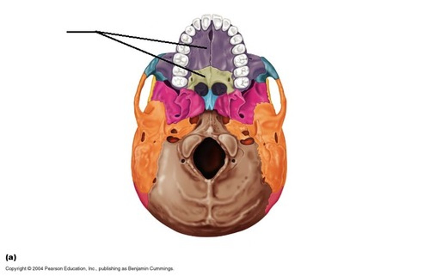
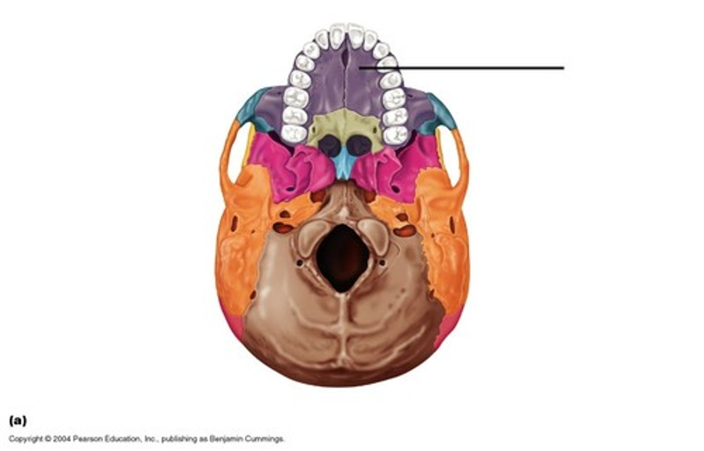
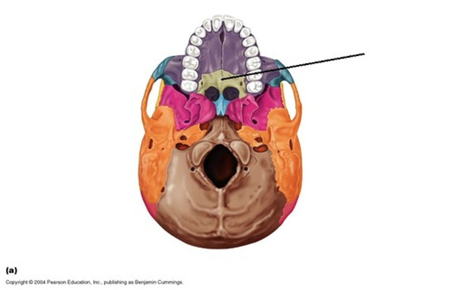
- Palatine process & maxilla
What makes up the “hard palate”?
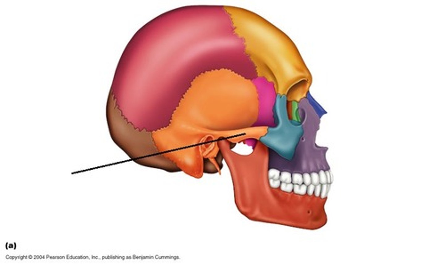
mandibular fossa
wehre Articulates with temporal bone
Alveolar process of maxilla
creates spaces so teeth fits into upper & lower jaw
temporal process
joins together with zygomatic process of temporal bone to form the zygomatic arch → cheekbone
Supraorbital margin
thickened region of frontal bone that protects region of orbit
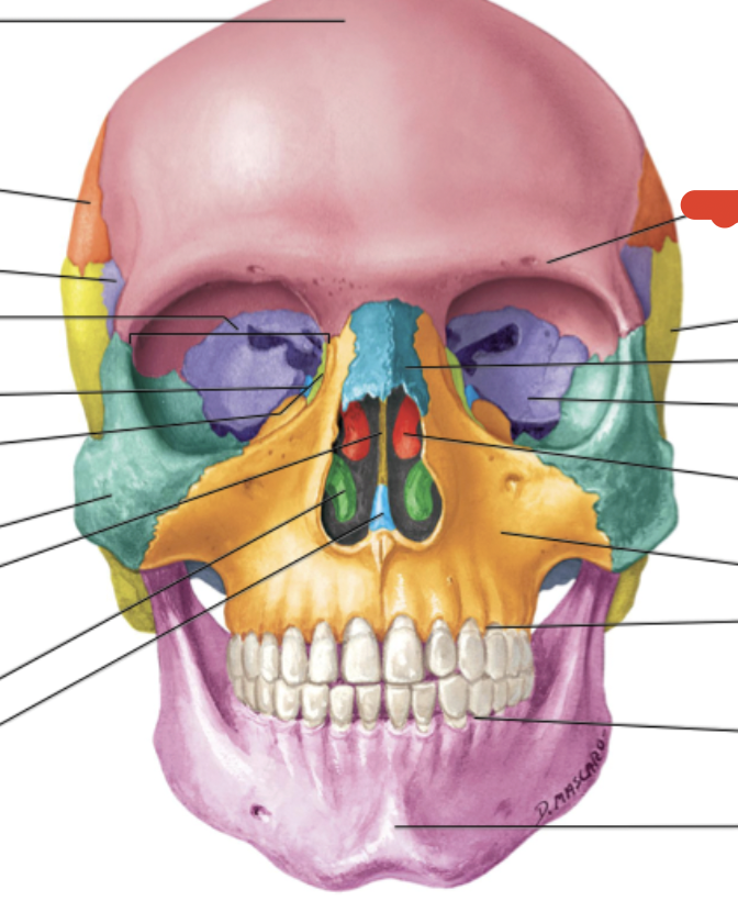
Inferior nasal conchae (2)
ridges - fhelps guide odorants to chemoreceptors & ridges trap air
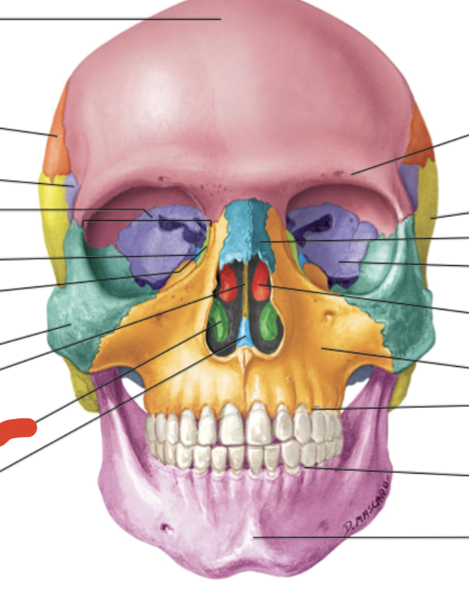
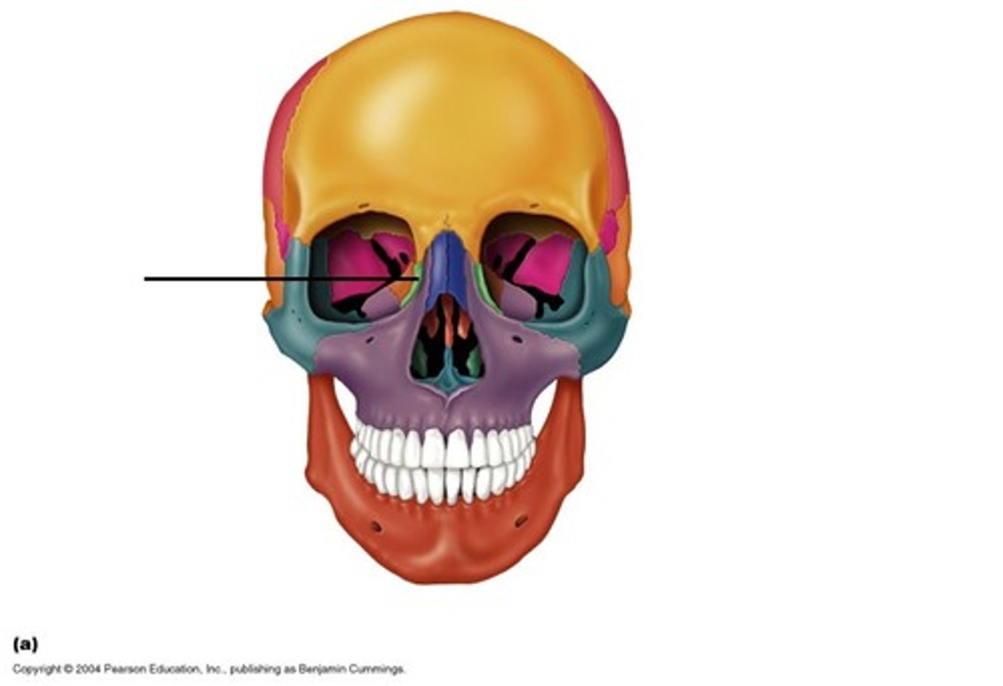
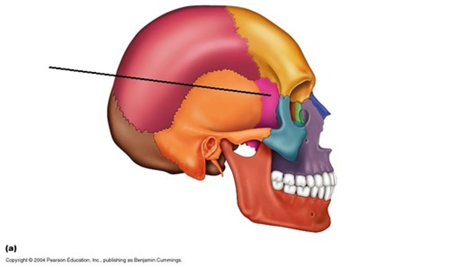
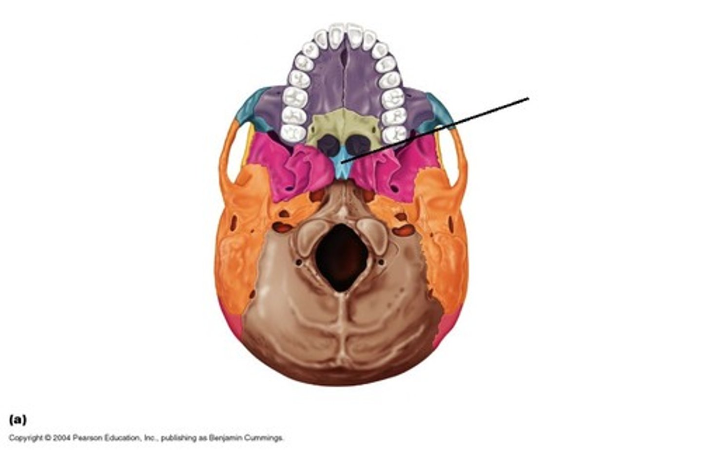
perpendicular plate of ethmoid bone
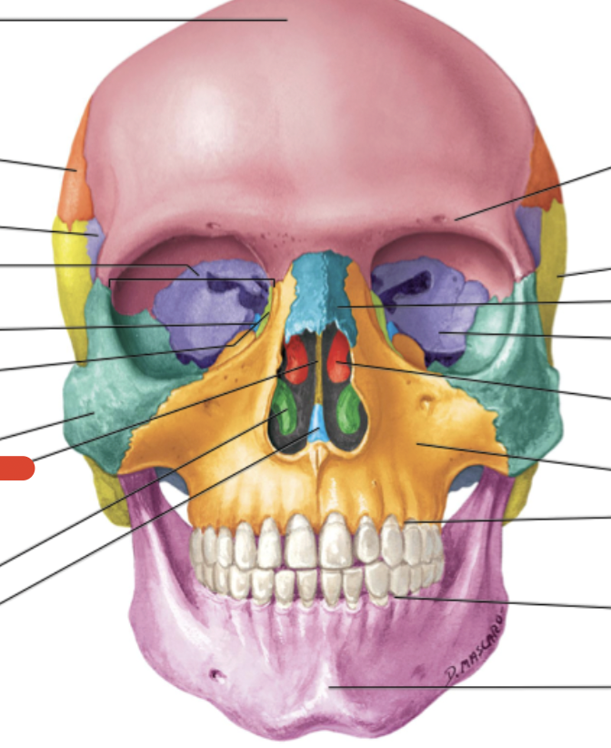
Middle nasal conchae & superior nasal conchae of ethmoid bone
Ethmoid bone
separates nasal & cranial cavities
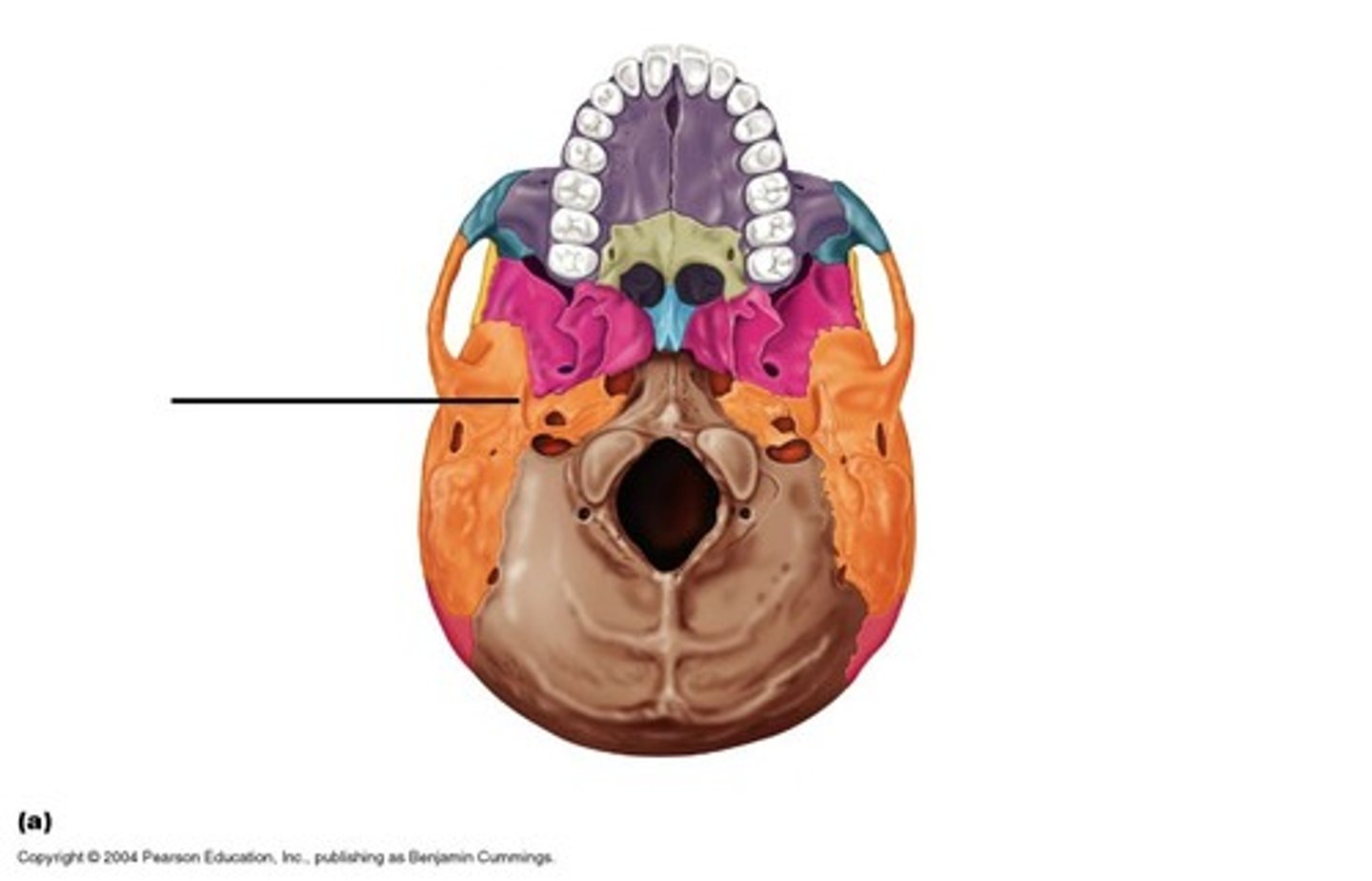
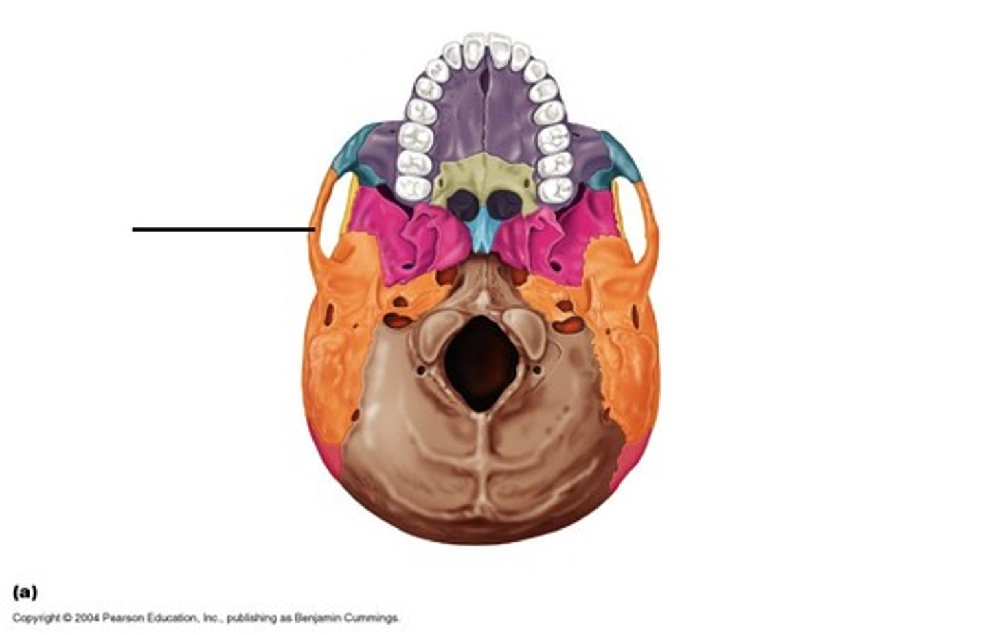
External Auditory Meatus

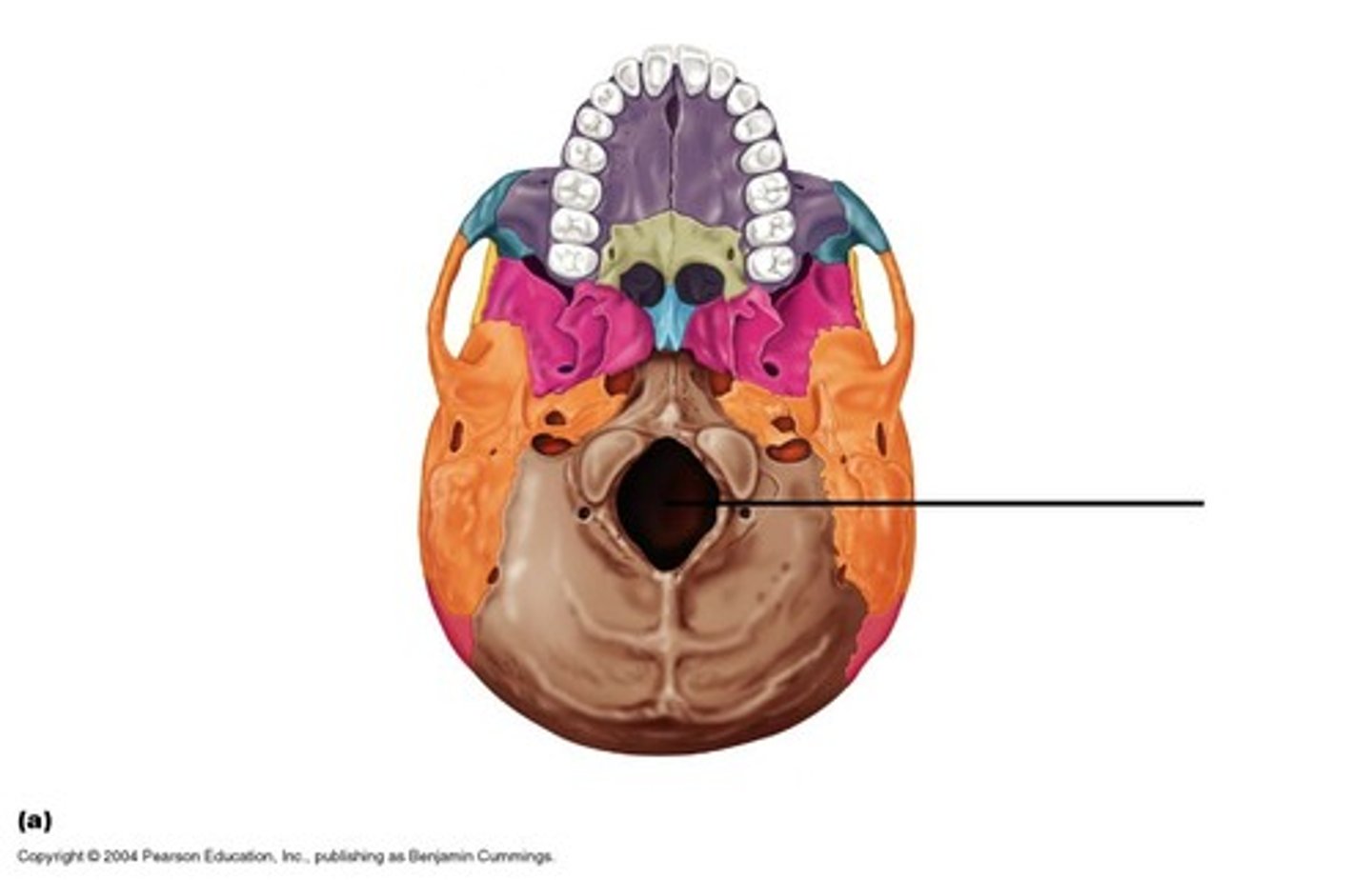
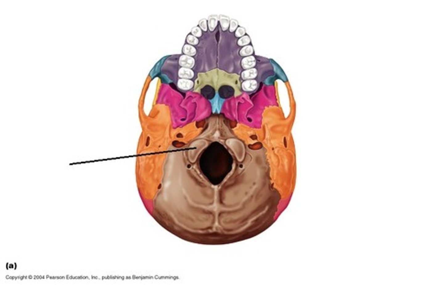
Foramen Magnum
spinal cord connects to brainstem in occipital
Frontal bone
Hard Palate
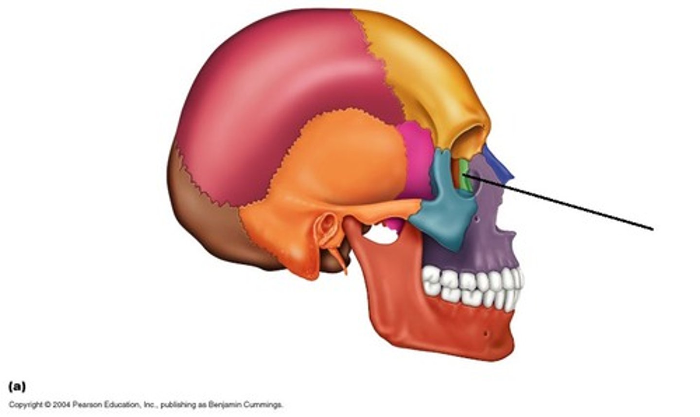
Lacrimal bone
smallest of facial bones & contain canal with lacrimal sac (eye orbit)
Lacrimal bone
smallest of facial bones & contain canal with lacrimal sac (eye orbit)

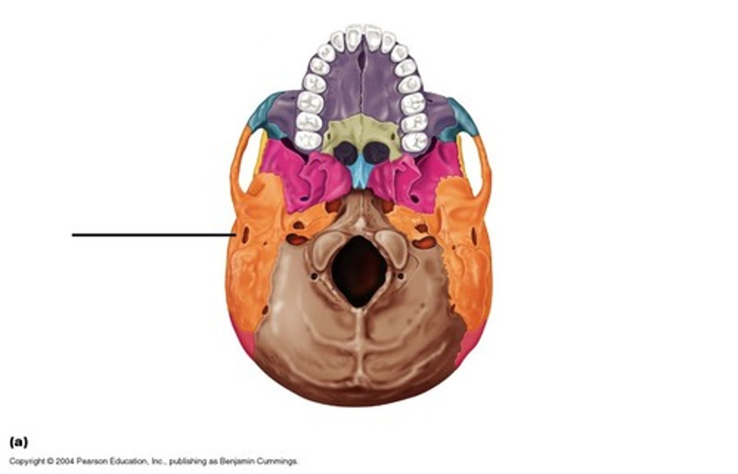
Mastoid process
attachment point for neck muscles

Maxilla
form upper jaw, floors of orbit, nasal cavity & hard palate (mouth roof)
Nasal bone
form the upper portion of bridge of nose & anterior roof of nasal cavity
Occipital condyle
two rounded bony features
Articulate with C1 vertebrae
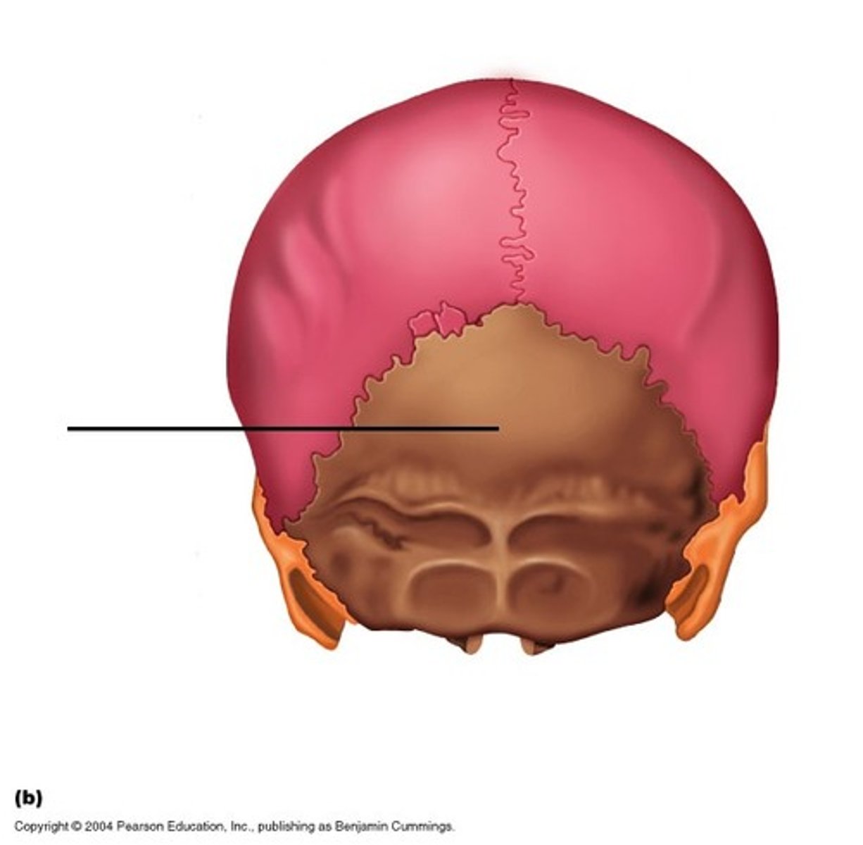
Occipital bone
posterior inferior portion – most base of cranium
Palatine bone
forms posterior portion of hard palate, floor of nasal cavity
L-shaped
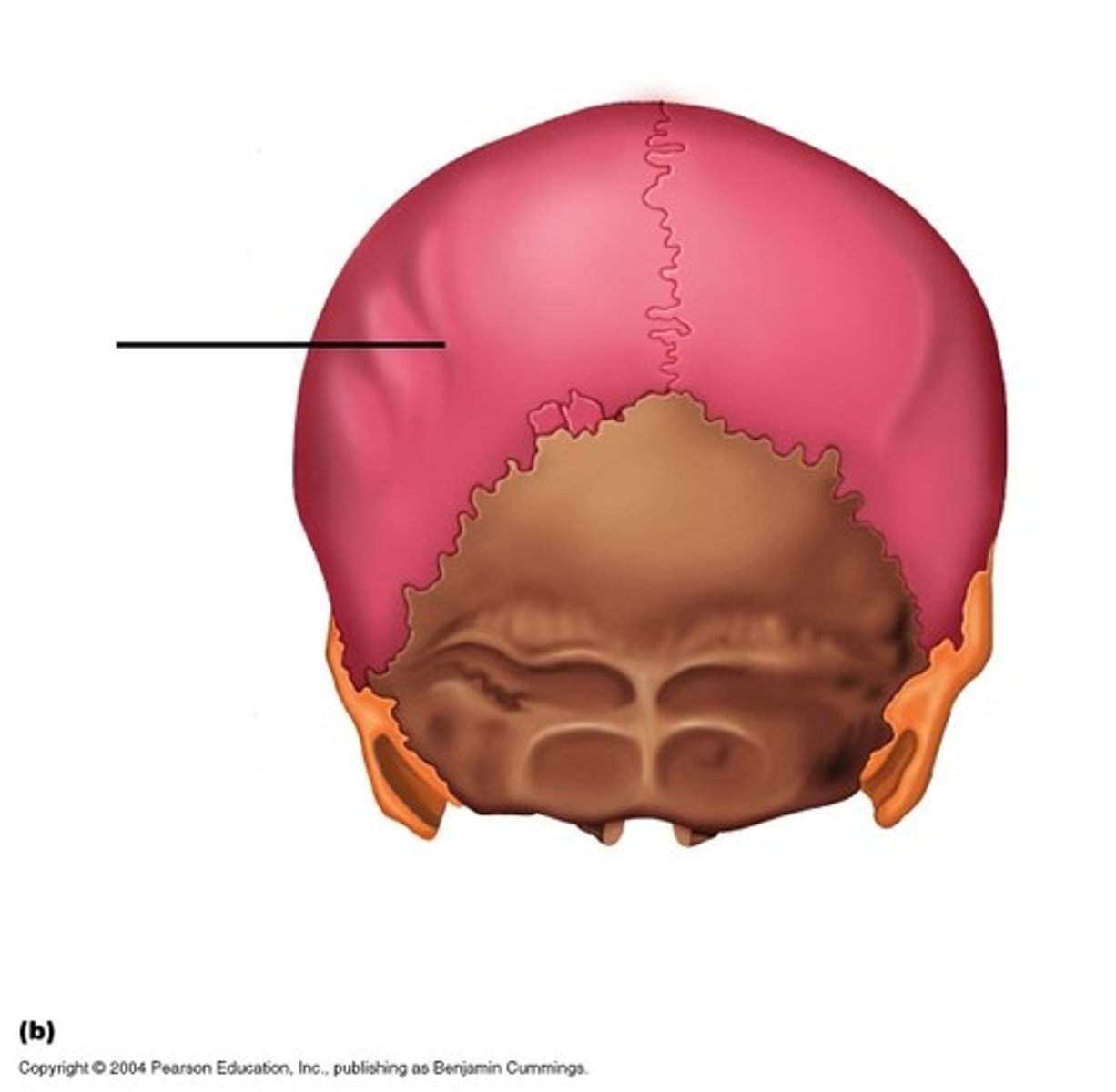
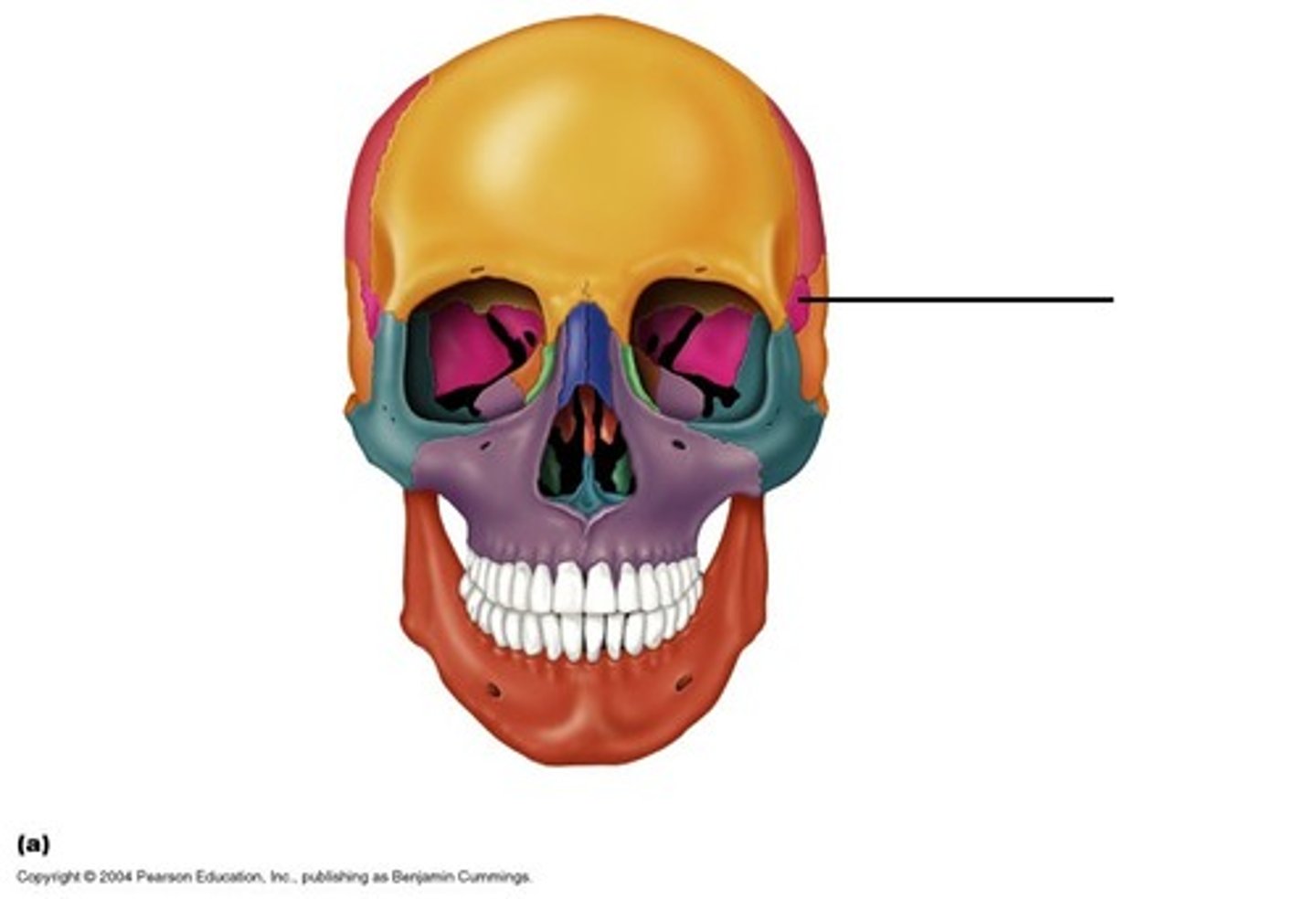
Parietal bone
most of sides & roof of cranial cavity
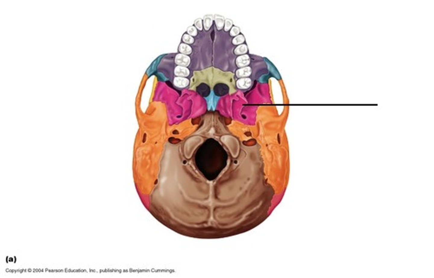
Sphenoid bone
portion of lateral skull & cranial floor & portion of orbit
Sphenoid bone
portion of lateral skull & cranial floor & portion of orbit
Sphenoid bone
portion of lateral skull & cranial floor & portion of orbit
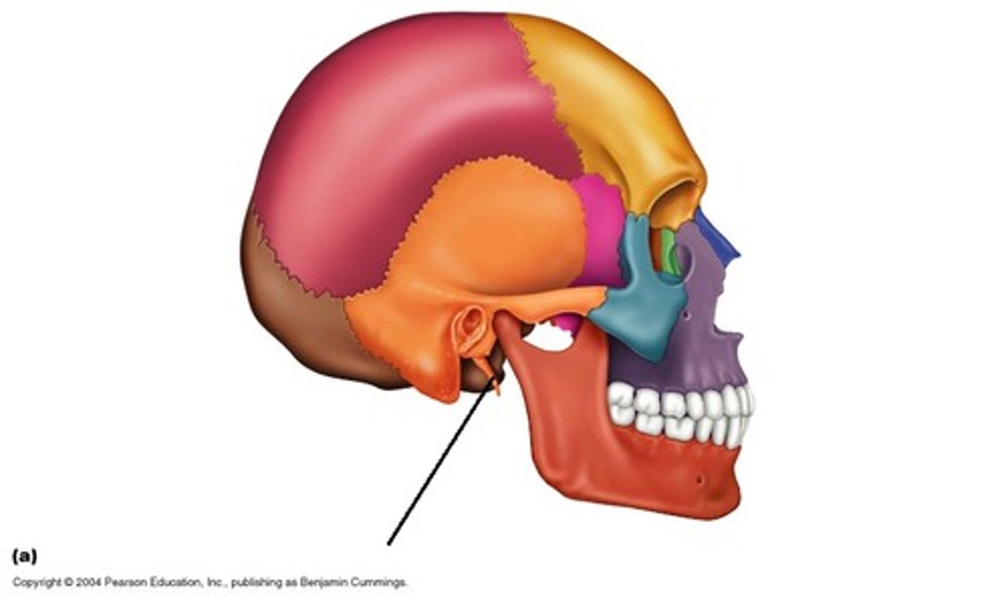
Styloid process
attachment point for neck & tongue muscles
Styloid process
attachment point for neck & tongue muscles
Temporal bone
Temporal bone

Vomer
forms inferior posterior region of nasal septum
Zygomatic process
makes up region of cheek bone
Zygomatic process
makes up region of cheek bone
Zygomatic bone
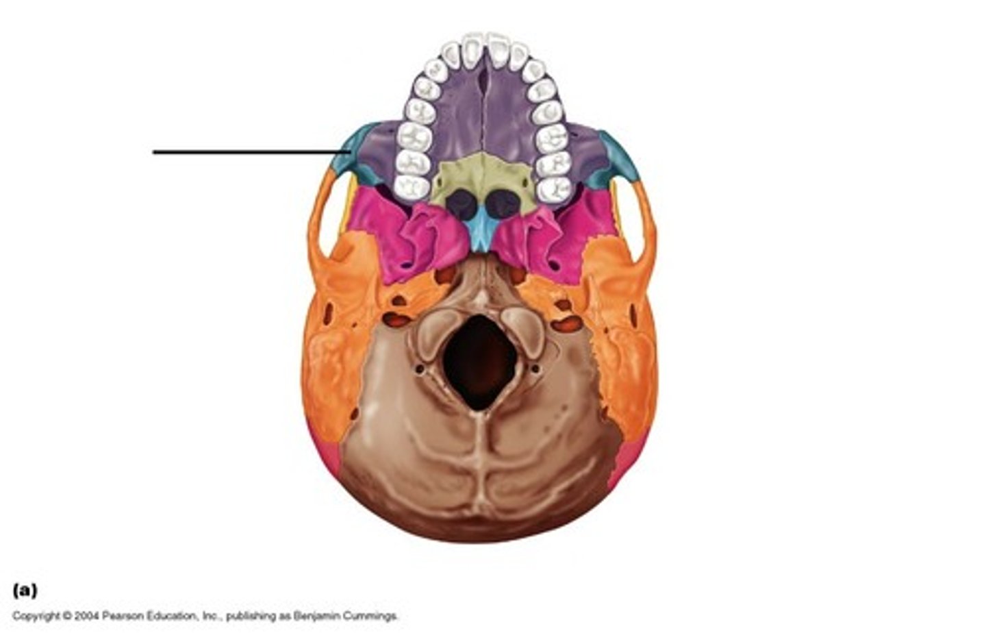
Zygomatic bone
Zygomatic bone

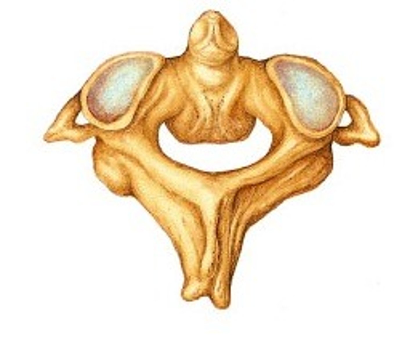
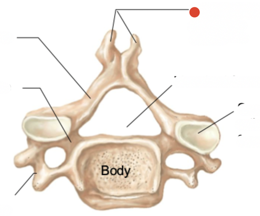
Axis vertebrae
second
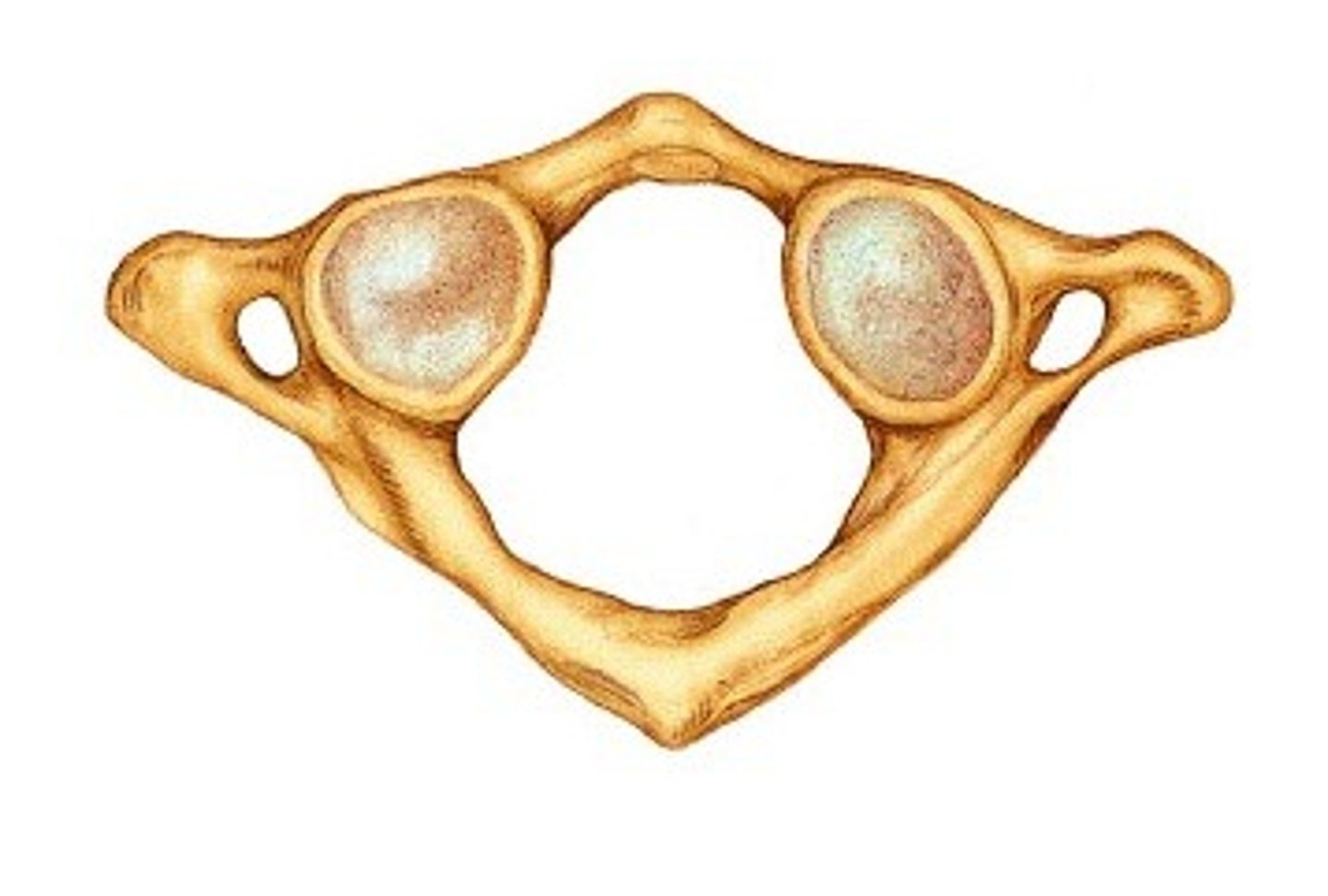
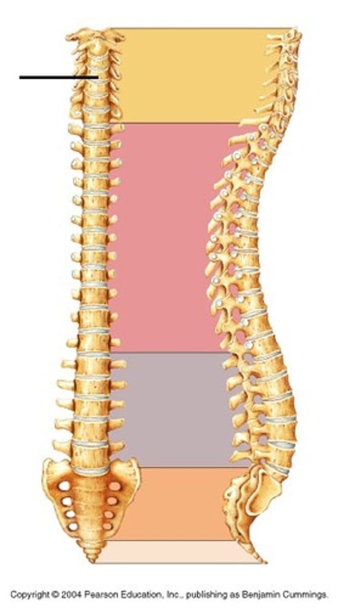
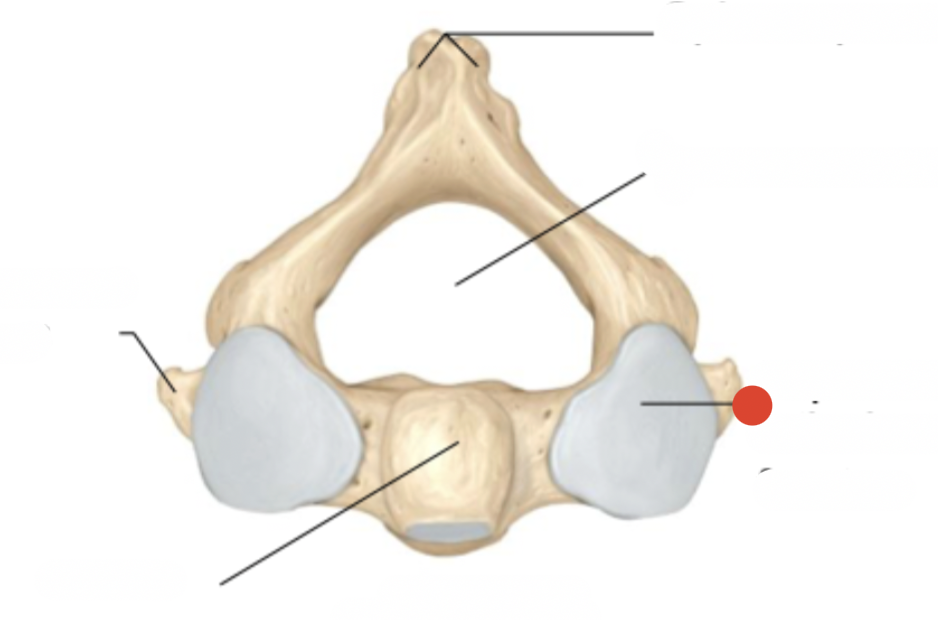
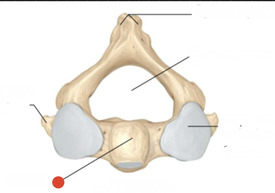
Atlas vertebrae
first cervical vertebra
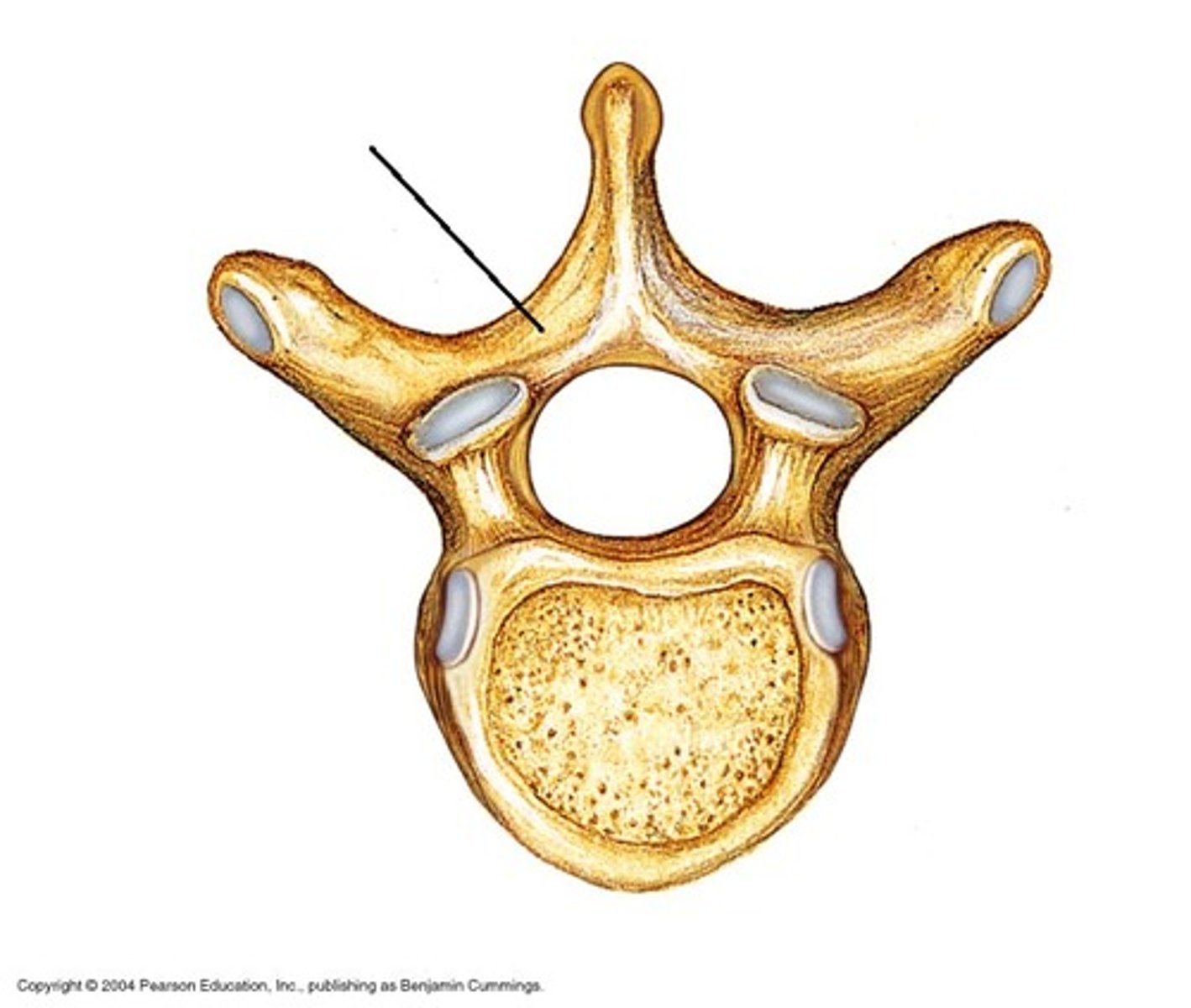
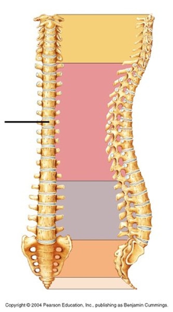
Thoracic vertebrae
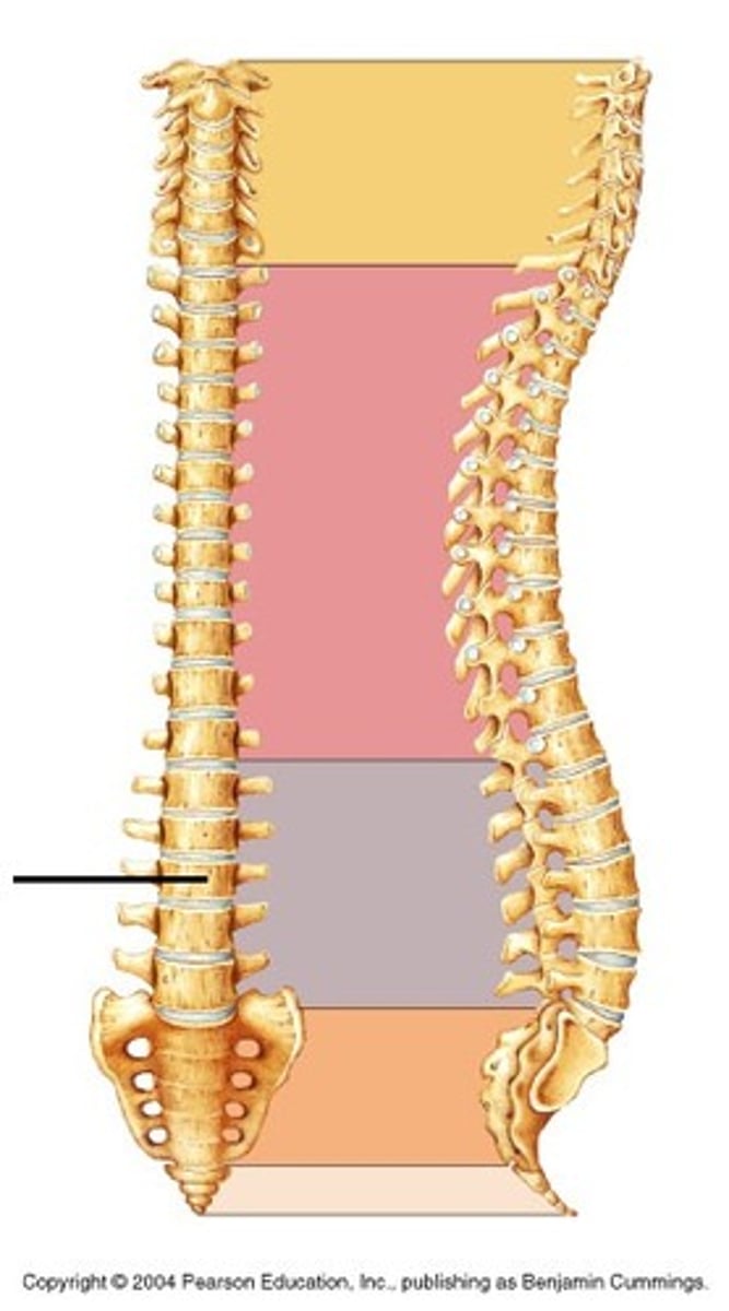
Lumbar vertebrae
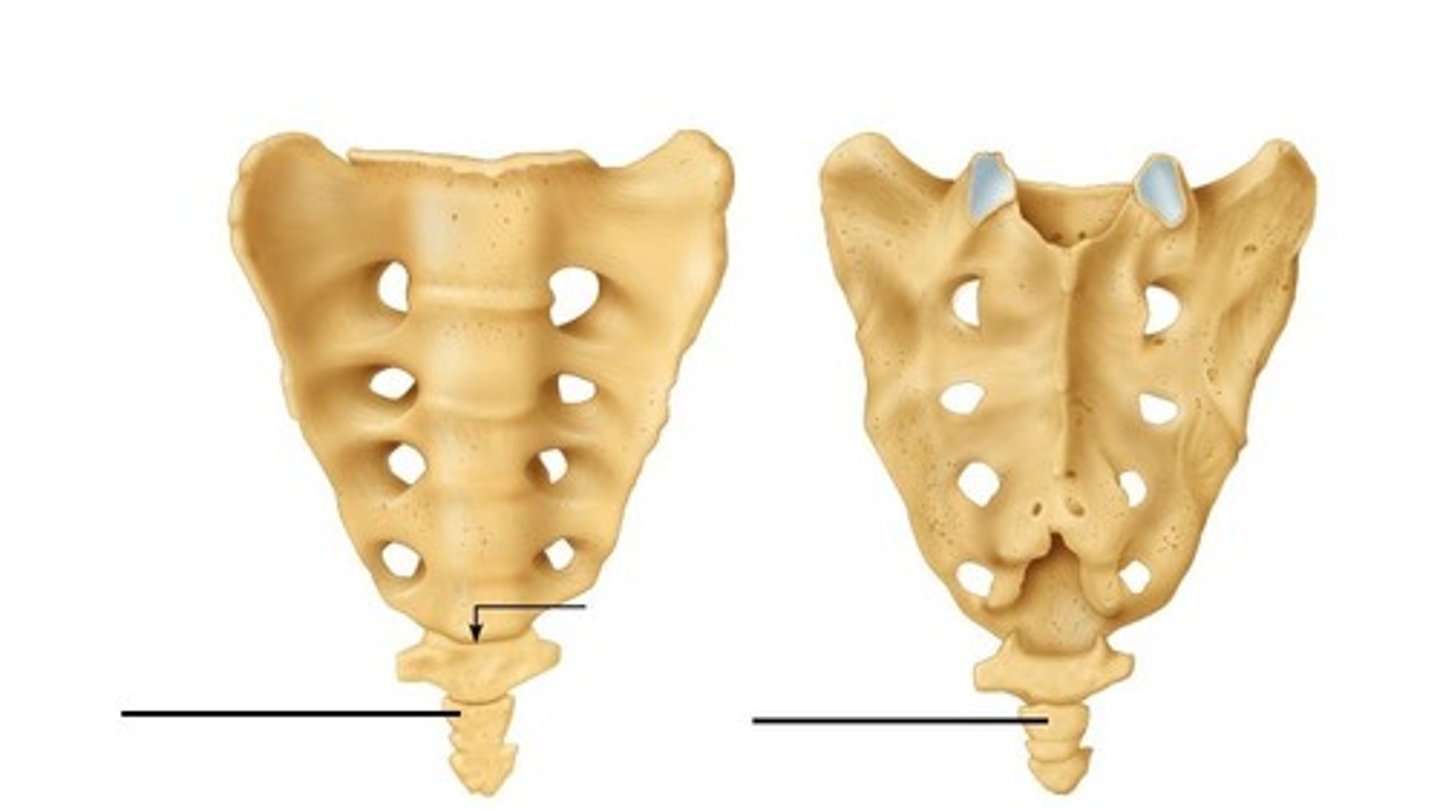
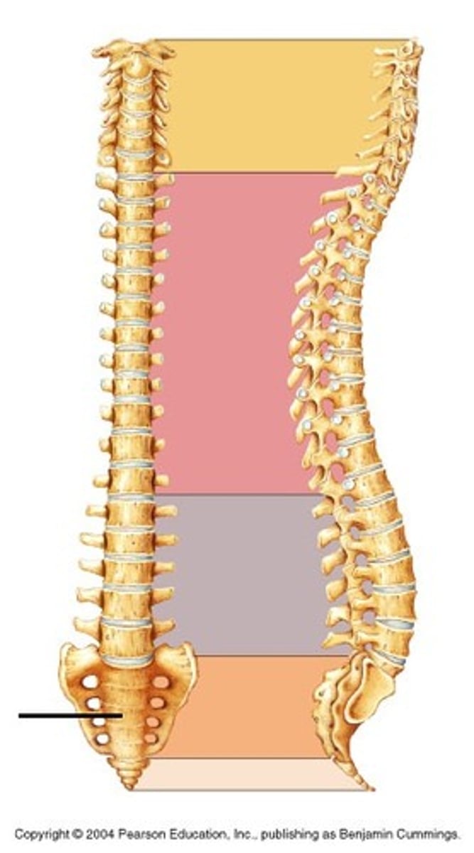
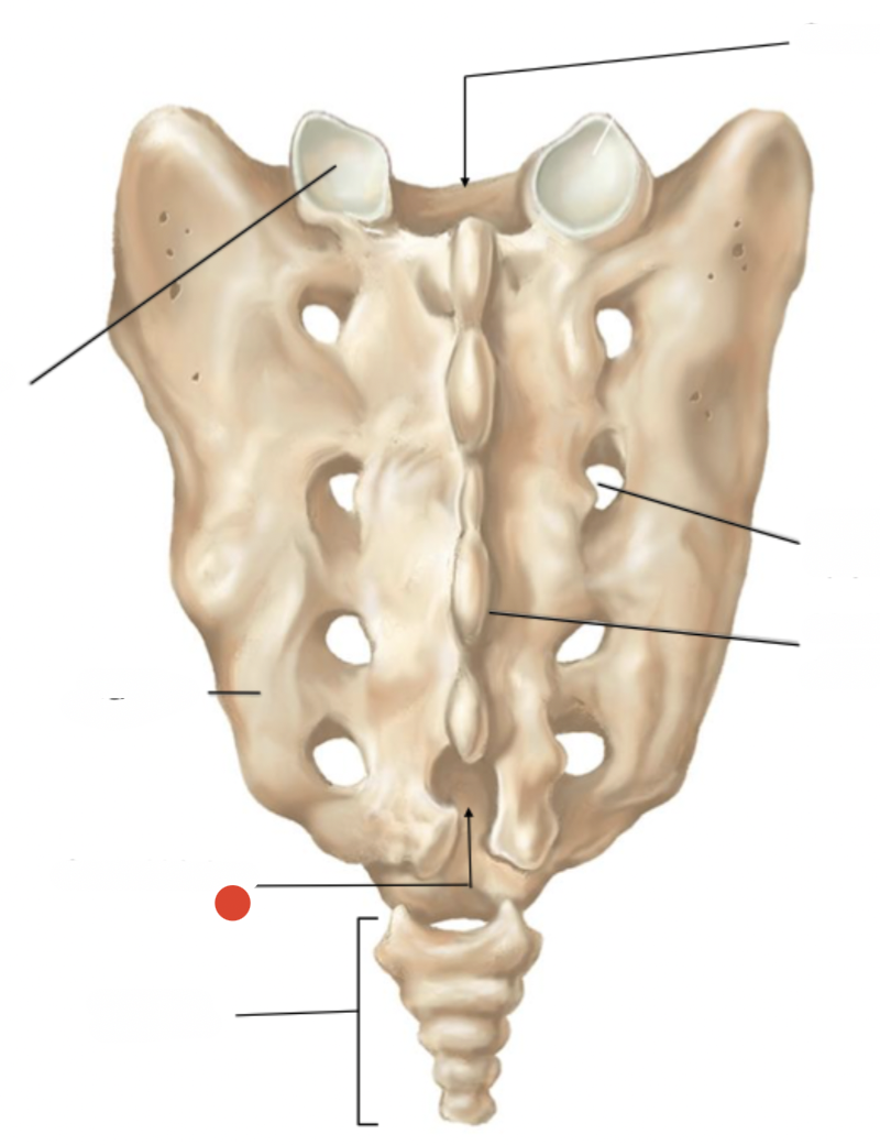
Sacrum
fusion of 4-5 by age 16-18
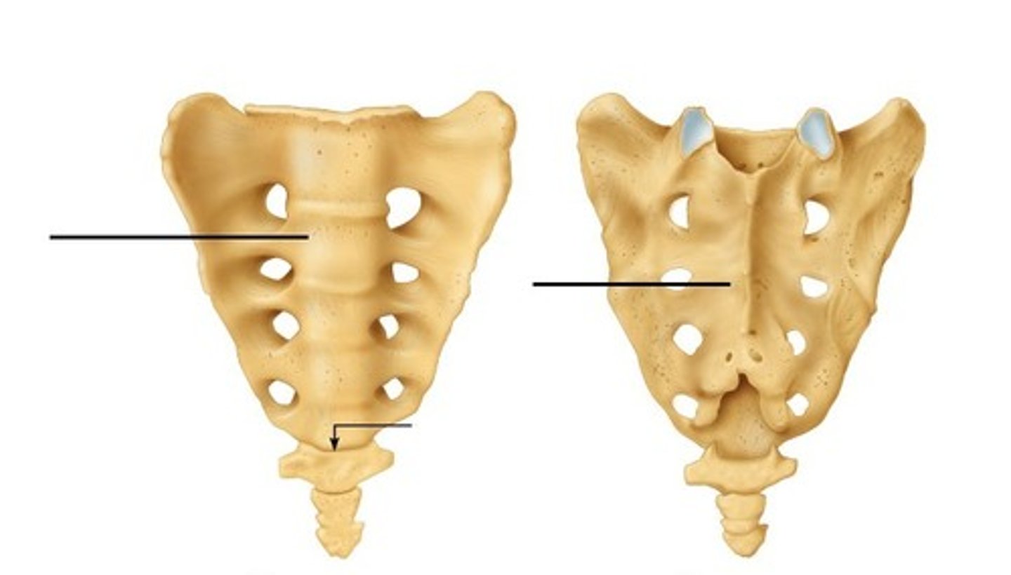
Coccyx
bones fused together. complete by age 20-30
attachement for filum terminale
Lamina
Spinous process

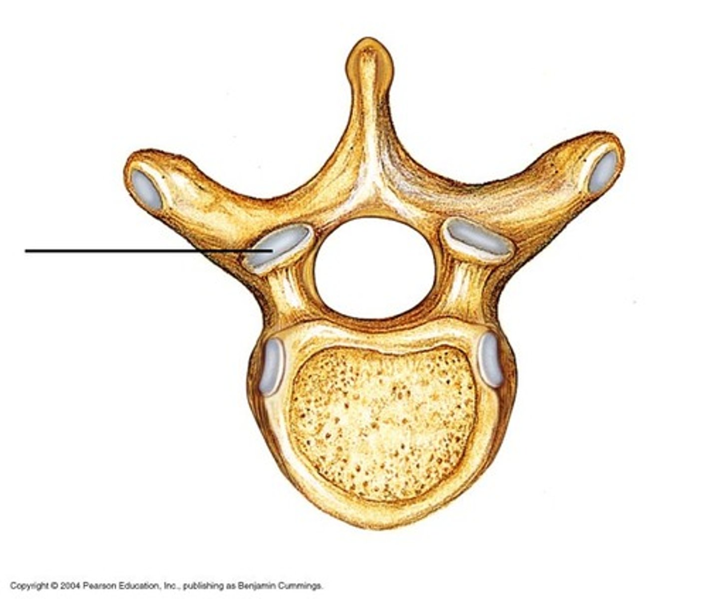
Superior Articular Cartilage
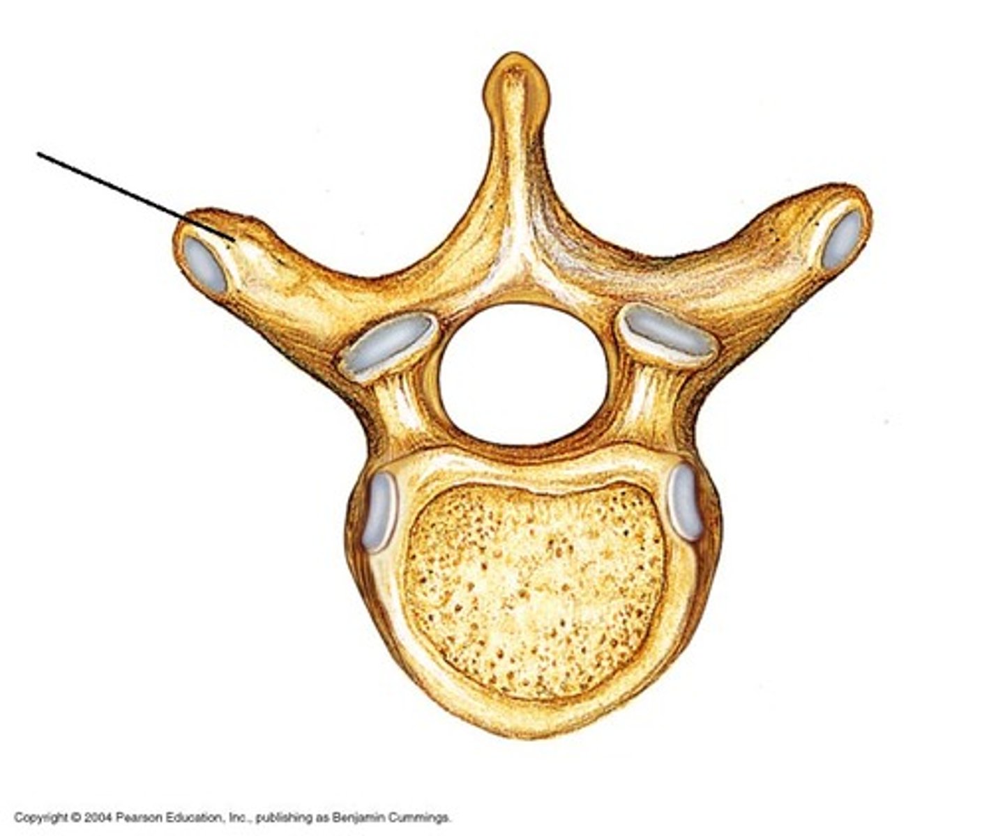
Transverse process
Vertebral foramen
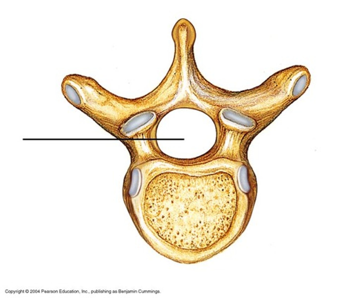
Vertebral arch

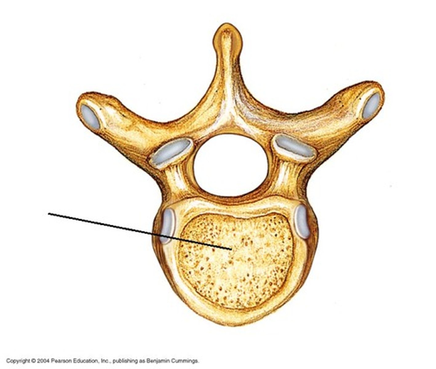
Body
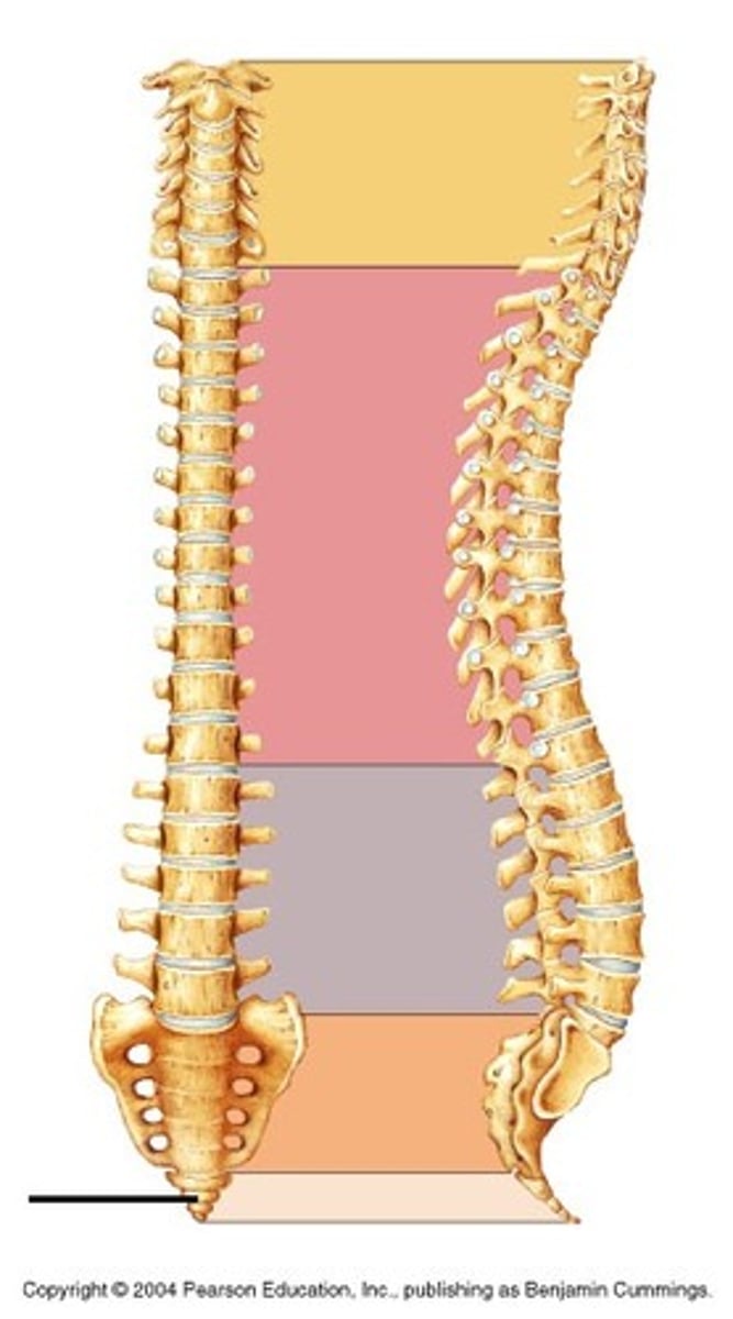
Cervical vertebrate (curvature)
Thoracic vertebrae (curvature)
Lumbar vertebrae (curvature)
Sacrum (sacral curvature)
Coccyx
Superior articular
facet (articulates
with occipital condyle)
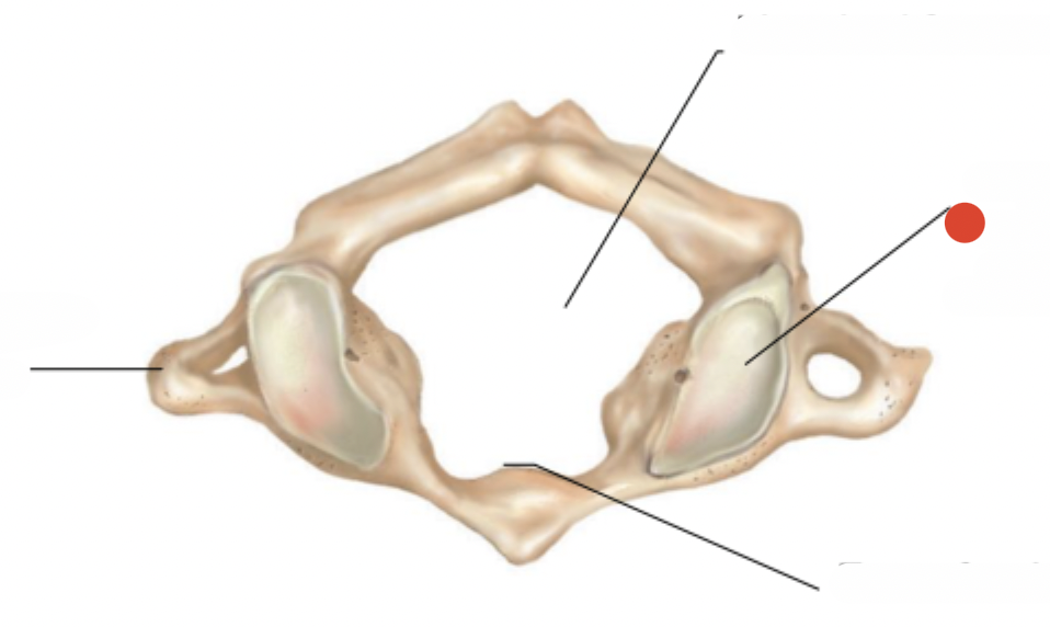
transverse of atlas
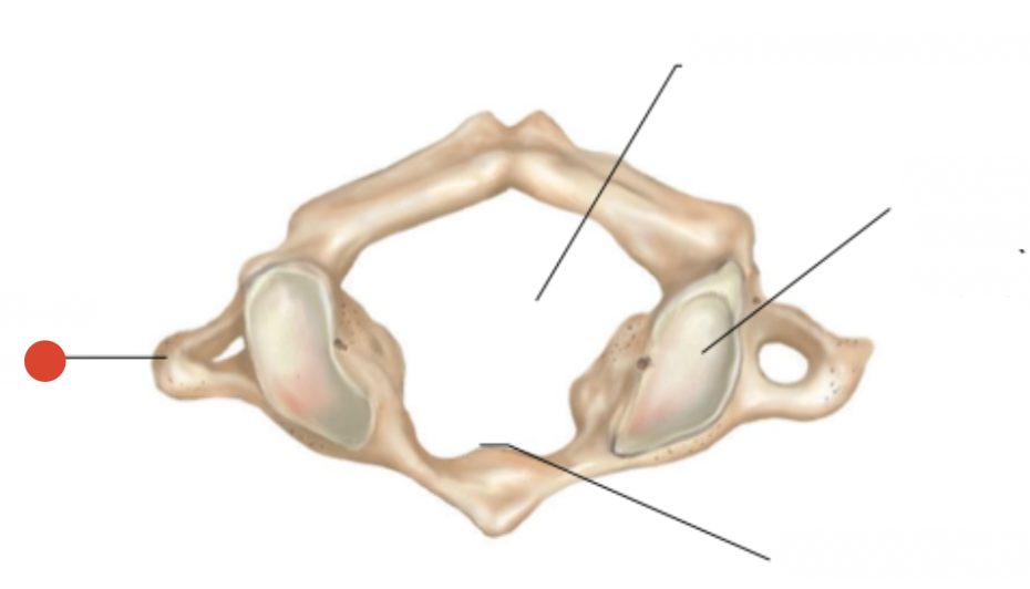
Vertebral foramen of atlas
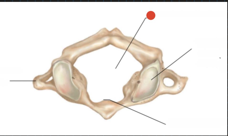
atlas facet for dens
forms joint with 2nd cervical vertebra

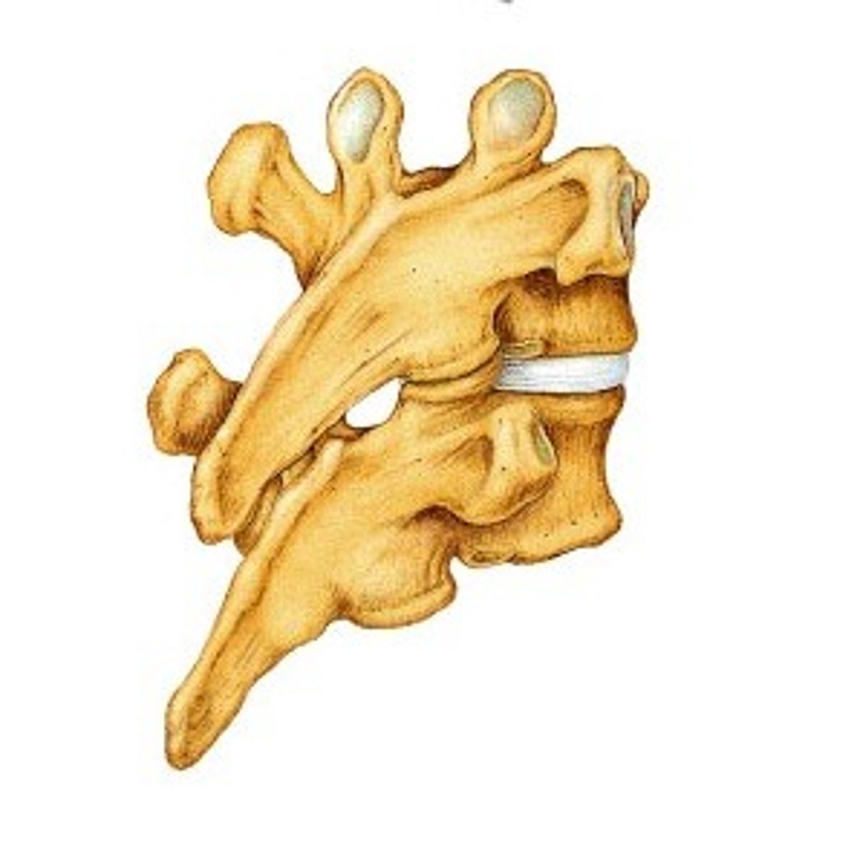
superior articular facet and under inferior
point directly up and down
flat because sit on top of each other
allows for large degree of movement
dens'
what atlas is connected to
so it can swivel
spinous process - bifid
unique to cervical
positioned at oblique angles to
lock together when stacked & provide greater stability
superior & inferior articular facet of thoracic
superior oriented medially
post laterally
movement limited
superior & inferior articular facet of lumbar
sacral canal
initially the vertebral foramen of the original 4-5 bones that make sacrum
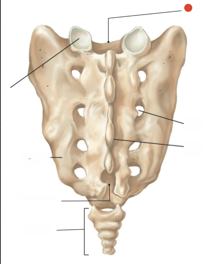
median sacral crest
orignally spinous processes
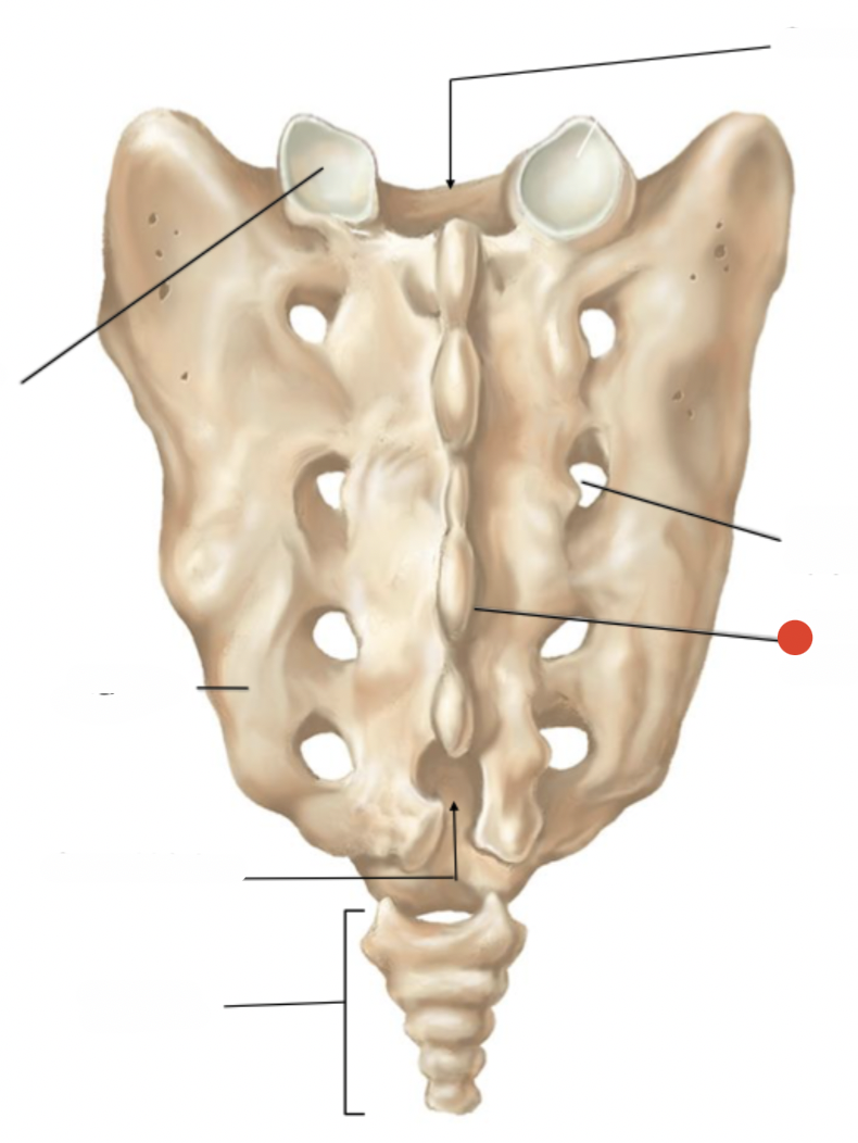
posterior sacral foramen
openenings to allow for exit of spinal nerves
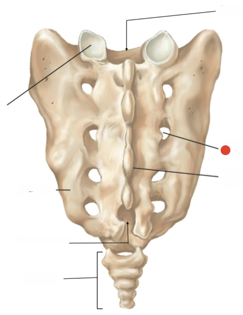
sacral hiatus
last bone has no spinous process
exit for last of spinal nerves
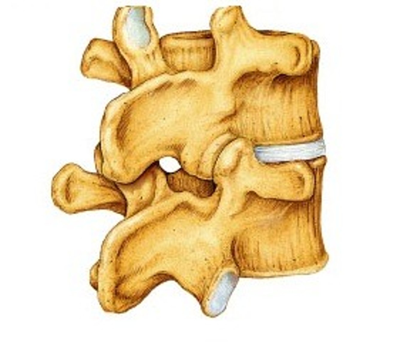
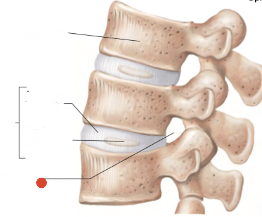
intervertebral foramen
space between bones
where spinal nerves exit
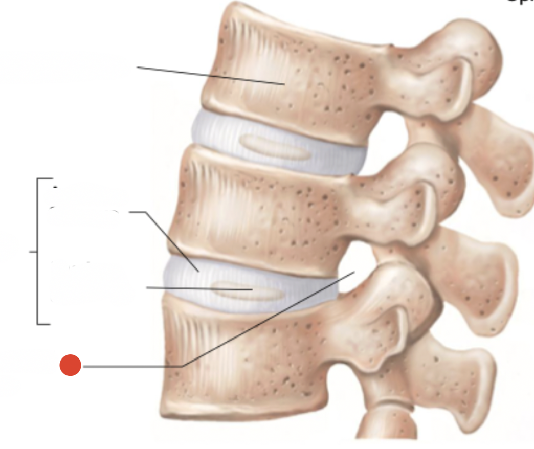
annulus fibrosus
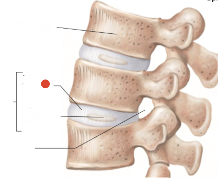
interverebral foramen
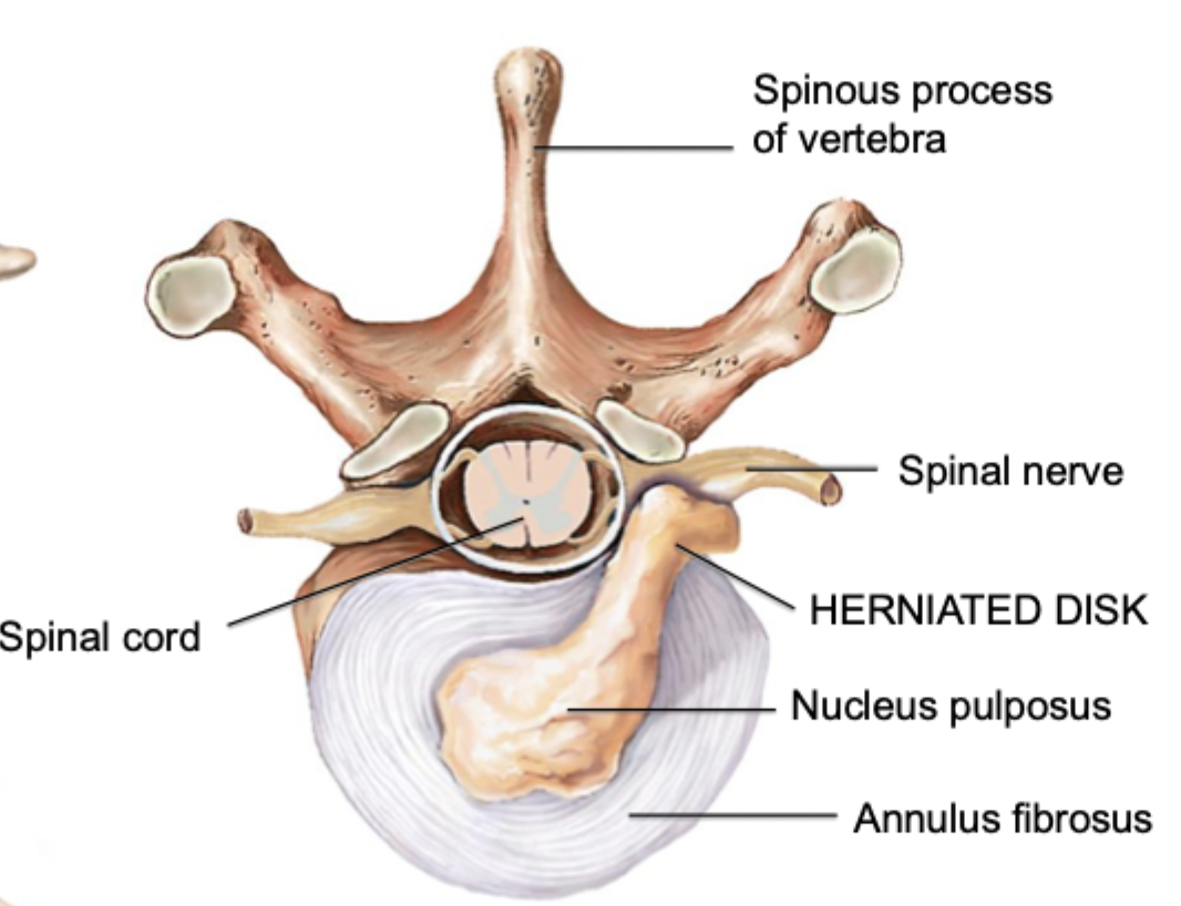
herniated disc
cartilage breaks and nucleus pulposus comes out of intervertebral foramen
towards back side
pushes on spinal nerve
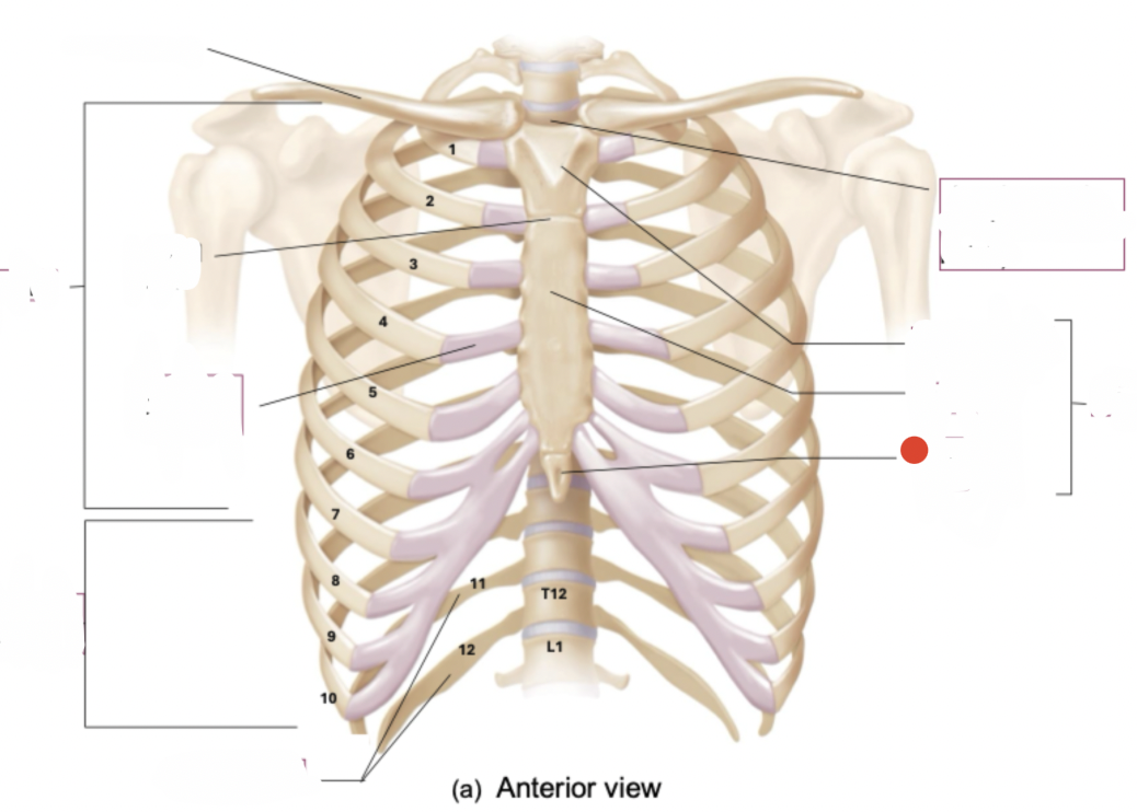
protect heart and lungs
semi rigid bcs bones in it are semi moveable
bcs they form joints with other bones
have cartilage
Thoracic vertebrae, ribs and costal cartilages, and sternum
thoracic cage
12 pairs
7 pairs true ribs
ribs
7 pairs are true ribs - vertebrosternal ribs
vertebrae to sternum
true ribs
5 pairs - “false ribs”
3 pair are vertebrochondral ribs (connect to sternum indirectly by connecting to costal cartilage of rib 7)
2 pair are “floating” or vertebral ribs
false ribs
manubrium and sternal angle (angled towards from front as move from top to bottom)
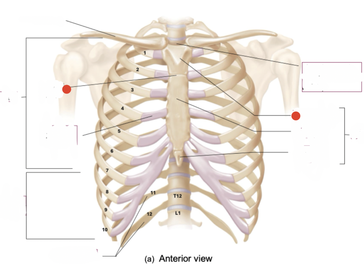
body
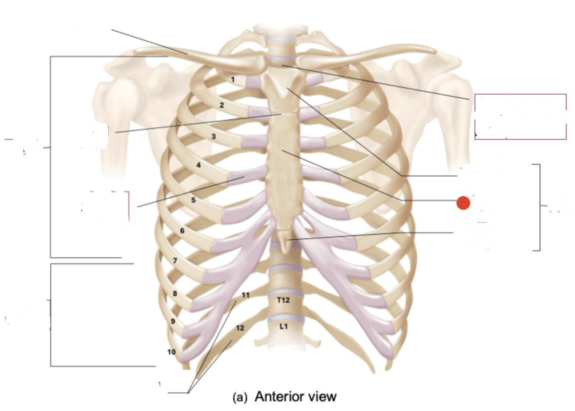
xiphoid process
made of cartilage until youre 40
bump on back of your skull that joins to ligaments which hold your head upright
External occipital protuberance