Micro Lab Quiz: Staining
1/47
There's no tags or description
Looks like no tags are added yet.
Name | Mastery | Learn | Test | Matching | Spaced |
|---|
No study sessions yet.
48 Terms
Why do we stain cells?
To increase contrast, bacteria have a refractive index similar to water, making them nearly invisible.
To identify different types of cells or different cells (differential stains).
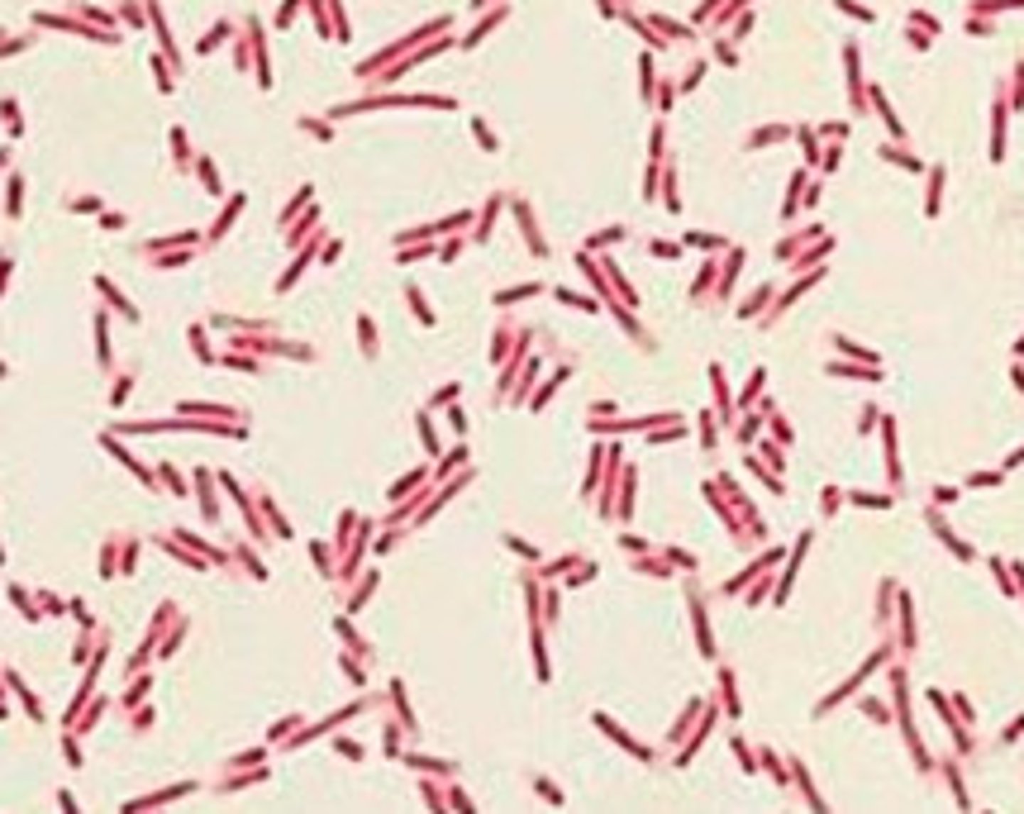
What is the shape of this cell?
Bacillus (Rod)

What is the shape of this cell?
Coccus (Sphere)
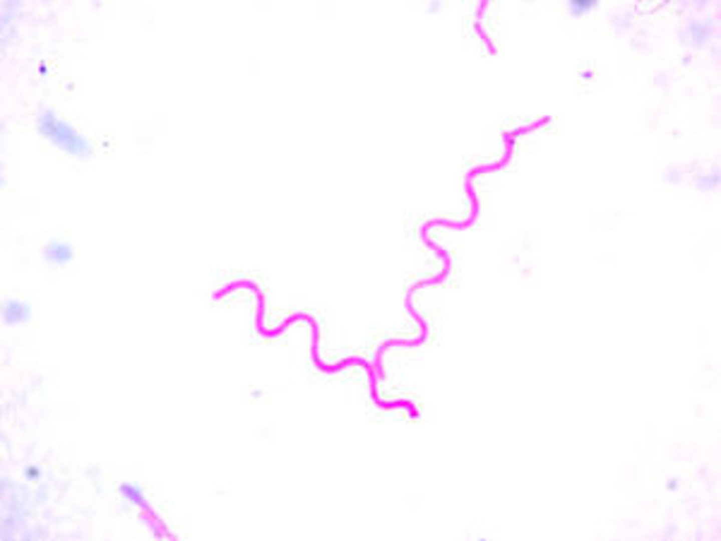
What is the shape of this cell?
Spirilla (Spiral)
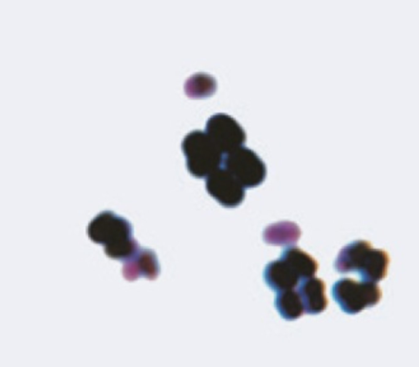
What is the arrangement of this cell?
Diplococci
Divides in one plane and remains attached after cell division.

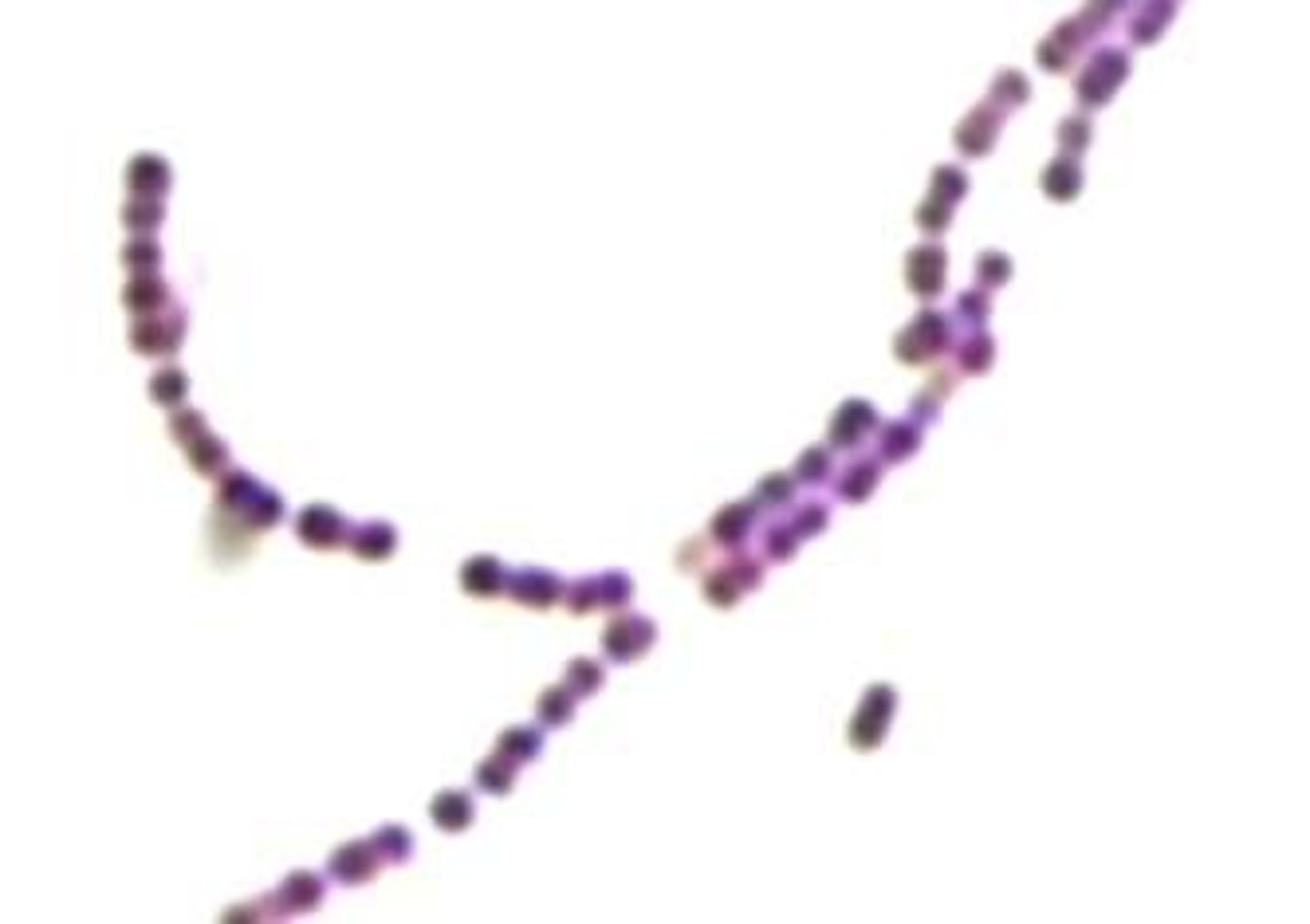
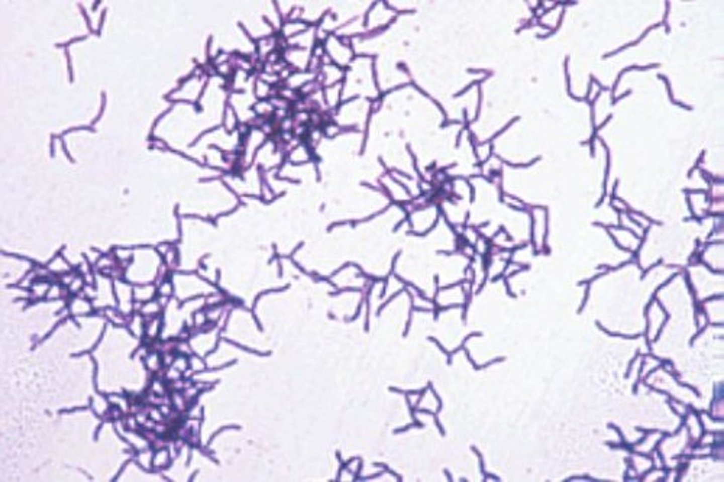
What is the arrangement of this cell?
Streptococci
Divides in one plane and forms long chains of attached cells.
What is the arrangement of this cell?
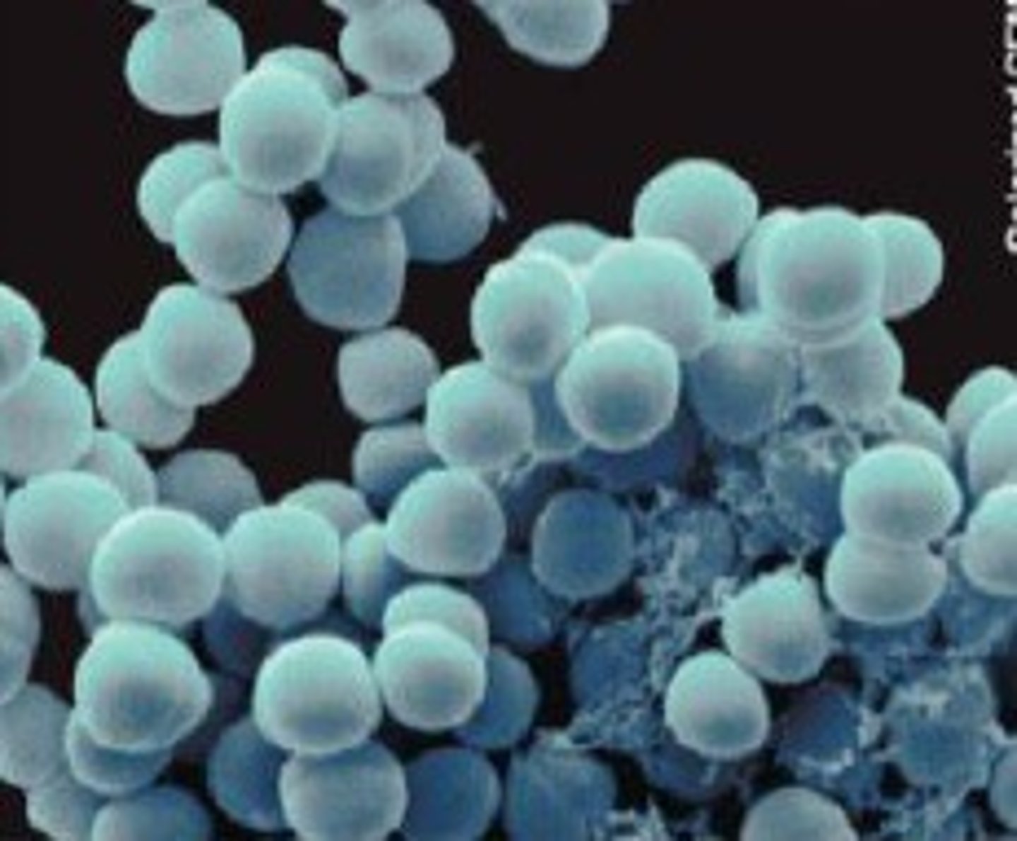
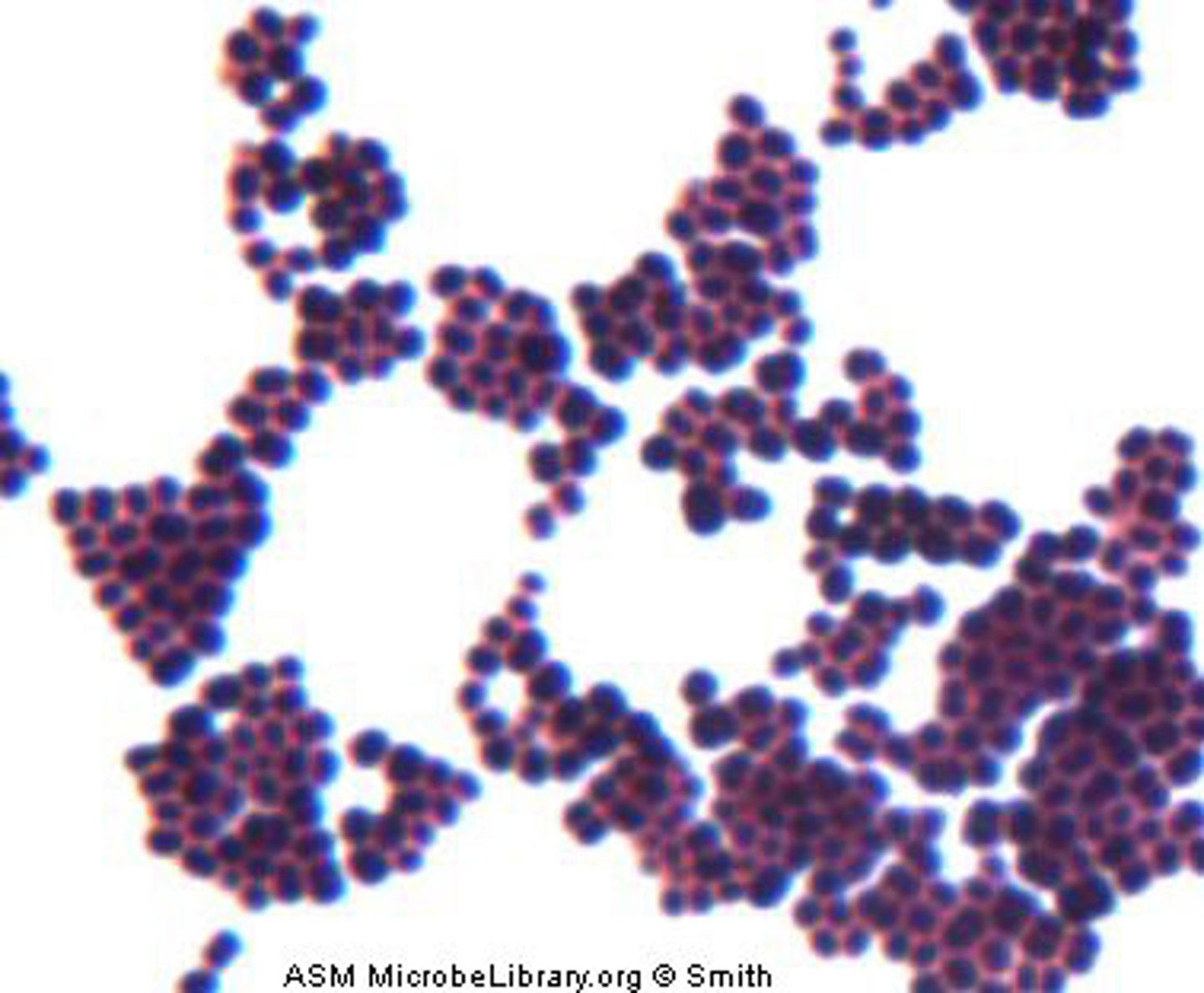
Staphylococci
Divides in many planes and remains attached, forming "grape like clusters".
What is the arrangement of this cell?
Tetrad
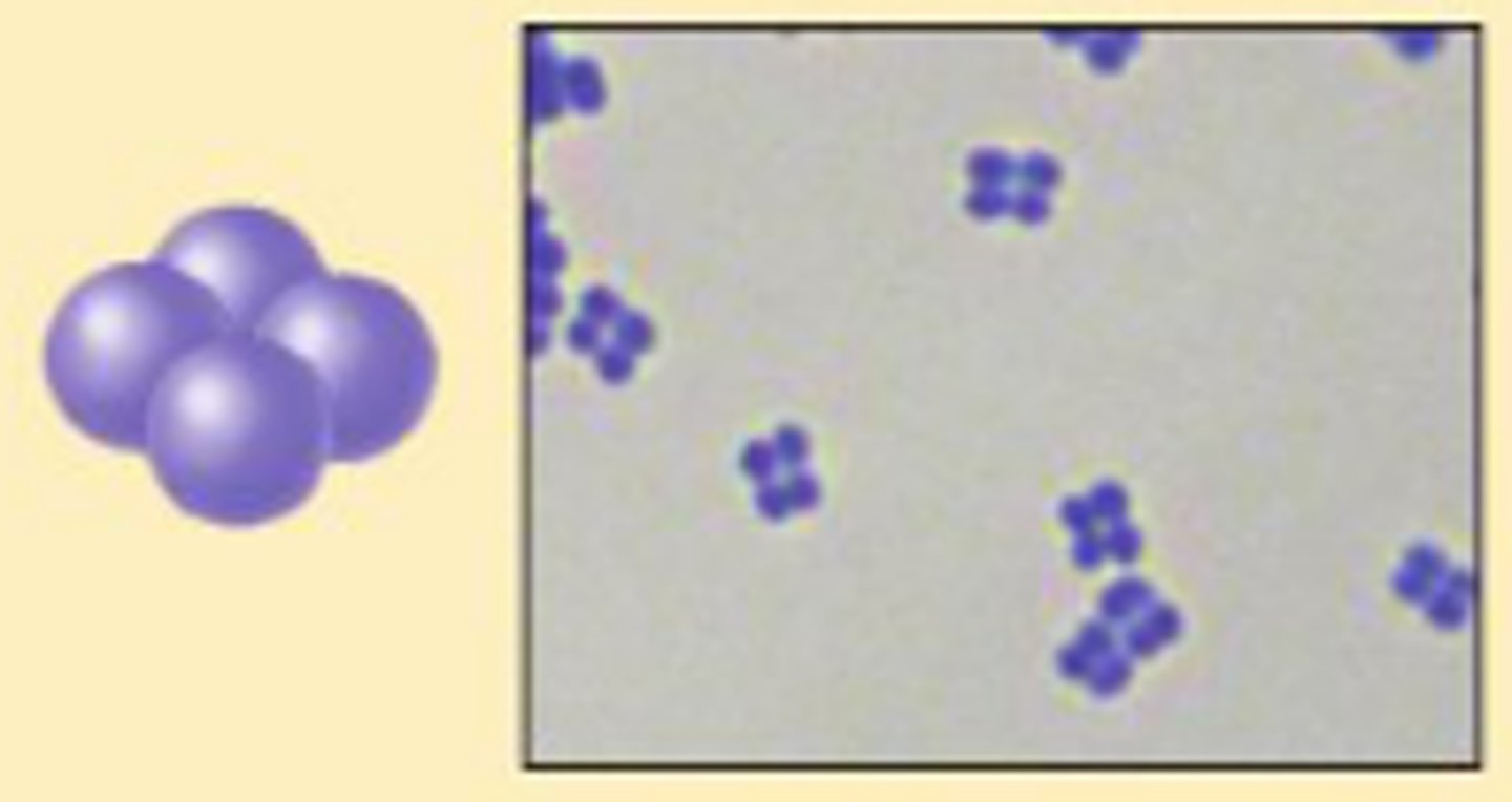
What is the arrangement of this cell?
Sarcina
Pleomorphism
Some bacterial species can show slight deviations in their overall shape and/or size.
Some bacteria alter their morphology, biological functions, or reproductive modes in response to environmental conditions.
What is the charge of bacteria's cell surface?
Negative
Negative Stain (aka acidic stain)
The auxochrome has a negative charge causing like charges to repel.
The auxochrome cannot bind to the bacterial cell surface, causing there to be no association between the bacteria and the chromophore.
The bacteria are left colorless, and the background is stained.
What are the pros and cons to positive stains?
Pros- only requires one dye, uncomplicated procedure.
Cons- cannot differentiate unique cell structures or features between bacteria.
Positive Stain
The auxochrome has a positive charge (opposites attract).
The auxochrome binds to the bacterial cell surface. The chromophore is attached to the auxochrome.
The bacteria are stained, and the background stays white.
What are some examples of basic dyes (positive)?
Methylene blue, safranin, and crystal violet.
What are some examples of acidic dyes (negative)?
Eosin, acid fuchsin, and nigrosin (also india ink).
Simple Stain
1. Prepare a heat-fixed smear of the bacteria.
2. Flood the slide with a basic dye.
3. Stain for 1 minute.
4. Briefly rinse the stain from the slide using a slow stream of water from the squirt bottle.
5. Carefully blot the slide dry using blotting paper.
What are the characteristics of a good smear of bacteria for staining?
White space between cells to make arrangement obvious.
A thin, even layer of bacteria. No clumping. Looks like "dried shower scum".
What do you need to do to make a good smear (also positive stain protocol)?
1. Label a slide.
2. Place a drop of water on the slide.
2. Sterilize a culture loop.
3. Pick up bacteria from an isolated colony (a small portion of a large colony or all of a small colony).
4. Mix the bacteria with the water using the sterile loop.
5. Spread the mixture over the surface of the slide in a thin layer.
6. Air Dry
7. Heat Fix
8. Stain and dry.
Negative Stain Procedure
1. Use ethanol on a paper towel to wipe the slide clean of dust.
2. Place a drop of stain on one end of the slide.
3. Add a small amount of bacteria to the drop of stain and mix well until the bacteria is evenly dispersed.
4. Rest one end of a clean slide on the center of the sldie with the stain. Tilt the clean slide at an acute angle and draw the slide towards the stain.
5. Once the clean slide touches the drop of stain it will cause it to spread along the edge of the clean spreader slide.
6. Keeping the spreader slide at an acute angle push the spread slide towards the other end of the slide to be stained.
7. Once the spreader slide has been pushed all the way across the slide, the thin film on the stained slide should be left to air dry.
8. Once dried the slide is ready to be examined under the microscope, the slide should NOT be heat fixed.
9. Discard slide into the pokey bucket.
Why do we heat fix? What important things does heat fixing do for us?
It kills the bacteria and secures the dead organisms to the slide (can distort the bacterial shape).
How long should you heat fix a slide for?
20-30 seconds
What are the common shapes of bacterial cells?
Coccus, bacillus, and spirilla.
What are the common arrangements of bacterial cells?
Singles, pairs, clusters, chains, and palisades.
Do certain types of cells only have certain types of arrangements?
Yes?
What is a stain?
A solution in which a dye or chromogen (color that it produces) has been added to a liquid.
What are the two components of a chromogen?
Chromosphere- gives the stain its color.
Auxochrome- the charged portion of the stain.
The chromosphere and auxochrome are connected by a chemical link.
What's the difference between a positive simple stain and a negative stain?
Explain this in terms of how we set up and stain the samples, and in terms of what the end products look like.
Positive stains require a bacterial smear to be heat fixed prior to staining.
Heat fixation secures the organisms on the slide.
Negative stains do not allow heat fixing.
When you do not heat fix there is minimal cell distortion so you can better discern the size and cell arrangements/patterns.
What information can you get from viewing a simple stain of a bacterial sample.
You can identify cell shape and arrangement.
In a negative stain you can better view the size of the cell.
What are the steps of a gram stain?
1. Make bacterial smear, air dry.
2. Heat fix the sample.
3. Stain the sample with Crystal Violet for 1 minute.
4. Wash with water.
5. Stain the sample with Iodine for 1 minute.
6. Wash with water.
7. Decolorize the cell with alcohol (KOH) until the drainage becomes colorless.
8. Wash with water.
9. Counterstain with Safranin for 1-2 minutes.
10. Blot or air dry the slide.
11. Exame slides at 1000x (oil-immersion)
What occurs during the process of a gram stain if the bacterial cell has a gram-positive cell wall?
1. The crystal violet turns the cell purple.
2. The iodine turns the cell a blue-purple color.
3. The cell remains the blue-purple color after the alcohol wash.
4. The safranin does not affect the color of the cell, the cell remains dyed blue-purple.
What occurs during the process of a gram stain if the bacterial cell has a gram-negative cell wall?
1. The crystal violet turns the cell purple.
2. The iodine turns the cell a blue-purple color.
3. The alcohol wash causes the cell to lose its stain.
4. The safranin dyes the cell an orange-red (pink) color.
What are some examples of gram-positive bacteria?
Streptococcus, bacillus, and corynebacterium.
What does a gram stain test?
It tests the ability of bacteria to hold crystal violet, which is dependent on cell wall structure.
Why are gram stains so important?
They can be used to differentiate between bacteria, which makes the identification of the organism a much easier task.
It allows one to better examine the size of the bacterium and identify characteristics unique to that bacterium.
True or False: The gram stain is a differential stain.
True
What is a differential stain?
A stain that uses two dyes, a primary stain and a counterstain.
A stain that distinguishes different cell types or structures.
What is a differential stain based on the ability of?
It is based on the ability of the cell to resist decolorization.
What are some examples of differential stains?
Spore Stain, Acid Fast Stain, and Gram Stain.
Compare and contrast gram-positive and gram-negative cell walls.
Both gram negative and positive cell walls have a cytoplasmic membrane.
Gram-positive cell walls have a THICK layer of peptidoglycan, and NO outer membrane (teichoic acid).
Gram-negative cell walls have a THIN layer of peptidoglycan, and they HAVE an outer membrane (LPS).
What is a capsule stain?
A differential stain that uses acidic and basic dyes to stain the background and bacterial cells, allowing the capsule to be easily visualized.
What does an endospore stain show?
Endospores
What does an acid-fast stain show?
A waxy cell wall (nearly impermeable).
Used to identify mycobacterium (and more).
Using appropriate colony morphology terms, explain how the typical shape of a mold colony differs from that of a yeast or bacteria colony.
Mold colonies typically exhibit filamentous or fuzzy growth due to their multicellular structure composed of hyphae, which extend outward to form a network called a mycelium. This gives mold colonies an irregular, spreading shape with a textured or powdery surface.
In contrast, yeast colonies resemble bacterial colonies, appearing smooth, moist, and round due to their unicellular nature and budding reproduction. Yeast colonies are usually convex or raised and lack the filamentous structure seen in molds.
Bacterial colonies also tend to be round with well-defined edges, but they vary in texture, opacity, and elevation. Depending on the species, bacterial colonies may be smooth, rough, mucoid, or wrinkled, but they do not exhibit the filamentous growth characteristic of molds.
When making slides of fungi versus bacteria, how would your staining techniques differ?
Fungal Stain:
1. Add a small drop of lactophenol blue to a fresh glass slide.
2. Using a sterilized probe, gather some fungi from a plate and mix them into the drop of lactophenol blue.
3. Add a coverslip gently and view on the compound microscope.
What is the best range of total magnifications to use when viewing fungal samples under the microscope? Bacterial samples? Do these ranges differ? If so, why?
Fungal samples are best viewed at 100x to 400x magnification.
Bacterial samples typically require 1000x magnification with oil immersion.
The ranges differ because bacteria are significantly smaller in size.
How does the composition fungal cell walls differ from bacterial cell walls?
Fungal cell walls are primarily composed of chitin while bacterial cell walls are mainly composed of peptidoglycan.
What are some examples of foods that we use fungi to produce for us?
Bread (saccharomyces), blue cheese and other cheeses (penicillium), beer, wine, soy sauce, etc.