Cell Biology (Notes 35)
1/14
Earn XP
Description and Tags
Final Exam
Name | Mastery | Learn | Test | Matching | Spaced | Call with Kai |
|---|
No analytics yet
Send a link to your students to track their progress
15 Terms
What are the hierarchy of events?
Cell growth leads to cell division, which can lead to cell growth, and then asymmetric division
Cells give birth to each other and one line of cells along the SAME line is a cell lineage
Alternatively, a cell can die
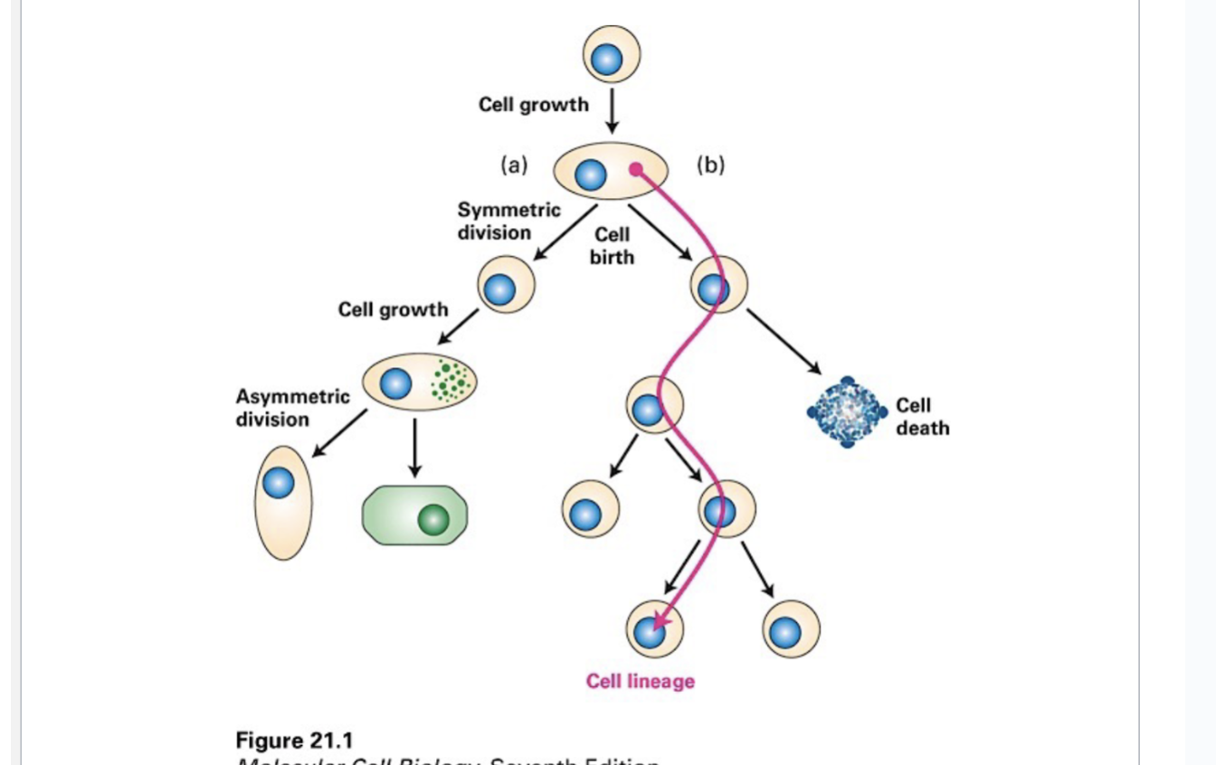
Symmetric Division
Cells divide equally
Asymmetric Division
Cells divide unequally
Metaphase plate is closer to the left or the right, leading to one daughter cell being much smaller than the other
Cells can lead to two different cell populations - this is a process of DIFFERENTIATION through gene expression
Cells can acquire certain characteristics or distinguishing functions
Ex. intestinal epithelial cells, with the microvilli on the apical surface, those microvilli are specialized to absorb nutrients from the intestinal tract
Where are the cells born? From Stem Cells
We start as a zygote — this cell is referred to as TOTIPOTENT
Cells are born from stem cells, which originate from a zygote that is totipotent, meaning it can develop into any cell type necessary to form an entire organism.
The zygotę has everything it needs to produce an ENTIRE organism
The cell will NOT make a copy of itself - can become any cell in the body
After the first embryonic division, there are a population of cells that are pluripotent — generic cells
They have the great capacity to differentiate into all/most cell types
There are adults that can either be multipotent or unipotent
Multipotent: can only differentiate into multiple cell types.
Unipotent: can only differentiate into one type of cell
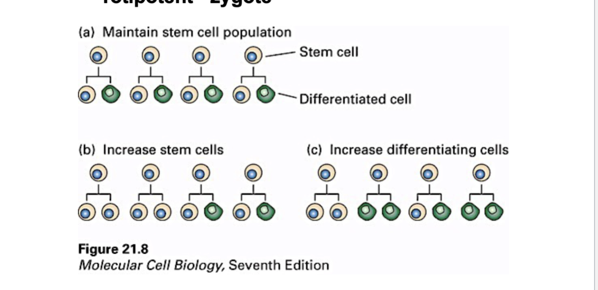
Stem cells are self-renewing - making the same population of themselves and NOT differentiating
Stem cell over here can produce two other stem cells - in other contexts, some of the stem cells will actually produce two differentiated stem cells
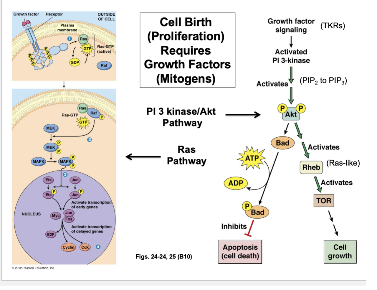
What does cell birth (proliferation) require? Growth Factors (Mitogens)
One signaling pathway —> Ras pathway
Growth factor or mitogen binding the receptor on the cell surface
This is an ENZYME-LINKED RECEPTOR — undergoing an auto-phosphorylation, signaling complex formed
Activated Ras that is GTP bound, which activates another protein — MAP KINASE
Begins to phosphorylate transcription factors and activate them - Myc, Jun, and Fos
These transcription factors promote cell division and proliferation by promoting transcription gene expression needed in S phase where DNA replication occurs, once activated
These transcription factors enable the synthesis of E2F — Cyclins and Cdks are also being transcribed and subsequently translated
Another pathway —> Act pathway
Growth factor/mitogen signaling activates PI 3-kinase
What this kinase does is it converts PIP2 to PIP3
That conversion of a lipid to another type of lipid through the action of this kinase results in the activation of the protein, Act
Akt, when phosphorylated, will activate two subsequent pathways
Activation of Bad protein —>
When Bad is activated, it will become phosphorylated
Phopshorylated Bad inhibits cell death (apoptosis)
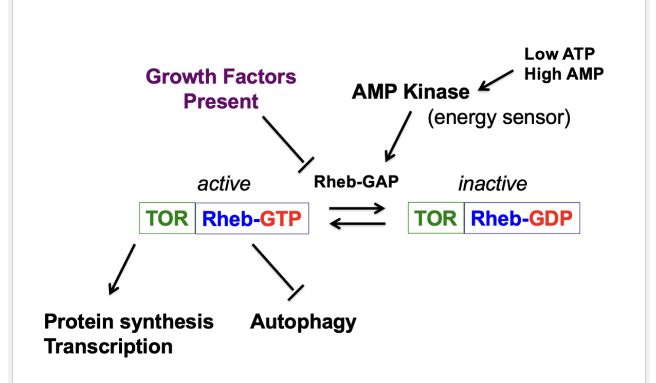
Activation of Rheb protein —>
Belongs to the Ras family of GTPases
When Rheb is GTP-bound, it’s going to activate TOR
When TOR is active, many things happen and the result is cell growth
What does TOR Kinase do? Central Regulator of Cell Growth/Proliferation
When GTP is bound to Rheb, it’s ON
When GDP is bound to Rheb, it’s OFF
There’s a GAP and a GEF that controls the GTP-bound state of the protein
GAP makes it inactive
Growth factors inhibit it
AMP kinase (low ATP, high AMP - energy sensor) activates it
Rheb-GTP interacts with TOR and when active, there will be a series of events that will allow fro transcription and protein synthesis
Rheb-GTP will also inhibit autophagy - process by which cells start consuming its own organelles under nutrient-starved conditions
Rheb-GAP shifts the balance
When growth factors present (signaling pathway with PI 3-kinase and Akt kinase), those growth factors are sending signals that are inhibiting the Rheb GAP
GTP is not being converted to GDP
Balance between Rheb GTP and Rheb GDP is part of an energy sensing mechanism
If you want to undergo cell proliferation, you have to make sure there’s energy
If you’re cellular ATP levels are low and your AMP levels are high, the AMP kinase is activated and sensing the high amount of AMP
AMP kinase is the energy sensor — AMP kinase will ACTIVATE the Rheb GAP, converting the GTP to GDP — now TOR kinase is off - protein synthesis, transcription can’t occur
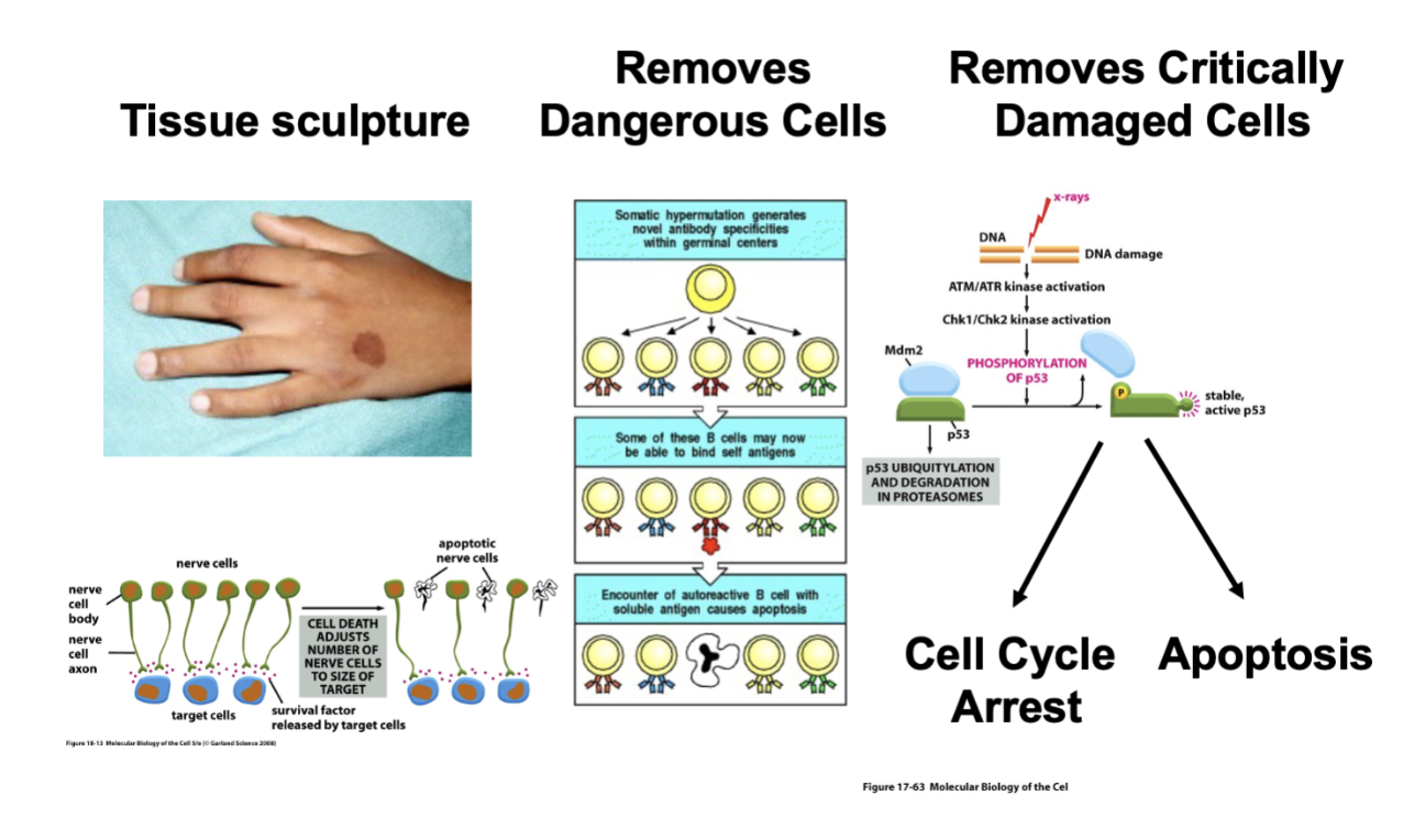
What are the diverse functions of cell death?
Essential process
Needed to sculpt tissue
Cell death takes away tissue when there’s too much (extra webbing between the fingers)
As our brains develop in the late teenage years, early 20, it is a natural process for certain neurons in the brain to undergo death
Removed from the cell population as a general principle — what you are doing is creating a more stable neural network, one with the proper connectivity
Normal developmental process
Cell death removes dangerous cells
Immune systems recognize our own cells and attack our own cells to develop an auto-immune disease '
Early in development, cells in the immune system that attack other cells are removed through the process of cell death
It is important not just in developmental process but a mechanism for dealing with damage
Cell death removes critically damaged cells
Activation of kinases lead to the phosphorylation of transcription factor of p53
The activation of p53 leads to the expression of p21, which is an inhibitor of S phase cyclin CDK complexes
Cell with damaged DNA could not proceed into S phase
The other thing that p53 is important for is the activation of apoptosis (cell death removing damaged cells from a population)
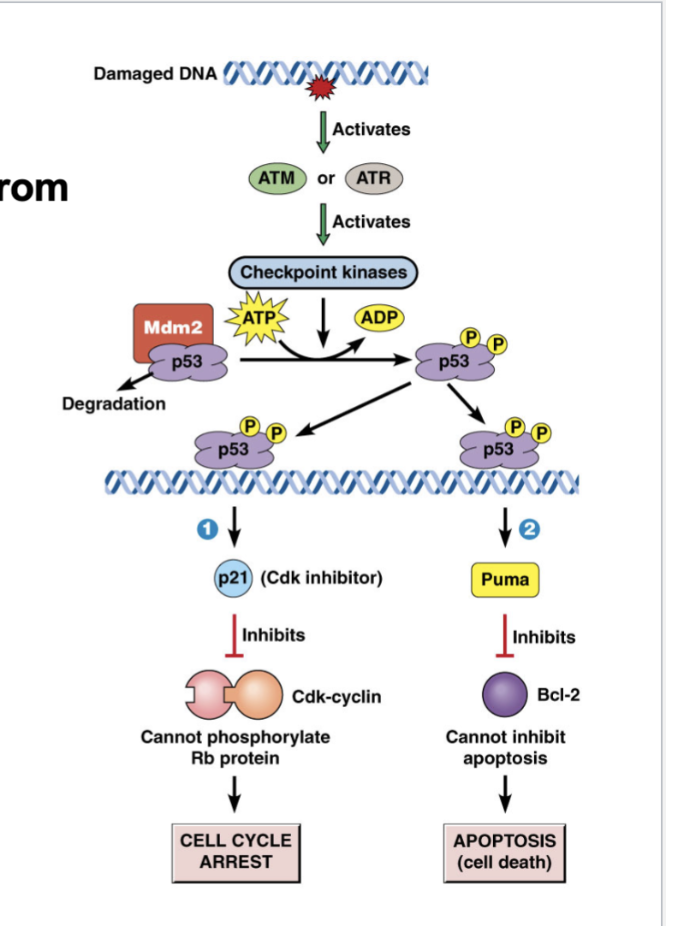
How does cell death remove critically damaged cells from population?
Damaged DNA activates ATM or ATR, which activates checkpoint kinases
Activated p53 goes down two pathways
Activates Puma, which inhibits Bcl-2, which cannot inhibit apoptosis (cell death) — Bcl-2 is an inhibitor of apoptosis or cell death
Activates p21 (Cdk inhibitor), which inhibits Cdk-cyclin, which cannot phosphorylate Rb protein, which arrests the cell cycle
Do these pathways happen simultaneously? The pathway will arrest the cell cycle. .. if damage can’t be repaired, that’s when the other pathways leading to cell death kick in
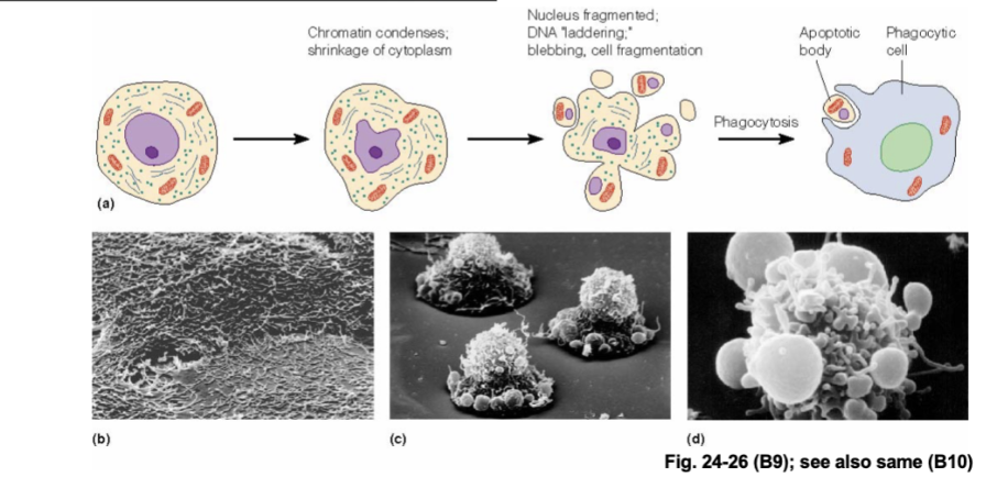
Morphological, Biochemical, Electrical Changes Mark Apoptosis
A whole cascade of events occur
Chromatin will condense
Nucleus will fragment
Membrane will bleb - apoptotic bodies will be released
Apoptotic bodies will be cleaned by cells of the immune system
Patterned DNA cleavage (after chromosomes condensed)
Phoshotidylserine flipping from inner to outer leaflet of plasma membrane
Removal of “don’t eat me” signals from PM
Loss of electrical potential across IMM — mitochondria is important!
Leakage of cytochrome c into cytosol — protein that leaks out of the mitochondria
Cytochrome c is normally embedded in the inner mitochondrial membrane and is part of the electron transport chain
C elegans: an important MODEL FOR TRACING CELL LINEAGES
Why is it a good model?
Small
Many genetic techniques can be applied to it
Multicellular
Translucent, allowing observation of cells during development
Visualization is so good you can see every cell in the worm
Depending on the sex of the cell, there’s about 969 to 1031 cells
Biologists can watch the worm divide into different cells and can watch every zygote can be mapped out and traced
See how each cell is related to each other
In the process, there is programmed cell death that removes some cells from the final population of cells
Extremely important organism for being able to track cell lineages
That, and the ability to track mutant worms, led to discovery of proteins involved in apoptosis
C elegans Mutants Revealed a Gene Required for Cell Death
ced-1 (mutation) worm developed little buttons along its body
What are those little buttons? As the worm developed and the tissue is being sculpted, there’s cell death occurring
In this mutant worm, the dead cells and the apoptotic bodies were not cleared by other cells - debris is NOT phagocytose and taken up by other cells
Cell debris accumulated in little round buttons
ced-1 and ced-3 mutant worm had little button-like structures that disappeared
Those little buttons never formed in the first place because if you had a mutation in the ced-3 gene, you did not undergo cell death
No cell death, no debris, no accumulation of the larger structures
This is how the genes were first identified
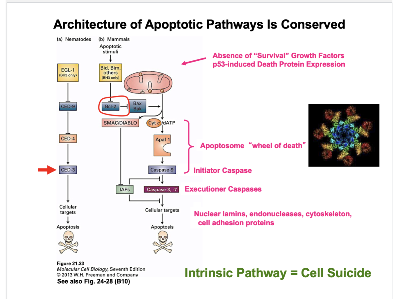
Architecture of Apoptotic Pathways is Conserved
Apopotic pathway in mammalian cells is similar to the pathway in worms
However, there are elaborations to it
Bcl-2 inhibits apoptosis by inhibiting proteins called Bax and Bak
When Bak and Bak are active, they form pores in the mitochondrial membrane, causing cytochrome C to leak out
When inactive, both proteins can’t do this
So when Bcl 2 is inhibited, which inhibits, Bax and Bak, this activates deoxygenated ATP
This activates Apaf1 and Caspase-9
Together, cytochrome C/dATP, Apaf1, and Caspase 9 form the apoptosome “wheel of death”
The reason it’s called the wheel of death is that the red parts are the caspase 9, which is a protease
It’ll degrade other proteins -→ caspase 9 is an initiator caspase that will proteolyzed other proteins or caspases
Caspase-9 activates secondary caspases or executioner caspases (caspase 3,-7), which degrades the other proteins
Those executioner caspases - go after the nuclear lamins
Destabilizes the nuclear envelope —> nuclear fragmentation
Will degrade cytoskeleton proteins and cell adhesion proteins — endonucleases and complexes forming
This is the intrinsic pathway —> events are being stimulated
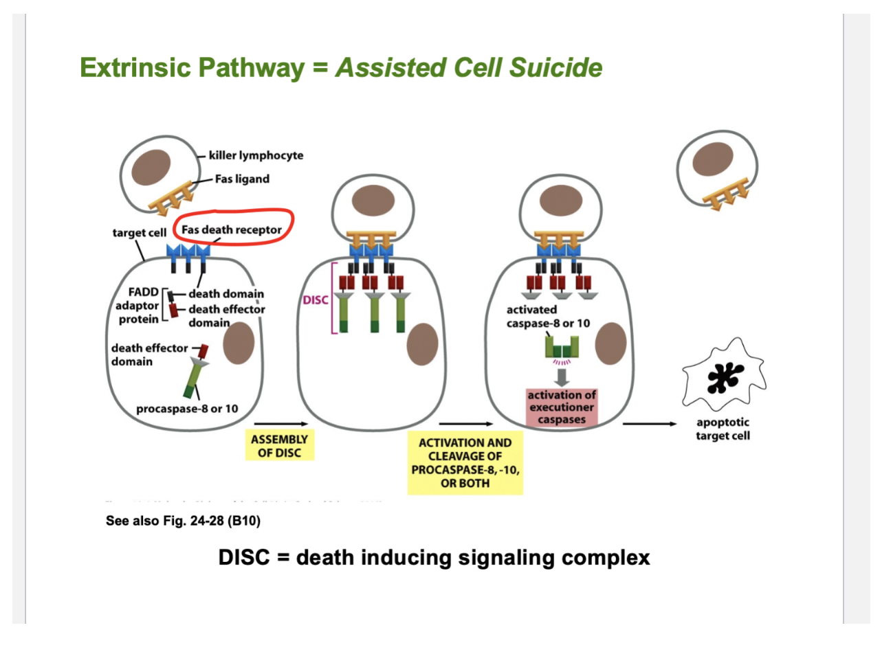
Is there another pathway?
Extrinsic pathway
Extrinsic Pathway = Assisted Cell Suicide
Another cell has to bind to the cell that has to die = killer lymphocyte, which has Fas ligand
The cells that are going to be targeted for death are expressing receptors for the Fas Death receptors
Thus this is juxtacrine signaling - two cells in contact with each other
Once those death cell receptors bind to the Fas presented by another cell, large complexes of proteins are forming
The proteins that associate with those proteins have enzymatic activities, which leads to the expression of caspases —> which destroy the rest of the cell
This is forming the death inducing signaling complex (DISC)
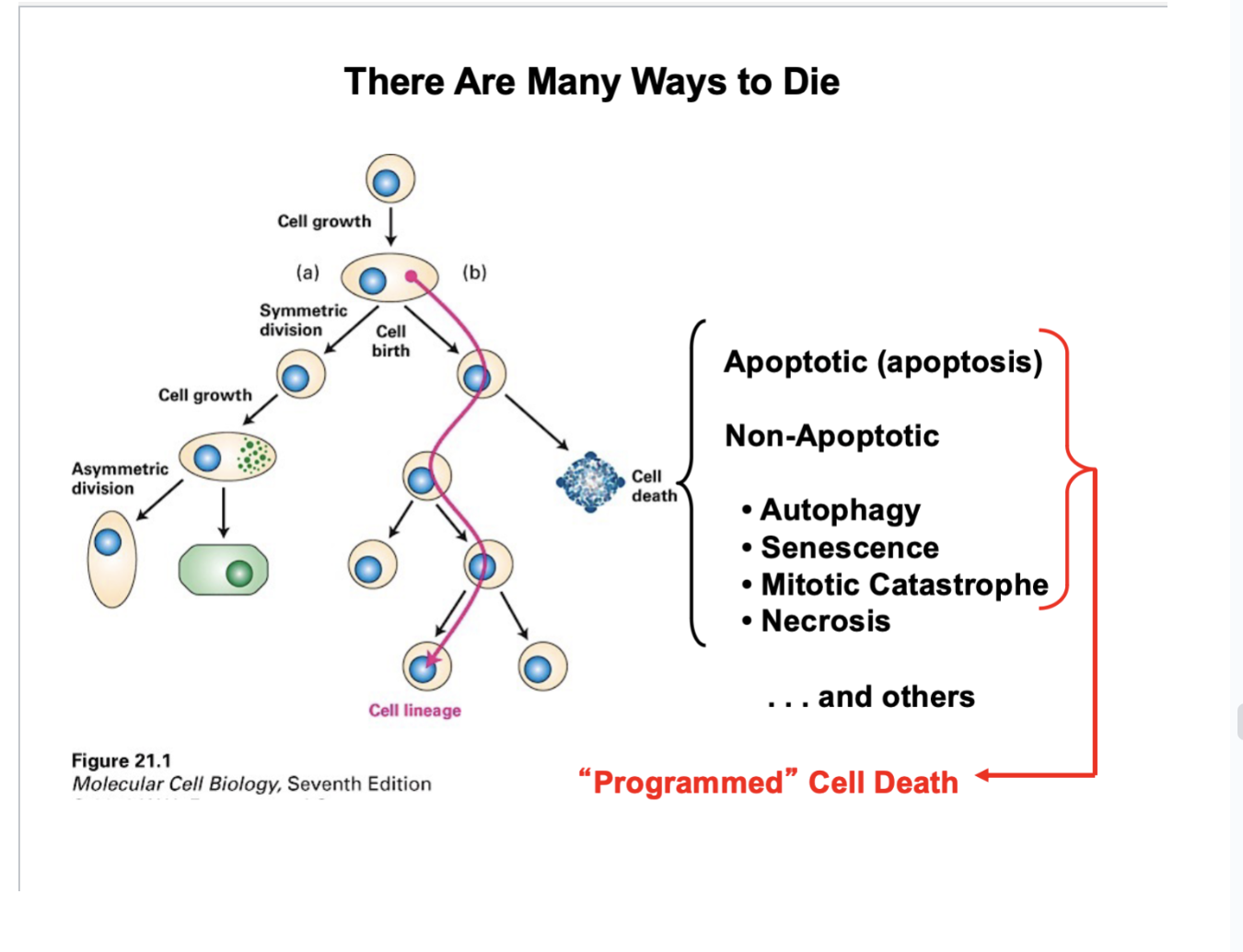
There are many ways to die
There Are Many Ways to Die
Apoptotic (apoptosis) —> programmed cell death (one form)
Non-Apoptotic
Autophagy
Senescence
Mitotic Catastrophe
Necrosis
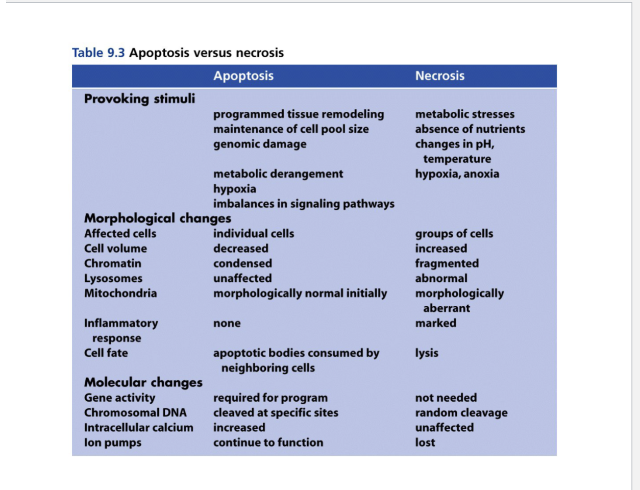
Apoptosis versus necrosis
Necrosis:
Doesn’t require expression of genes and proteins
Random cleavage of cells
Apoptosis
Requires expression of genes and proteins
Specific cleavage of cells