Ch. 15: Special Senses
0.0(0)
Card Sorting
1/202
Study Analytics
Name | Mastery | Learn | Test | Matching | Spaced |
|---|
No study sessions yet.
203 Terms
1
New cards
Special senses
Those senses with highly localized receptors that provide specific information about the environment.
2
New cards
How many primary odors are there that an average person can recognize?
Seven
3
New cards
How many different odors via the olfactory epithelium are there?
4000
4
New cards
What are the dendrites (ends) of an olfactory neuron called?
olfactory vesicles
5
New cards
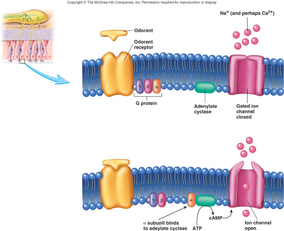
What do cilia (olfactory hairs) of olfactory neuron embedded in mucus do? (3 things)
1. Odorants dissolve in mucus
2. Attach to receptors
3. Cilia depolarize as a result of the action potential
2. Attach to receptors
3. Cilia depolarize as a result of the action potential
6
New cards
Can one olfactory receptor respond to more than one type of odor?
yes
7
New cards
What is unique about olfactory neurons compared to most other neurons?
olfactory neurons are replaced every two months, most neurons are permanent
8
New cards
Odorant Binding to Membrane of Olfactory Hair (figure)
9
New cards
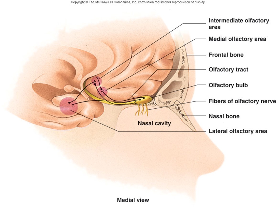
Olfactory neuronal pathway
bipolar olfactory neurons -> cribriform plate -> olfactory bulbs and synapse -> olfactory tract -> olfactory cortex (lateral olfactory area, medial olfactory area, intermediate olfactory area)
10
New cards
What is unique about olfactory information?
It goes directly to the olfactory cortex of the frontal/temporal lobes - it doesn't pass through the thalamus like all the other major sensations
11
New cards
Lateral olfactory area
conscious perception – smell
12
New cards
Medial olfactory area
visceral/emotional (instinctive) reactions to odors
13
New cards
Intermediate olfactory area
receives information from the medial and lateral olfactory areas (APs carried by axons in this area modulate the activity of the neurons in the olfactory bulb, enhancing rapid adaptation of the olfactory system)
14
New cards
Olfactory Neuronal Pathways and Cortex (figure)
15
New cards
How many primary odors have been identified?
A. eight
B. six
C. seven
D. eleven
A. eight
B. six
C. seven
D. eleven
C. seven
16
New cards
Olfactory neurons
A. Have projections called cilia.
B. Connect to the olfactory bulb.
C. Have receptors that react with odorants dissolved in mucus.
D. All of the above.
A. Have projections called cilia.
B. Connect to the olfactory bulb.
C. Have receptors that react with odorants dissolved in mucus.
D. All of the above.
D. All of the above.
17
New cards
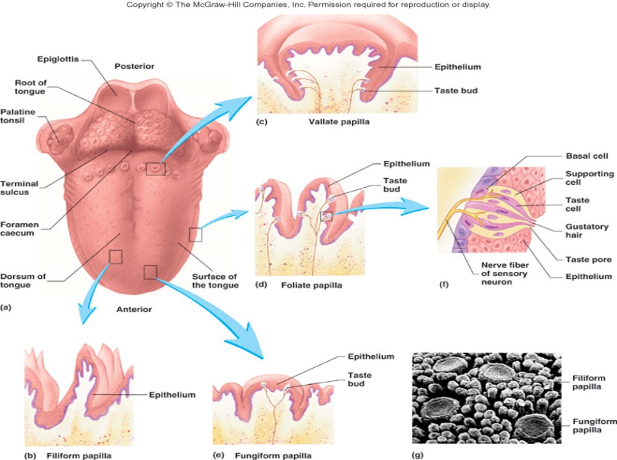
Where are taste buds primarily located?
on papillae (specialized tongue structures)
18
New cards
Vallate papillae
Largest but just 8-12 in V between anterior/posterior parts of tongue (have taste buds)
19
New cards
Fungiform papillae
Mushroom-shaped and scattered on superior surface of tongue - they look like "small red dots” interspersed among the filiform papillae (have taste buds)
20
New cards
Foliate papillae
Leaf-shaped and in folds on sides of tongue (have the most sensitive taste buds, but decrease in number with age)
21
New cards
Filiform papillae
filament-shaped and the most numerous (no taste buds)
22
New cards
How many tastebuds on the tongue?
approximately 10,000
23
New cards
The sensory cells of each taste bud are called
taste or gustatory cells
24
New cards
How often are gustatory cells replaced?
every 10 days
25
New cards
How many taste cells on each taste bud?
about 50
26
New cards
Taste cells have _____.
microvilli (gustatory hairs) extending into taste or gustatory pores
27
New cards
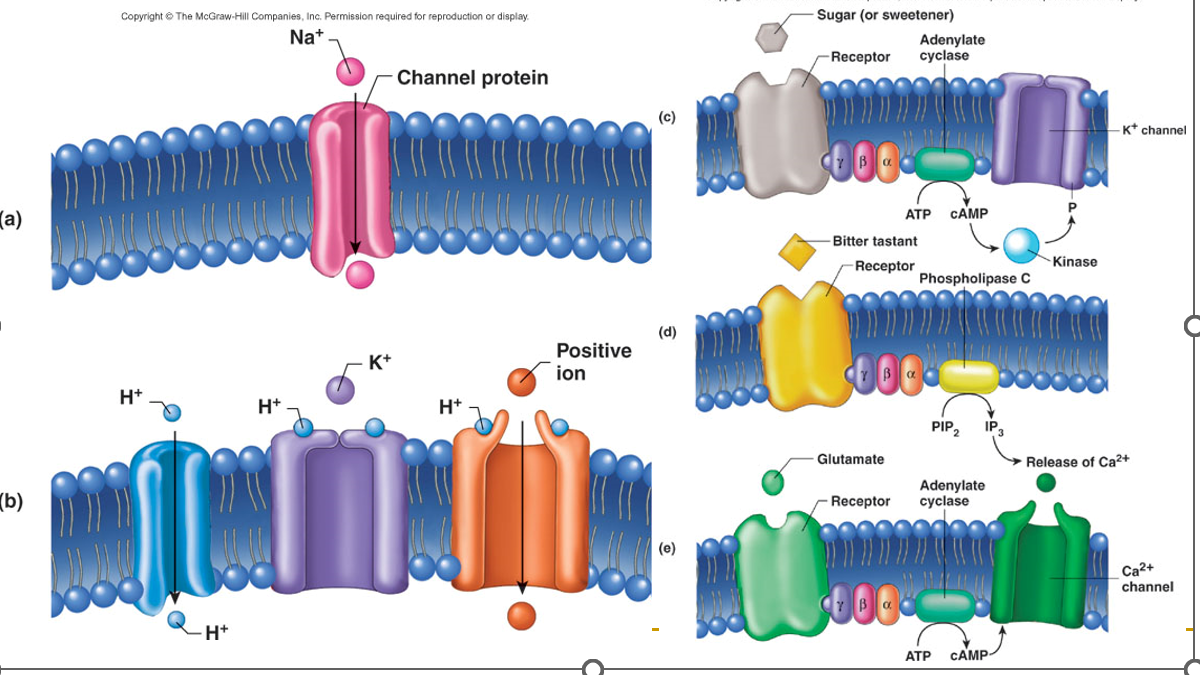
Tastants
chemical molecules that dissolve in saliva and enter the taste pore by various mechanisms, causing the taste cell to depolarize
28
New cards
Do taste cells have axons?
No, they don’t generate their own action potentials
29
New cards
What happens after the taste cells depolarizes?
Neurotransmitters are released from the taste cells and stimulate action potentials in the axons of sensory neurons associated with them
30
New cards
Taste Buds (figure)
31
New cards
Sour taste
Most sensitive on lateral aspects of tongue (produced by acids)
32
New cards
Salty taste
Most sensitive on the tip of tongue (produced by metal ions)
33
New cards
Bitter taste
Most sensitive on the posterior part of the tongue (produced by alkaloids, most of which are toxic)
34
New cards
Sweet taste
Most sensitive on the tip of the tongue (produced by sugars, some carbohydrates, and some proteins, but can be fooled - NutraSweet = aspartame)
35
New cards
Umami taste
Scattered sensitivity (produced by amino acids, like glutamate)
36
New cards
Which tastant has the highest threshold sensitivity?
salty and sweet
37
New cards
Which tastant has the lowest threshold sensitivity?
bitter
38
New cards
Which tastes do humans tend to crave?
sweet, salty, and umami tastes
39
New cards
Actions of the Major Tastants (figure)
(a) Salty
(b) Sour
(c) Sweet
(d) Bitter
(e) Umami
(b) Sour
(c) Sweet
(d) Bitter
(e) Umami
40
New cards
Which of these is not one of the basic tastes?
A. spicy
B. salt
C. umami
D. sour
A. spicy
B. salt
C. umami
D. sour
A. spicy
41
New cards
Which of these types of papillae have no taste buds associated with them?
A. circumvallate
B. filiform
C. foliate
D. fungiform
A. circumvallate
B. filiform
C. foliate
D. fungiform
B. filiform
42
New cards
What 3 things affect taste?
1. Texture (affects the perception of taste)
2. Temperature
3. Olfaction
2. Temperature
3. Olfaction
43
New cards
At what levels does taste very rapidly adapt?
at level of the taste buds and within CNS
44
New cards
Why is bitter the most sensitive threshold?
Many alkaloids are poisonous
45
New cards
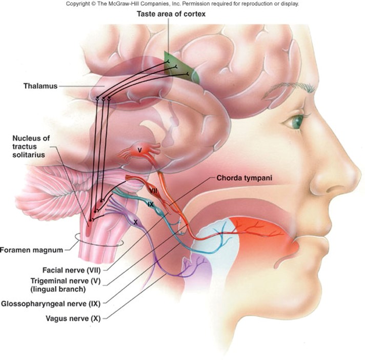
What are the 3 cranial nerves involved in sense of taste and what portion of the tongue do they innervate?
1. VII: Facial (anterior 2/3 tongue)
2. IX: Glossopharyngeal (posterior 1/3 tongue, the vallate papillae, and the superior pharynx.)
3. X: Vagus (posterior tongue)
2. IX: Glossopharyngeal (posterior 1/3 tongue, the vallate papillae, and the superior pharynx.)
3. X: Vagus (posterior tongue)
46
New cards
3 cranial nerves that make up the pathway for taste
1. Chorda tympani (part of facial nerve VII): sensation to anterior 2/3 of tongue, except from the vallate papillae
2. Glossopharyngeal nerve (IX)
3. Vagus nerve (X): carries a few fibers for taste from epiglottis and the root of the tongue
2. Glossopharyngeal nerve (IX)
3. Vagus nerve (X): carries a few fibers for taste from epiglottis and the root of the tongue
47
New cards
Neuronal pathway for taste
APs from the cranial nerves -> medulla oblongata (tractus solitarius) -> thalamus -> taste area of cortex (extreme inferior end of postcentral gyrus)
48
New cards
DO: Name the four types of papillae found on the tongue.
Circumvallate, filiform, fungiform, foliate
49
New cards
4 accessory structures of the visual system
eyebrows, eyelids, eyelashes, and conjunctiva
50
New cards
What is the function of eyebrows to the visual system?
provide shade, prevent sweat from running into eyes
51
New cards
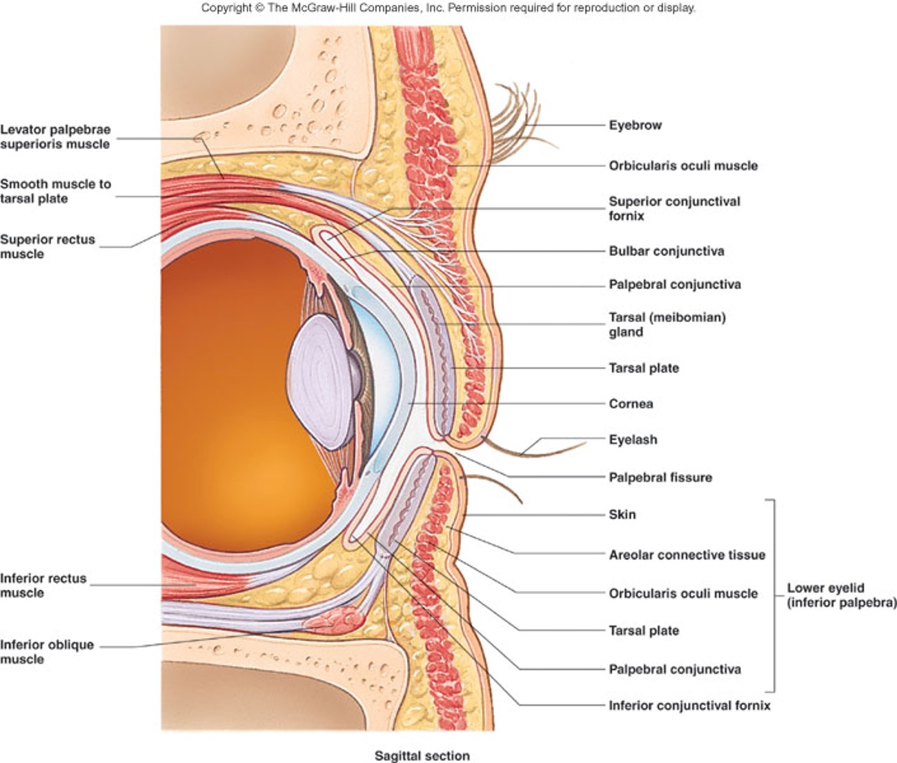
3 parts of the eyelids (palpebrae)
1. Palpebral fissure: space between eyelids
2. Canthi (lateral and medial): where eyelids meet
3. Caruncle: a reddish-pink mound housed by the medial canthus that consists of modified sweat & sebaceous glands
2. Canthi (lateral and medial): where eyelids meet
3. Caruncle: a reddish-pink mound housed by the medial canthus that consists of modified sweat & sebaceous glands
52
New cards
5 layers of the eyelid (outer -> inner)
1. a thin layer of skin on the external surface
2. a thin layer of areolar connective tissue
3. a layer of skeletal muscle consisting of the orbicularis oculi and levator palpebrae superioris muscles
4. the tarsal plate, which is a layer of dense connective tissue that maintains the shape of the lid
5. the palpebral conjunctiva
2. a thin layer of areolar connective tissue
3. a layer of skeletal muscle consisting of the orbicularis oculi and levator palpebrae superioris muscles
4. the tarsal plate, which is a layer of dense connective tissue that maintains the shape of the lid
5. the palpebral conjunctiva
53
New cards
Eyelashes
double/triple row of hairs on the external edge of the eyelid with Ciliary glands and Meibomian glands
54
New cards
Ciliary glands are
modified sweat glands that empty into the hair follicles of eyelashes
55
New cards
Meibomian glands are
sebaceous glands at the inner margins of the eyelids that produce sebum
56
New cards
Conjunctiva:
thin transparent mucous membrane
57
New cards
Palpebral conjunctiva:
covers the inner surface of the eyelids
58
New cards
Bulbar conjunctiva:
covers the anterior surface of the eye (except over pupil)
59
New cards
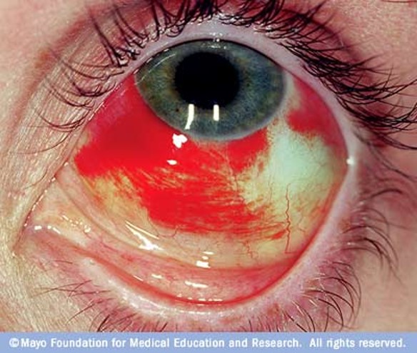
Subconjunctival hemorrhage
60
New cards
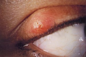
Sty
Ciliary gland
61
New cards
Chalazion
Meibomian gland
62
New cards
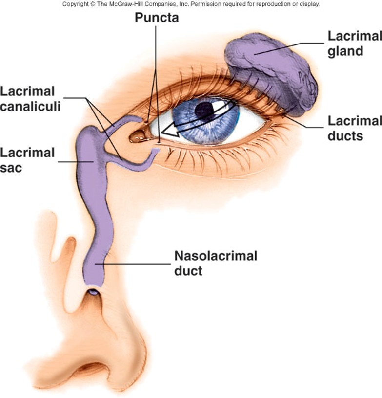
Lacrimal apparatuses (3 things)
1. Lacrimal gland
2. Lacrimal canaliculi
3. Lacrimal sac (leads to the nasolacrimal duct)
2. Lacrimal canaliculi
3. Lacrimal sac (leads to the nasolacrimal duct)
63
New cards
What does the lacrimal gland do?
produces tears to moisten, lubricate, and wash
64
New cards
Tears pass over the eye and contain what?
water, salts, mucus and lysozymes
65
New cards
What do the lacrimal canaliculi do?
collect excess tears via openings called puncta
66
New cards
Lacrimal sac leads to nasolacrimal duct, which
opens into nasal cavity
67
New cards
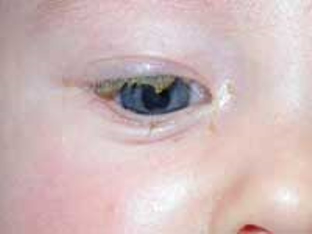
What does a blocked nasolacrimal duct look like?
68
New cards
Tears…
A. are released onto the surface of the eye near the medial corner of the eye.
B. in excess cause a sty.
C. pass through the nasolacrimal duct directly into the oral cavity.
D. contain water, salts, mucus and lysozymes.
A. are released onto the surface of the eye near the medial corner of the eye.
B. in excess cause a sty.
C. pass through the nasolacrimal duct directly into the oral cavity.
D. contain water, salts, mucus and lysozymes.
D. contain water, salts, mucus and lysozymes.
69
New cards
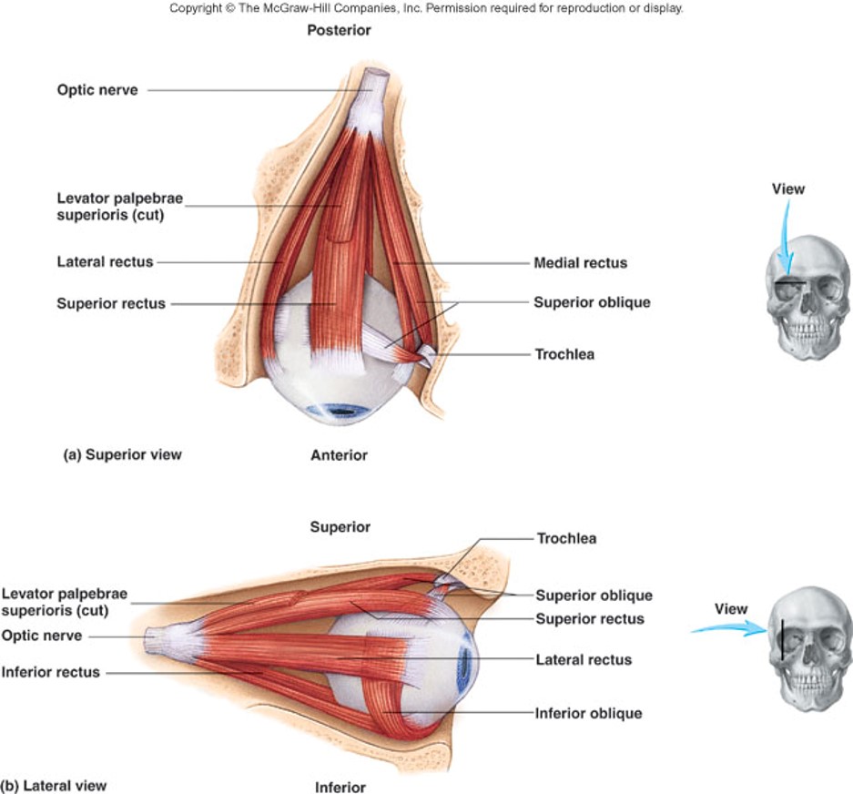
The 6 extrinsic eye muscles:
1. Superior rectus
2. Inferior rectus
3. Later rectus
4. Medial rectus
5. Superior oblique
6. Inferior oblique
2. Inferior rectus
3. Later rectus
4. Medial rectus
5. Superior oblique
6. Inferior oblique
70
New cards
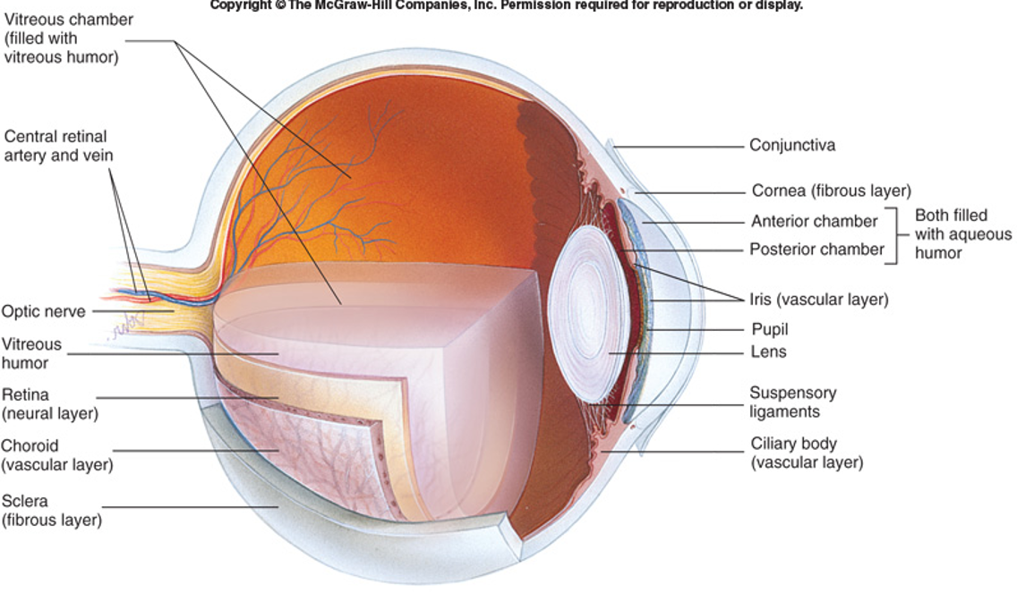
Anatomy of the Eye (figure)
71
New cards
3 layers/tunics of the eye and what they are made up of
1. Fibrous: sclera and cornea
2. Vascular: choroid, ciliary body, iris
3. Nervous: retina
2. Vascular: choroid, ciliary body, iris
3. Nervous: retina
72
New cards
Sclera:
Posterior (5/6) white outer layer (made up of opaque, dense collagenous CT/elastic fiber) that maintains shape, protects, and provides for muscle attachments
73
New cards
What is the sclera continuous with?
the cornea
74
New cards
Cornea:
an avascular, transparent CT layer made of small collagen fibers and proteoglycans (stratified squamous epithelium on outer surface) that refracts light
75
New cards
Why does the cornea have a low water content?
water scatters light
76
New cards
What was one of the first tissues to be transplanted?
the cornea
77
New cards
Vascular layer:
contains most of the blood vessels of the eye (from internal carotid artery) and melanin
78
New cards
Iris:
the colored part of eye that controls entering light with smooth muscles
79
New cards
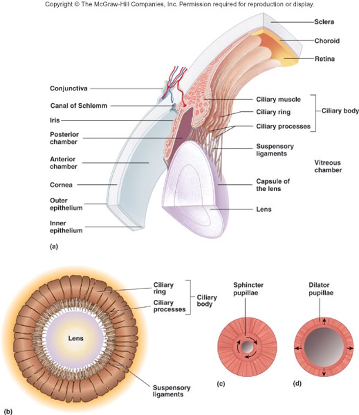
2 groups of the iris's smooth muscle
1. Sphincter pupillae
2. Dilator pupillae
2. Dilator pupillae
80
New cards
Sphincter pupillae:
constriction of the pupil (innervated by parasympathetic fibers from CN III - oculomotor)
81
New cards
Dilator pupillae:
dilation of the pupil (innervated by sympathetic fibers)
82
New cards
Ciliary body:
produces the aqueous humor that fills the anterior chamber
83
New cards
Ciliary muscles:
smooth muscles attached to suspensory ligaments of lens that control the lens shape
84
New cards
Choroid:
portion of the vascular tunic associated with the sclera (it is very thin and pigmented)
85
New cards
2 layers of the retina
1. Pigmented layer
2. Neural layer
2. Neural layer
86
New cards
Pigmented Layer:
outer layer of the retina that is pigmented simple cuboidal epithelium
87
New cards
What does the pigment of the pigmented layer and choroid help to do?
helps reduce light scatter
88
New cards
Neural Layer:
inner layer of the retina that contains the photoreceptor cells (rod and cone cells)
89
New cards
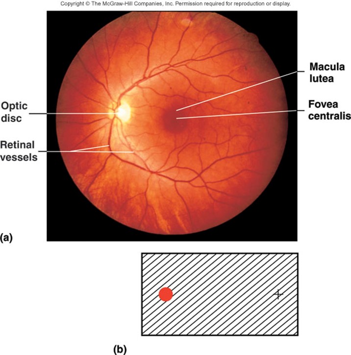
What does the lens focus light on?
macula lutea and fovea centralis
90
New cards
Macula lutea
small “yellow spot”
91
New cards
Fovea centralis:
area of greatest visual acuity where photoreceptor cone cells are tightly packed
92
New cards
Optic disc:
the blind spot in vision where blood vessels enter/exit eye and nerve processes from retina exit eye as optic nerve
93
New cards
What does the aqueous humor help to do? (3 things)
1. maintains intraocular pressure
2. provides nutrients to structures
3. refracts light
2. provides nutrients to structures
3. refracts light
94
New cards
What is the aqueous humor produced by?
ciliary processes
95
New cards
Where does the aqueous humor exit through?
canal of Schlemm/scleral venous sinus
96
New cards
Glaucoma
increase in intraocular pressure by aqueous humor buildup
97
New cards
What does the vitreous humor help to do? (3 things)
1. maintains intraocular pressure
2. holds lens and retina in place
3. refracts light
2. holds lens and retina in place
3. refracts light
98
New cards
What do the suspensory ligaments attached to the ciliary processes do?
change shape as ciliary muscles contract/relax
99
New cards
What is the Lens made of?
Cuboidal epithelial cells that produce lens fibers that lose their nucleus and accumulate proteins called crystallins
100
New cards
What is the crystalline lens is covered by?
a highly elastic, transparent capsule