Muscle Tissue Characteristics
1/112
There's no tags or description
Looks like no tags are added yet.
Name | Mastery | Learn | Test | Matching | Spaced | Call with Kai |
|---|
No analytics yet
Send a link to your students to track their progress
113 Terms
excitability(responsiveness)
ability to receive and respond to stimuli and generation potential
contractility
ability to shorten forcibly when stimulated
extensibility
ability to stretch
elasticity
ability to recoil to resting length
Prefixes for muscle
myo, mys, and sarco
muscle fibers
myocytes or muscle cells
sarcoplasm
muscle cell cytoplasm
Sarcoplasm contains
many glycogens storage as well as Myoglobin for O2 storage
Sarcoplasmic reticulum (modified endoplasmic reticulum)
stores calcium ions
Skeletal Muscle
attached to bones or (some fascial muscles) to skin
Cell shape appearance of skeletal muscles
single very long cylindrical multinucleate cells with obvious striations
Cardiac Muscle
Walls of the heart
Cell shape and appearance of the cardiac muscle
Branching chains of cells, uni, or binucleate; striations
Smooth Muscle
Unitary muscle in walls of hollow visceral organs (other than the heart); unit muscle in intrinsic eye muscles, airways, large arteries.
Cell Shape appearance Smooth muscle
single fusiform uninucleate no striations
Skeletal muscle tissue
is packaged into skeletal muscles organs that are attached to bones and skin
Skeletal muscle fibers (muscles cells)
longest of all muscle and have striations (stripes)
Skeletal muscles can also be called
voluntary muscle: can be consciously controlled
contract rapidly tired easily powerful
Key words for skeletal muscle
skeletal, striated and voluntary
Skeletal muscle is an organ made up of
different tissues
Nerve
consciously controlled skeletal muscle has nerves supplying every fiber to control activity
Cholinergic somatic
motoneuron axons form neuromuscular junctions
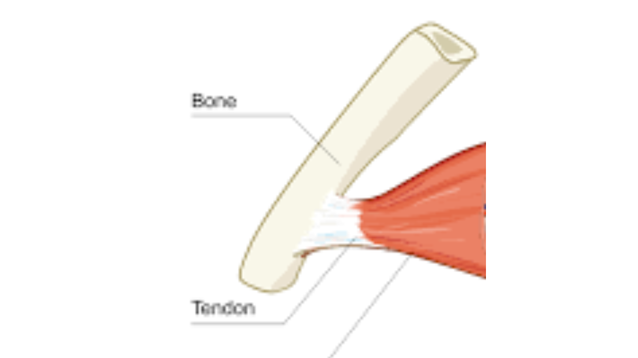
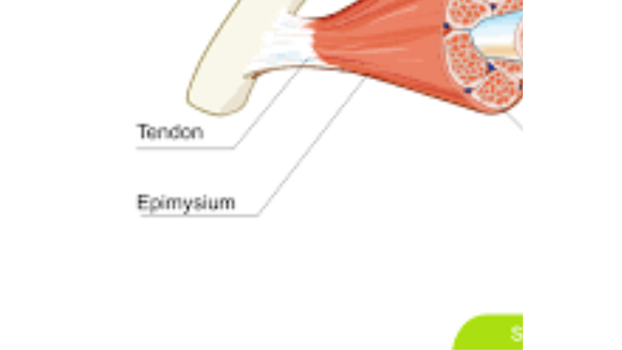
Tendons
attachments to bones
contracting muscles fibers requires
huge amount of energy oxygen and nutrients
Sarcoplasm
muscle cell cytoplasm contains many
glycosomes
myoglobin
mitochondria
glycosomes
for glycogen storage as well as
myoglobin for O2
storage mitochondria produce ATP
also, nee waste produces to removed quickly
Mitochondria produce
ATP
Muscle (organ)
a muscle consists of hundreds of thousands of muscle cells plus connective tissue wrappings blood vessels and nerve fibers
Muscle connective tissue wrappings
covered externally by the epimysium
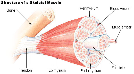
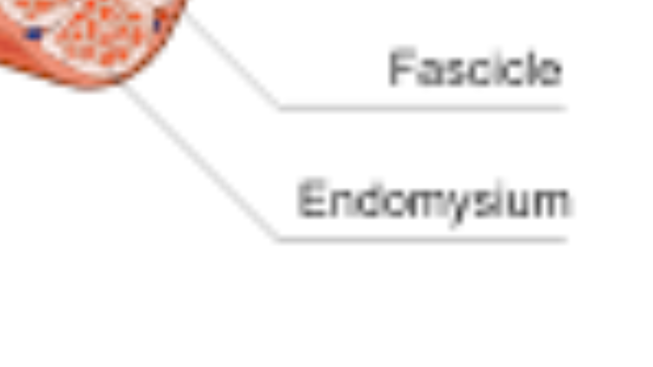
fascicle
is a discrete bundles of muscle cells segregated from the rest of the muscle by a connective tissue sheath
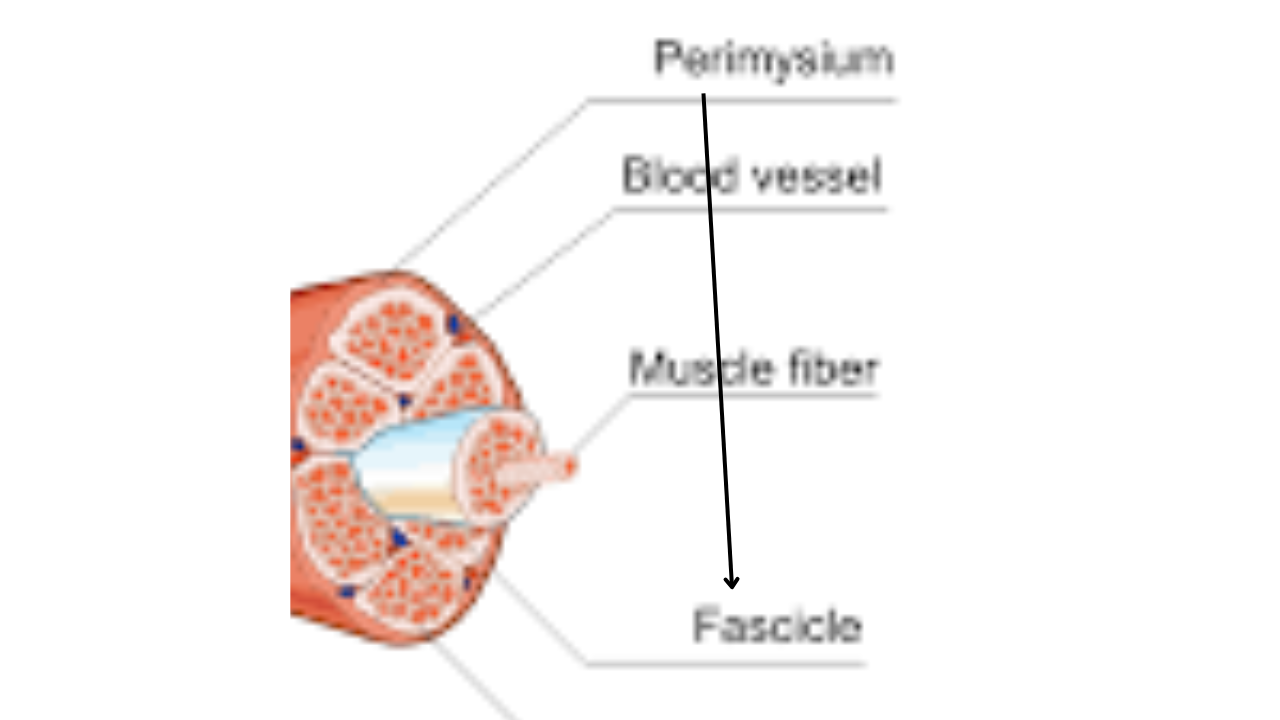
fascicle is surrounded by
perimysium
muscle fiber
an elongated multinucleate cell it has a banded (striated) appearance
Muscle fiber is surrounded by
endomysium
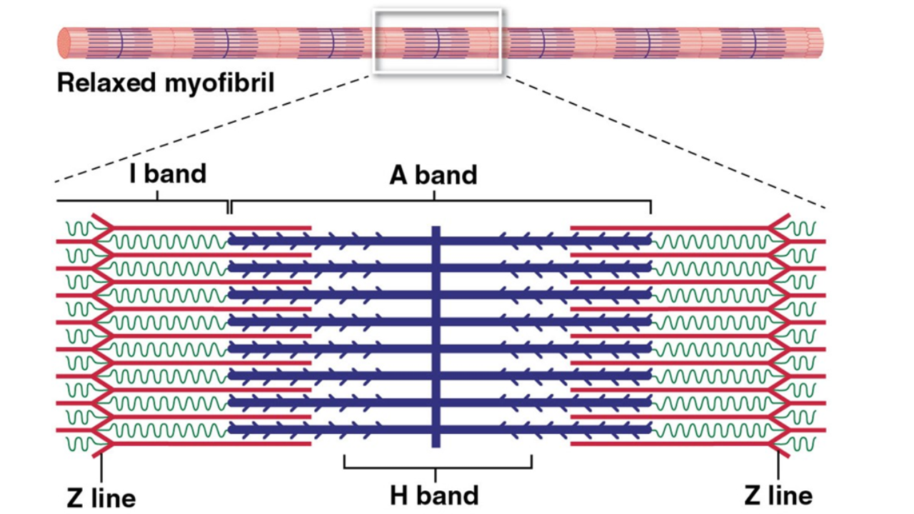
I bands are
light regions
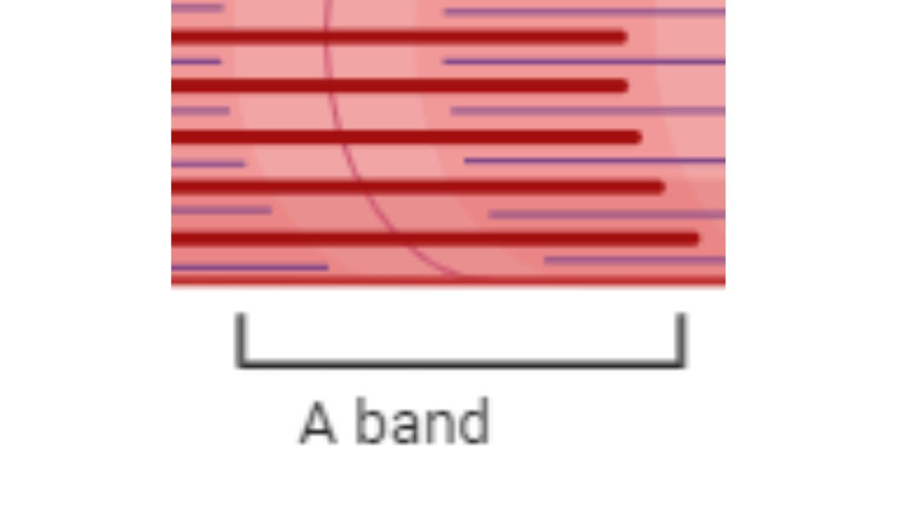
A Bands are
dark region
Myofibrils are composed of
long myofilaments proteins responsible for the banding pattern including:
Actin
Myosin
Titin
Actin
thin filaments anchored to Z disc with regularly protein Troponin and Tropomyosin
Myosin
thick filaments
Extend length of A band
Connected at M line
Titin
elastin and other proteins that hold them together
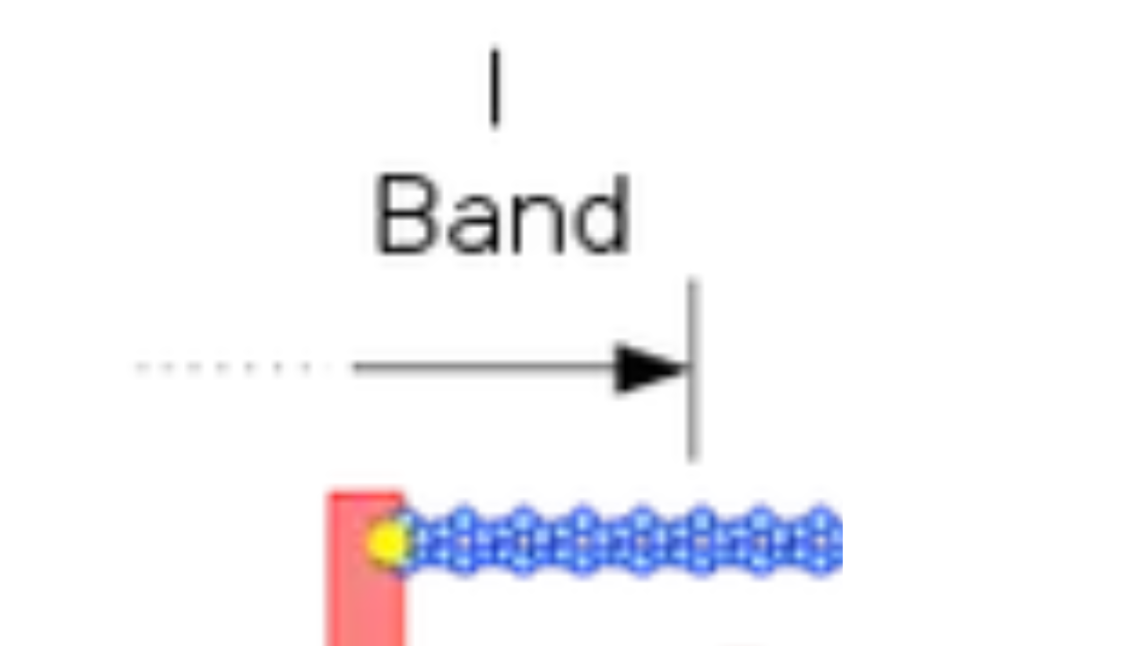
I Band
thin filaments only in Actin
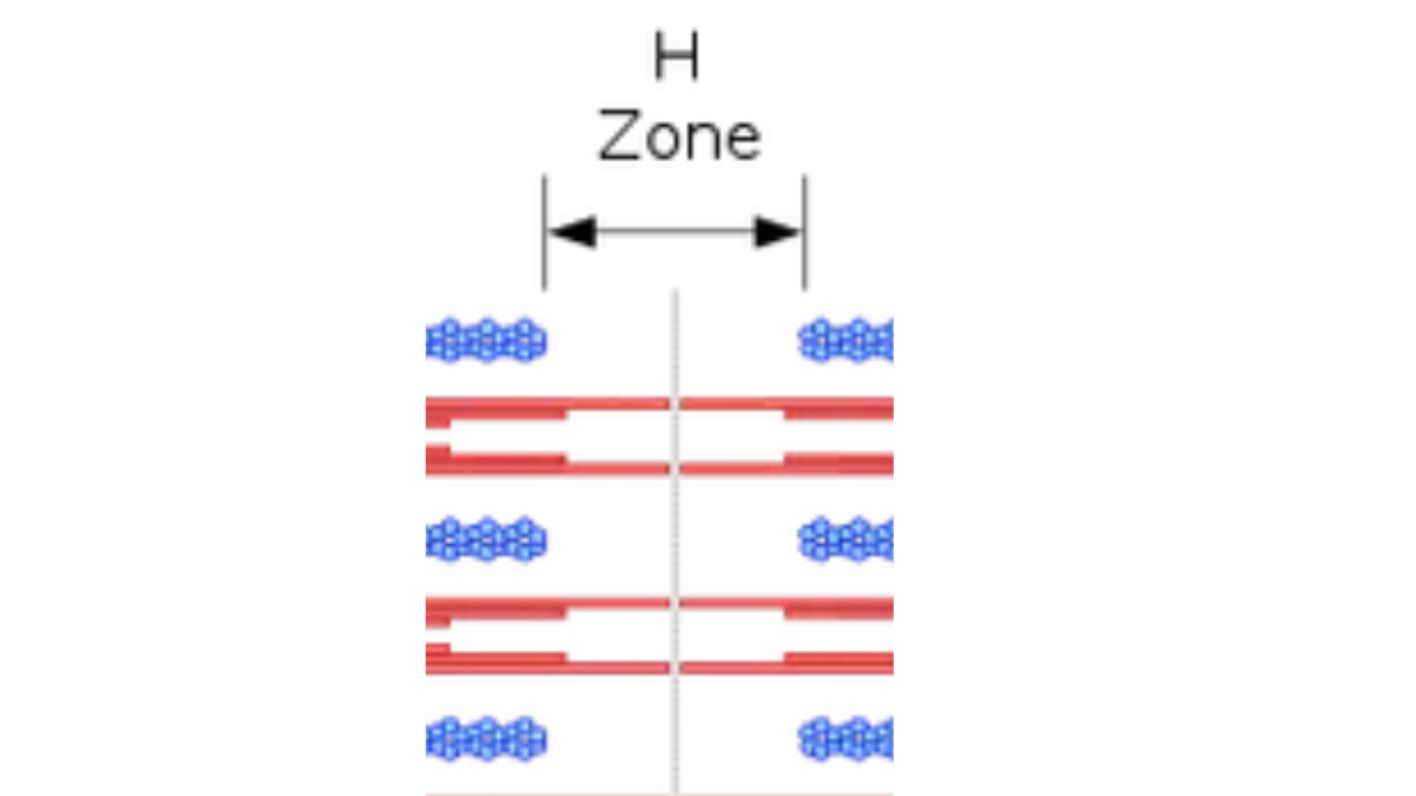
H Zone
thick filaments only myosin
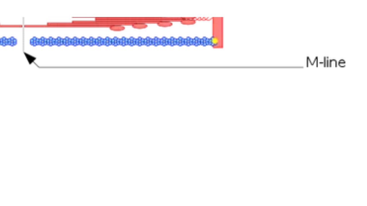
M line
thick filaments linked by accessory proteins
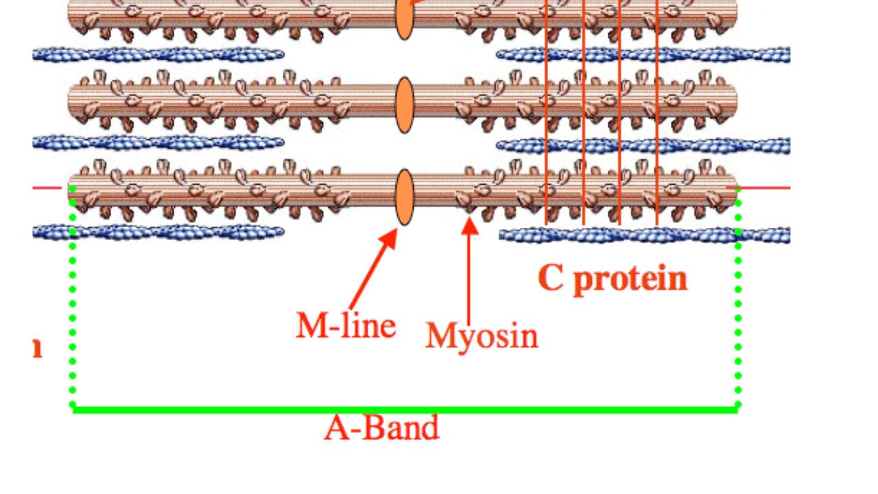
outer edge of A band
thick and thin filaments overlap
Dystrophin
links thin filaments to proteins of sarcolemma nebulin, myomesin (M line), C proteins bind filaments or sarcomeres together. Maintain alignment of sarcomere
During a contraction
A band stays the same width but the Z lines move closer together and the I band gets smaller
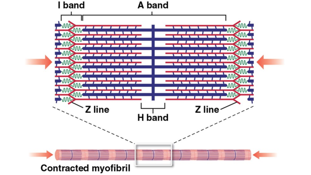
When the ends of a myofibril are to move
the sarcomeres shorten simultaneously, and the ends of a myofibril are pulled toward its center
In the relaxed state thin and think filaments
overlap only slightly at ends of A band
sliding filament model of contraction states that during the contraction
thin filaments slide past thick filaments causing actin and myosin to overlap more
Neither thick nor thin filaments change length just overlap more
When nervous system stimulates muscles fibers myosin heads are allowed
to bind to actin, forming cross bridges which causes sliding (contraction) process to begin
Sarcomere
smallest contractile unit (functional unit) of muscle fiber
Individual sarcomeres align
end to end along myofibril like boxcars of train
Each sarcomere extends from
one Z disc to the next
Striations
stripes formed from repeating series of dark and light bands along length of each myofibril
A bands
dark regions
H zone
lighter region in middle of dark A band
M line
line protein (myomesin) that bisects H zone vertically
I bands
lighter regions
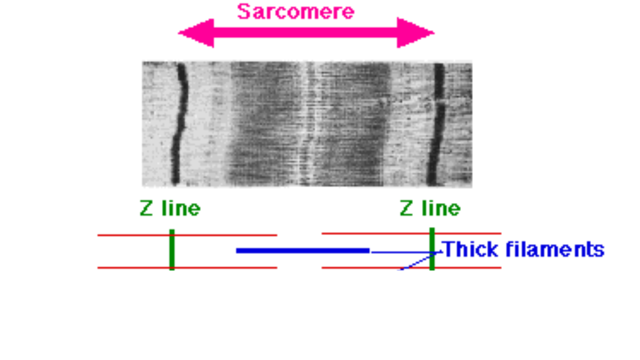
Z disc (line)
coined-shaped sheet of proteins on midline of light I band
Actin myofilaments
are anchors to the Z disc
Myofilaments orderly arrangement of
actin and myosin myofilaments within sarcomere
Actin myofilaments
extend across I band and partway in A band
anchored to Z discs
Myosin myofilaments
thick filaments
extend length of A band
connected at M line
Thin Filament consists of
two strands of actin subunits twisted into a helix plus two types of regulatory proteins (troponin and tropomyosin)
thick
thin filaments composed of
protein actin
actin
is polypeptide made up of kidney-shaped G actin (globular) subunits
G actin
subunits bears active sites for myosin head attachment during contraction
G actin subunits link together to form long fibrous F actin (filamentous)
Two F actins strands
twist together to form a thin filament
Tropomyosin and troponin
regulatory proteins to actin
Tropomyosin
covers active sites on G -actin
prevents actin-myosin interaction
troponin
globular protein
binds tropomyosin, G-actin and Calcium (Ca+2)
Sarcoplasmic reticulum (SR)
network of smooth endoplasmic reticulum tubules surrounding each myofibril
SR functions in the
regulation of intracellular Ca+2 levels
stores and releases Ca+2
T tubes
tube formed by the protrusion of sarcolemma deep into interior. Increase muscle fibers surface area greatly. Allow the action potential to reach deep into interior of each muscle fiber.
Tubules penetrate cell’s interior at ach A-I band junction between terminal cisterns
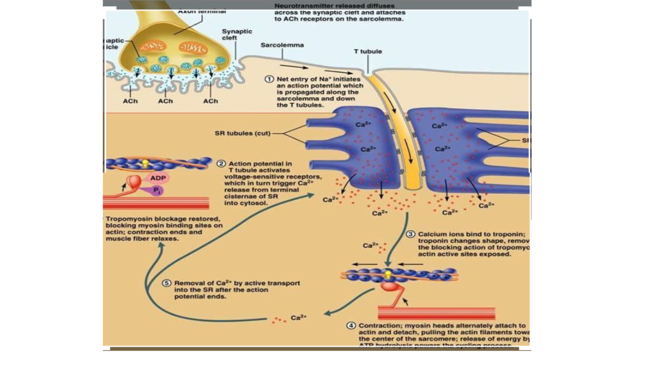
action potential generated by a
nerve stimulation along the sarcolemma and down the tubules
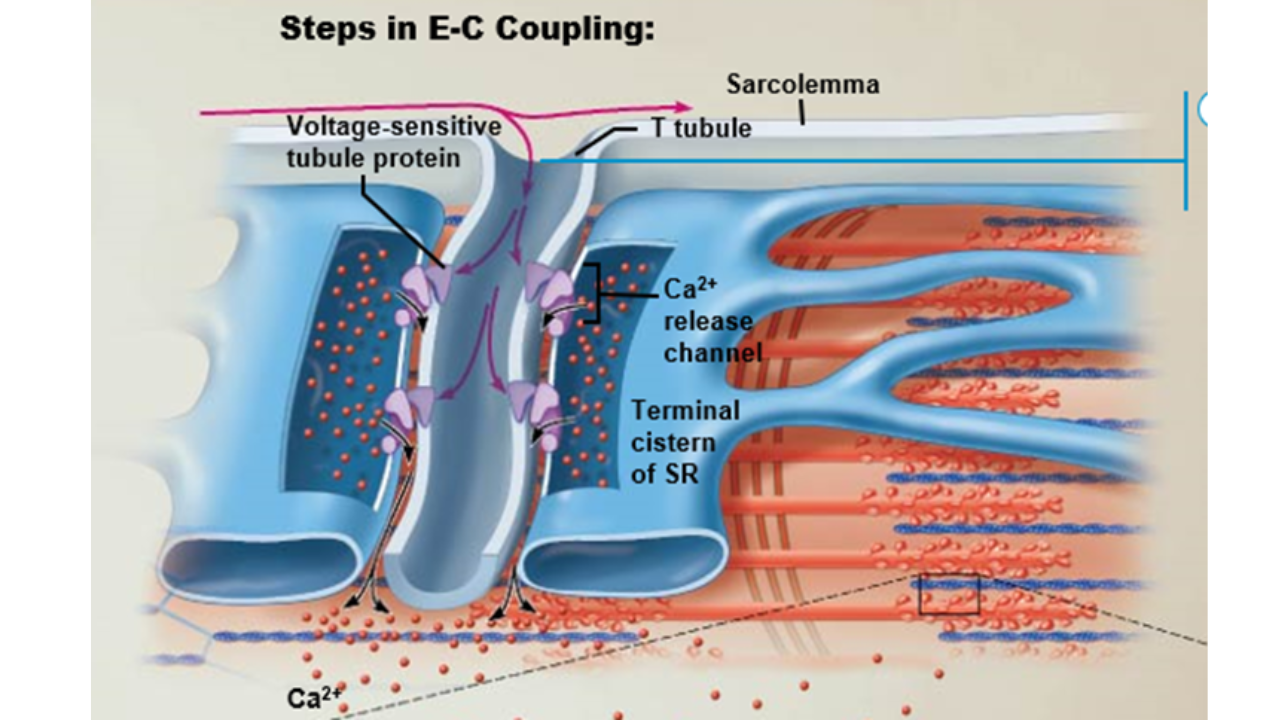
transmission of the AP along the T tubules of the triads causes
the voltage-sensitive tubule proteins to change shape. This shape change opens the Ca+2 release channels in the terminal cisterns of the sarcoplasmic reticulum (SR) allowing Ca+2 to flow into the cytosol
A motor unit consists of
one motor neuron and all the muscle fibers it innervates
Branching axon terminals forms neuromuscular junctions one per muscle fiber
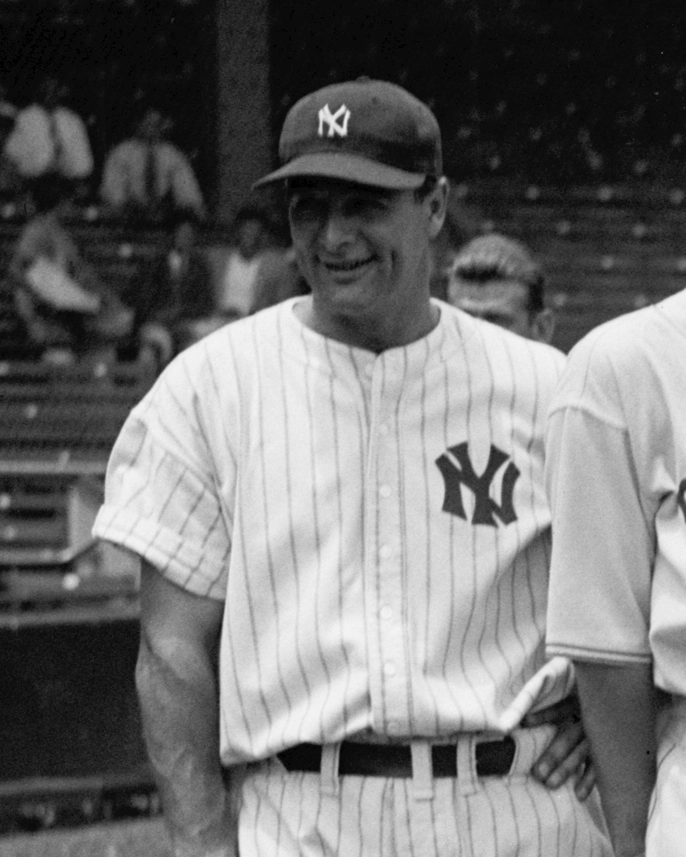
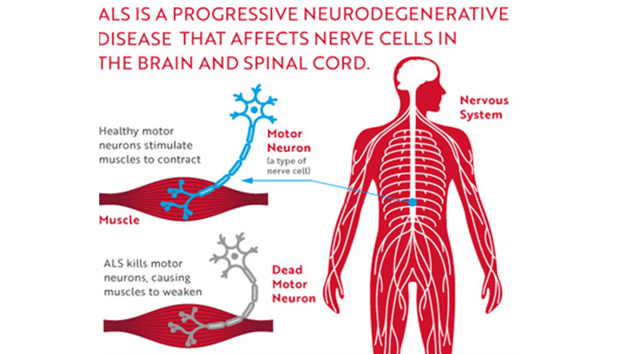
ALS (AKA Lou Gehrig’s disease)
a progressive neurodegenerative disease that affects nerve cells in the brain and spinal cord.
Healthy motor neurons stimulate muscles to contract
ALS kills motor neurons causing muscles to weaken
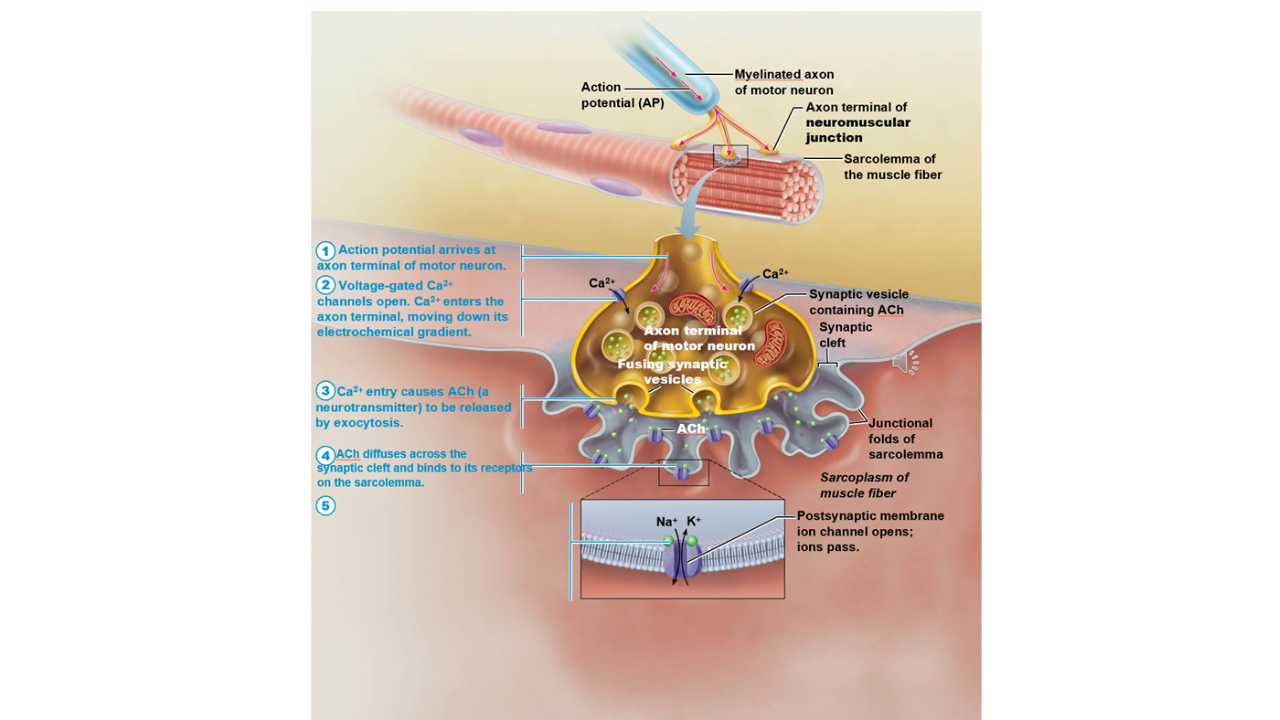
Neuromuscular junction or Synapse
between a neuron (motoneuron) and a skeletal muscle fiber at the level of the motor end plate.
Skeletal muscles are stimulated by somatic motor neurons
That are cholinergic (release Acetylcholine)
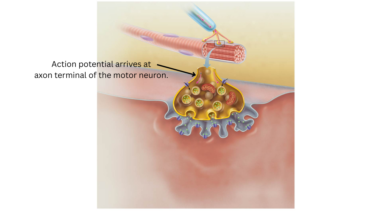
Action potential arrives at
axon terminal of motor neuron
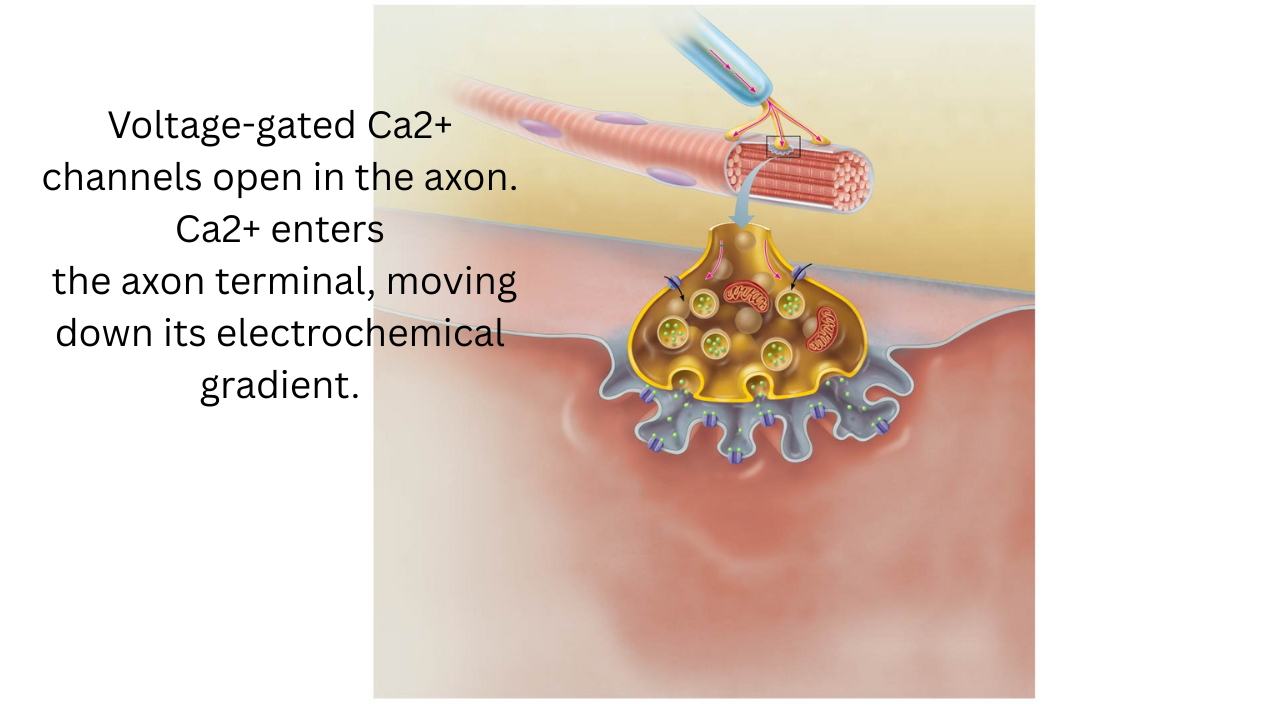
Voltage-gated Ca+2 channels open in the
axon Ca+2 enters the axon terminal, moving down its electrochemical gradient
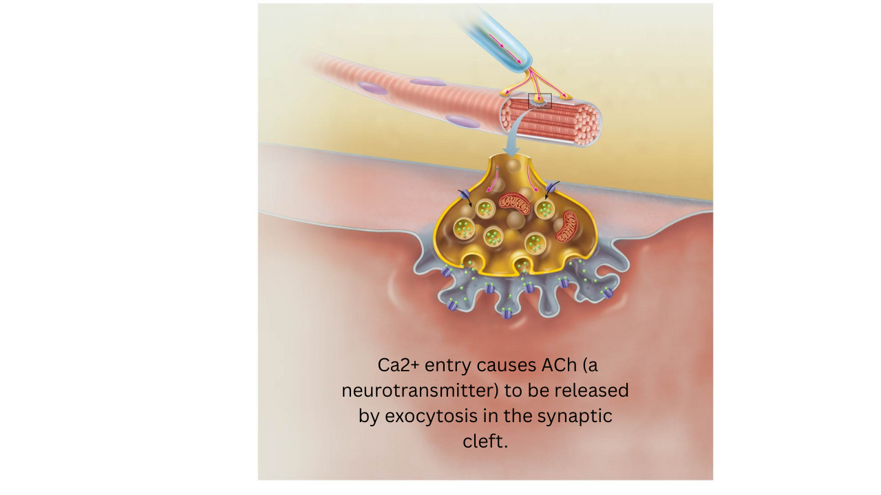
Ca+2 entry causes
ACh (a neurotransmitter) to be released by exocytosis in the synaptic cleft
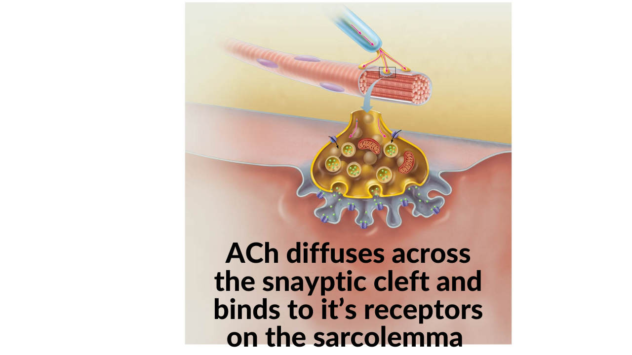
ACh diffuses across the
the synaptic cleft and binds to its receptors on the sarcolemma
ACh binds opens ion channels in the receptors that allow
simultaneous passaged of Na+ into the muscle fiber and K+ out of the muscle fiber. More Na+ ions enter than K+ ions exit, which produces a local depolarization of post synaptic cell.
In the muscle fiber this depolarization is called end late potential
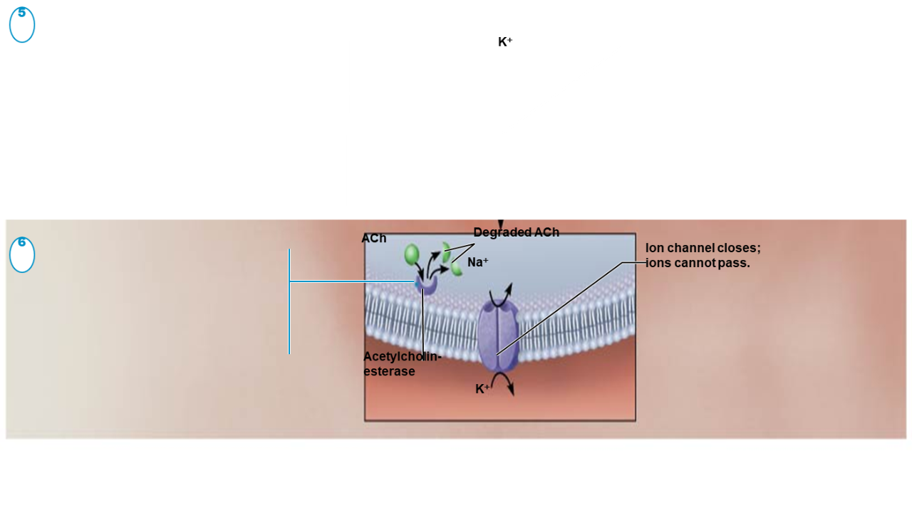
ACh effects are terminated by it’s breakdown in the
synaptic cleft by acetylcholinesterase into choline (taken by the axon) and acetate that diffuses away from the junction
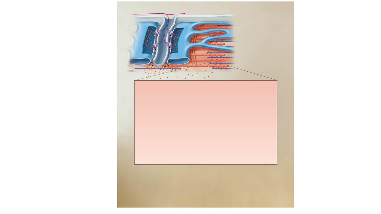
Action potential is generated on the
muscle plasma membrane and reaches the T-tuble
Excitation-Contraction Coupling
describes the rapid communication between electrical events occurring in the plasma membrane of skeletal muscle fibers and Ca+2 releases from the sarcoplasmic reticulum (SR) which leads to contraction
Action Potential propagates along the
sarcolemma and down the T tubules
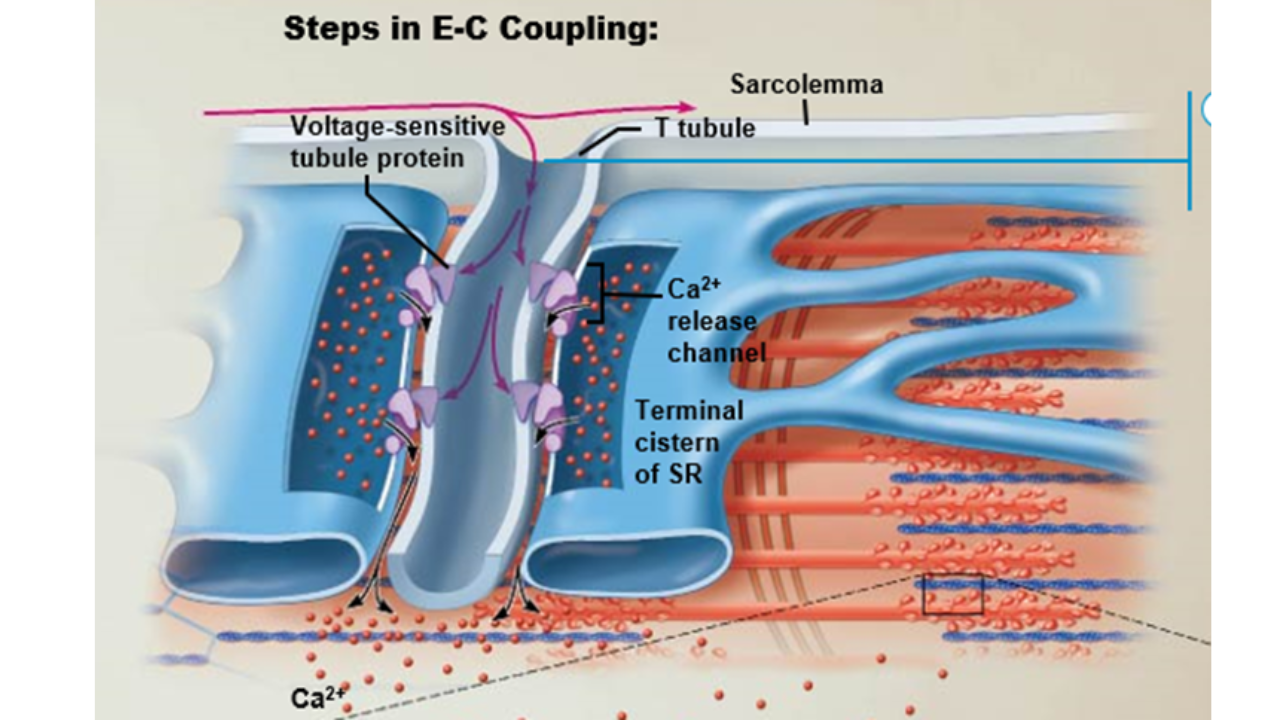
transmission of the AP along the T-tubles of the triads causes the
voltage-sensitive tuble proteins to change shape. This shape change opens the Ca+2 release channels in the terminal cisterns of the sarcoplasmic reticulum (SR) allowing Ca+2 to flow into the cytosol
calcium binds to troponin and removes the blocking action of tropomyosin
when Ca binds troponin changes shape, exposing binding sites for myosin (active sites) on the thin filaments
4) contraction begins
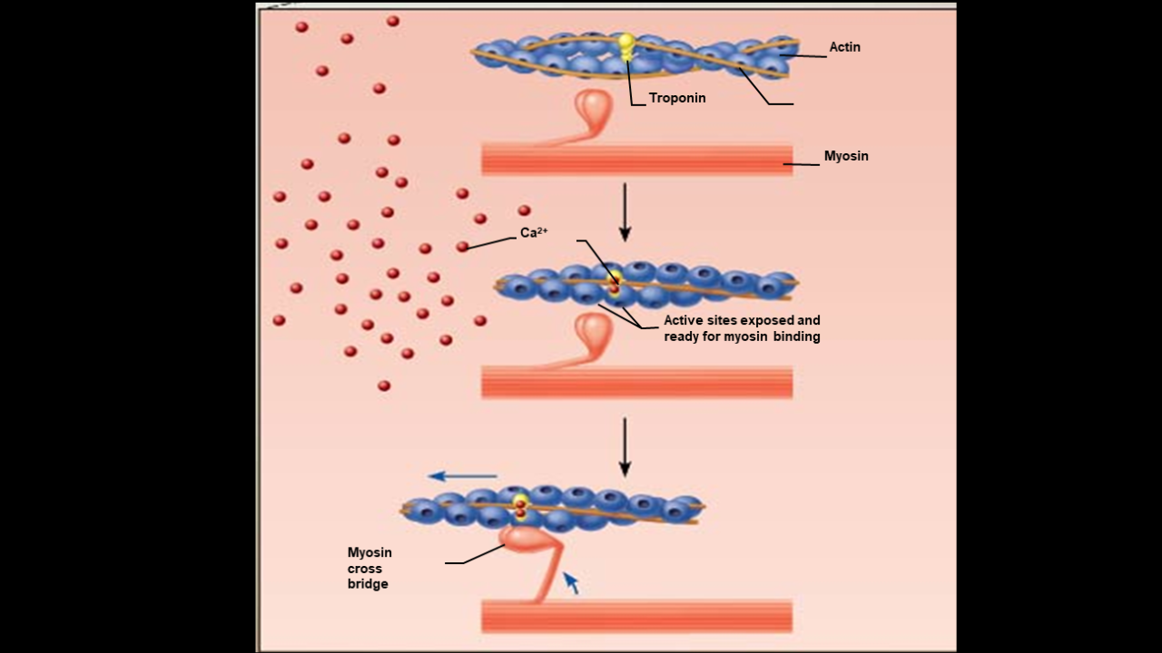
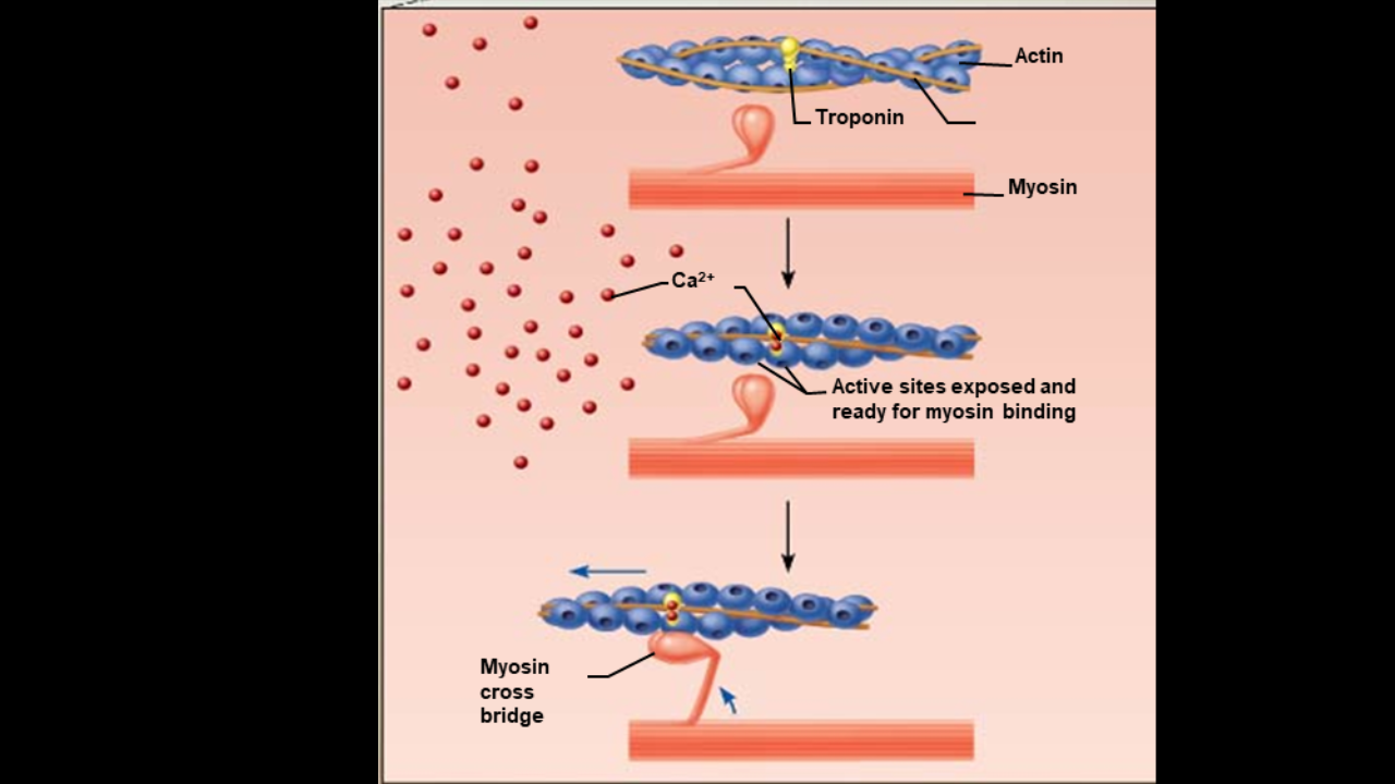
Contraction begins:
Myosin binding to actin forms cross bridges and contraction (cross bridge cycling) begins at this point E-C coupling is over
EC-coupling Excitation-Contraction Coupling
describes the rapid communication between electrical events occurring in the plasma membrane of skeletal muscle fibers and Ca+2 release from the sarcoplasmic reticulum (SR), which leads to contraction
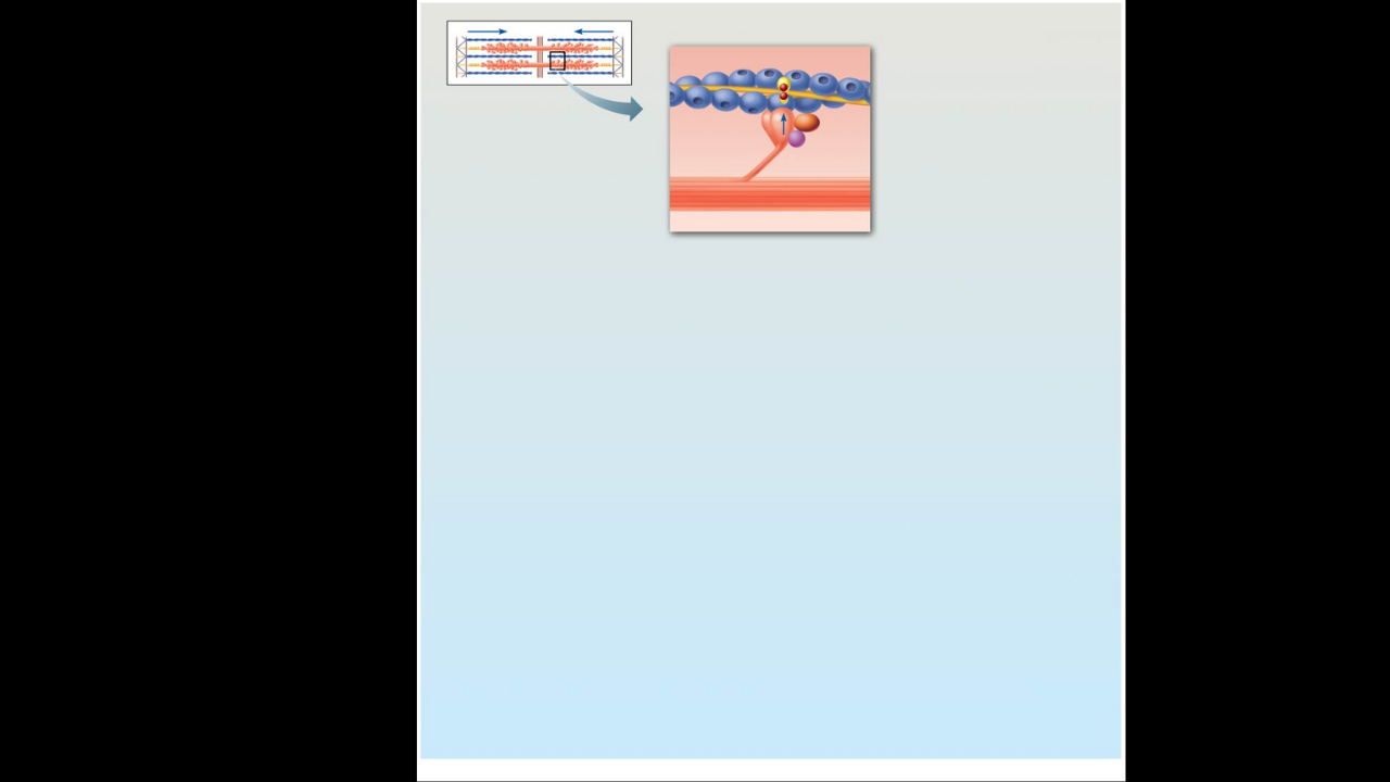
1) Cross Bridge formation
energized myosin head attaches to an actin myofilament forming cross bridge
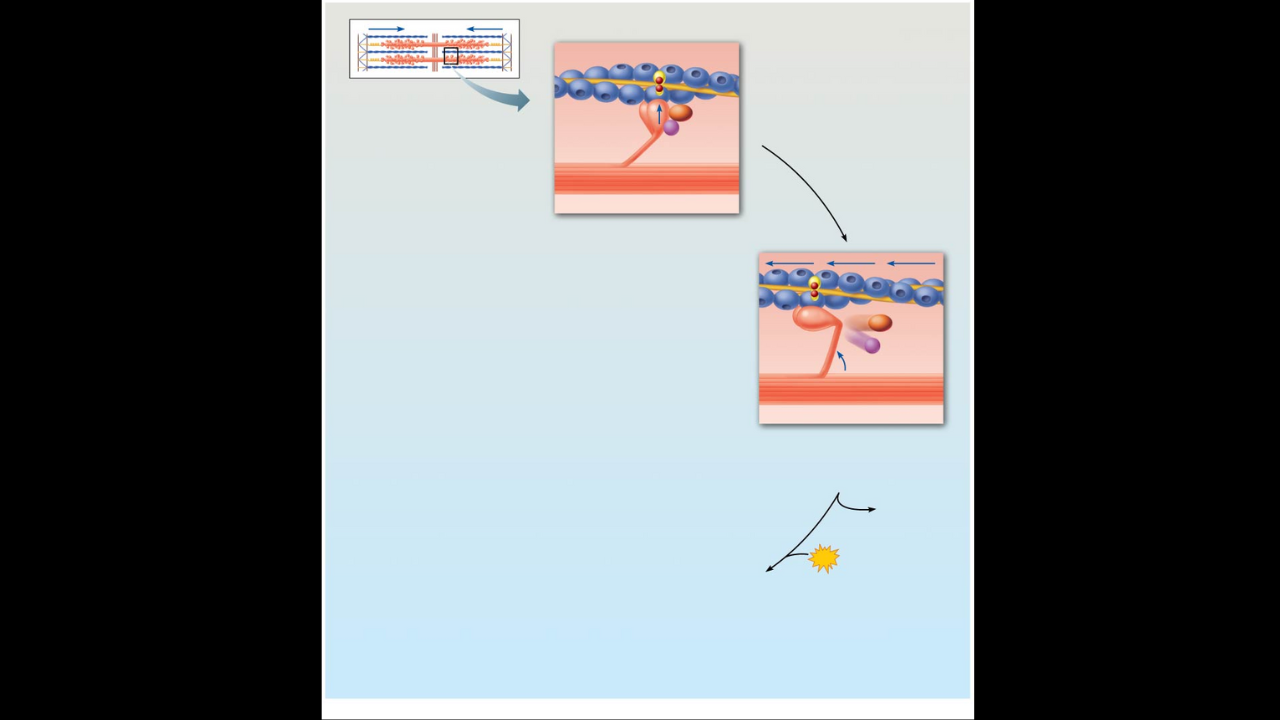
2) Power working stroke
ADP and P are released and the myosin head pivots and bends changing to its bent low-energy state. As a result it pulls the actin filament toward the M line.
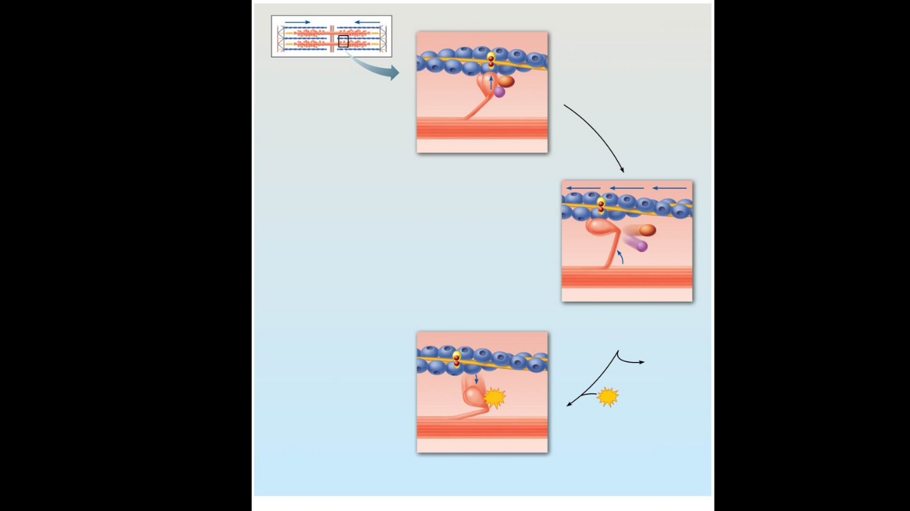
3) Cross bridge detachment
After ATP attaches to myosin the link between myosin and actin weakens and the myosin head detaches (cross bridge breaks)
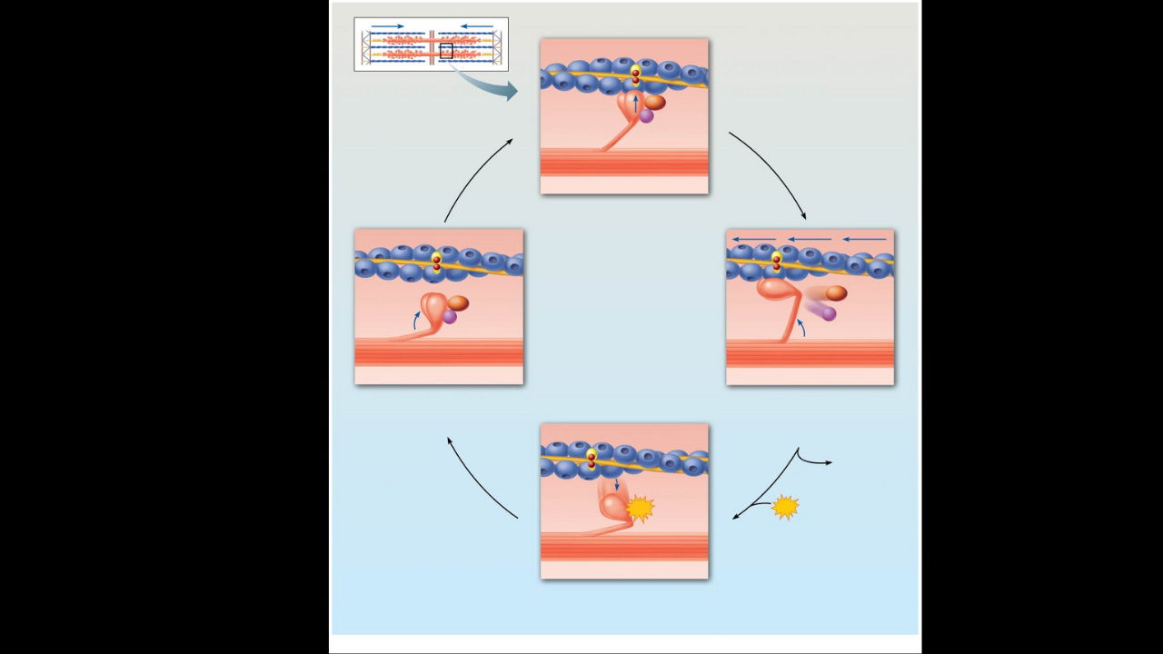
4) Cocking of the myosin head
As ATP is hydrolyzed to ADP and P the myosin returns to its prestroke high energy or cocked position which is ready to bind again to actin.
Rigor mortis
-34-4 hours after death, muscles begin to stiffen
-Peak rigidity occurs about 12 hours and postmortem
Intracellular calcium levels increase because
ATP is no longer being synthesized, so Calcium cannot be pumped back into SR
results in cross bridge formation