heart
1/61
There's no tags or description
Looks like no tags are added yet.
Name | Mastery | Learn | Test | Matching | Spaced | Call with Kai |
|---|
No analytics yet
Send a link to your students to track their progress
62 Terms
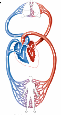
What is the heart’s role in the cardiovascular system?
The heart functions as a transport pump, moving blood through two main circuits:
Pulmonary circuit
Systemic circuit
Pulmonary circuit
Right side pumps oxygen-poor blood to the lungs to release CO₂ and pick up O₂.
Systemic circuit
Left side pumps oxygen-rich blood to the body tissues to deliver oxygen and nutrients.
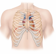
Where is the heart located in the human body?
in the mediastinum, the central area of the thoracic cavity
Between the second rib and fifth intercostal space
Where does the heart rest?
on the superior surface of the diaphragm
Anterior to the vertebral column and posterior to the sternum
What is the size of a heart?
a fist and weighs less than 1 pound
What is the purpose of the serous fluid in the pericardial cavity?
To reduce friction between the heart layers as the heart beats, allowing smooth, frictionless movement during contraction and relaxation.
What is the functional significance of the mediastinum?
It protects and anchors the heart in place while allowing room for the heart to expand and contract during each heartbeat.
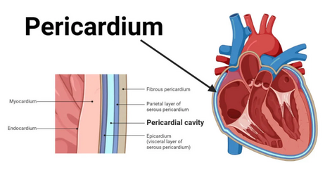
What is the pericardium?
A double-walled sac that surrounds and protects the heart.
What are the functions of the fibrous pericardium?
It protects, anchors the heart to surrounding structures, and prevents overfilling with blood.
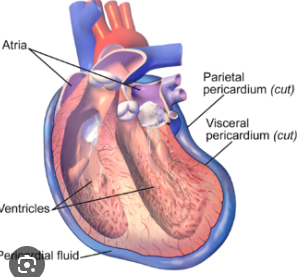
What are the deep two layers of the serous pericardium?
Parietal layer
Visceral layer (epicardium)
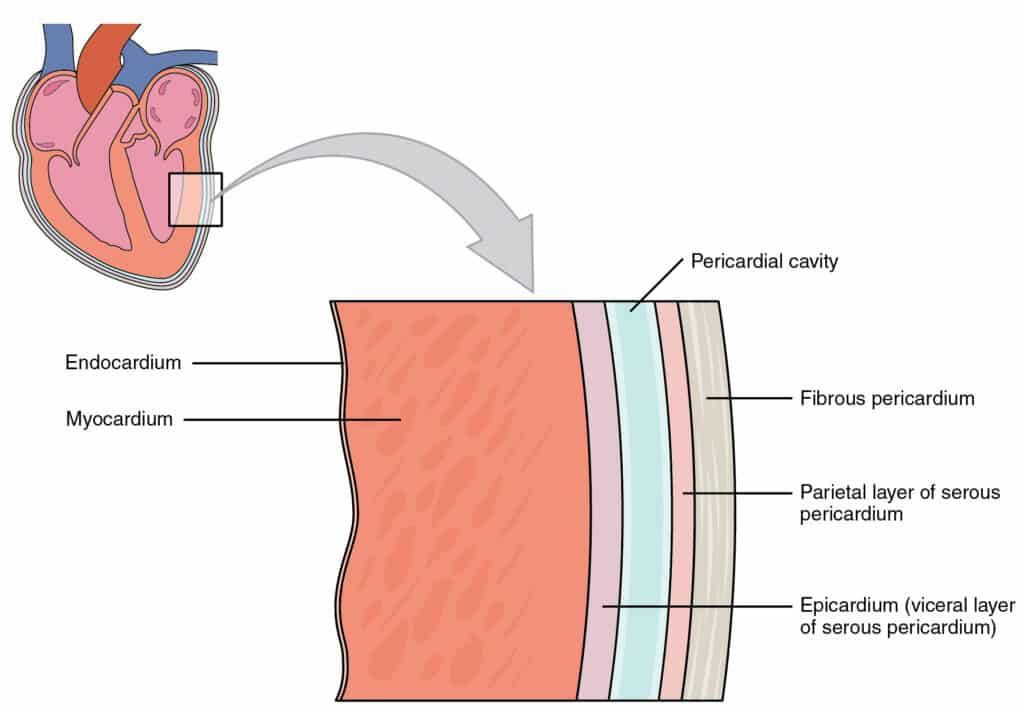
Parietal layer
lines the internal surface of the fibrous pericardium
Visceral layer (epicardium)
covers the external surface of the heart
What is the function of the pericardial cavity?
It contains serous fluid that reduces friction between heart layers during movement.
Layers of the Heart wall
Epicardium
Myocardium
Endocardium
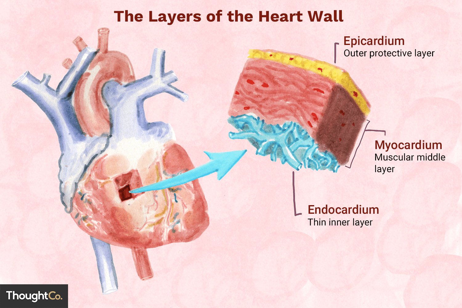
What is the epicardium?
The outermost layer of the heart; it is the visceral layer of the serous pericardium.
What is the myocardium and its function?
The middle layer, made of circular or spiral bundles of contractile cardiac muscle cells
it is responsible for heart contractions.
What is the cardiac skeleton?
A connective tissue framework that anchors cardiac muscle fibers and supports valves.
What is the endocardium?
The innermost layer of the heart that lines the heart chambers and covers the cardiac skeleton of the valves.
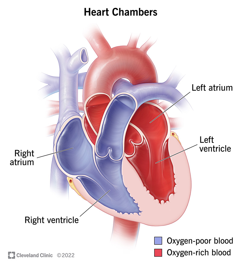
How many chambers does the heart have, and what are they?
Two atria (superior)
Two ventricles (inferior)
What does the interatrial septum do?
It separates the right and left atria.
What does the interventricular septum do?
It separates the right and left ventricles.
What is the coronary sulcus (atrioventricular groove)?
A groove that encircles the junction between the atria and ventricles.
What are the anterior and posterior interventricular sulci?
Grooves that mark the boundary between the left and right ventricles on the front and back of the heart.
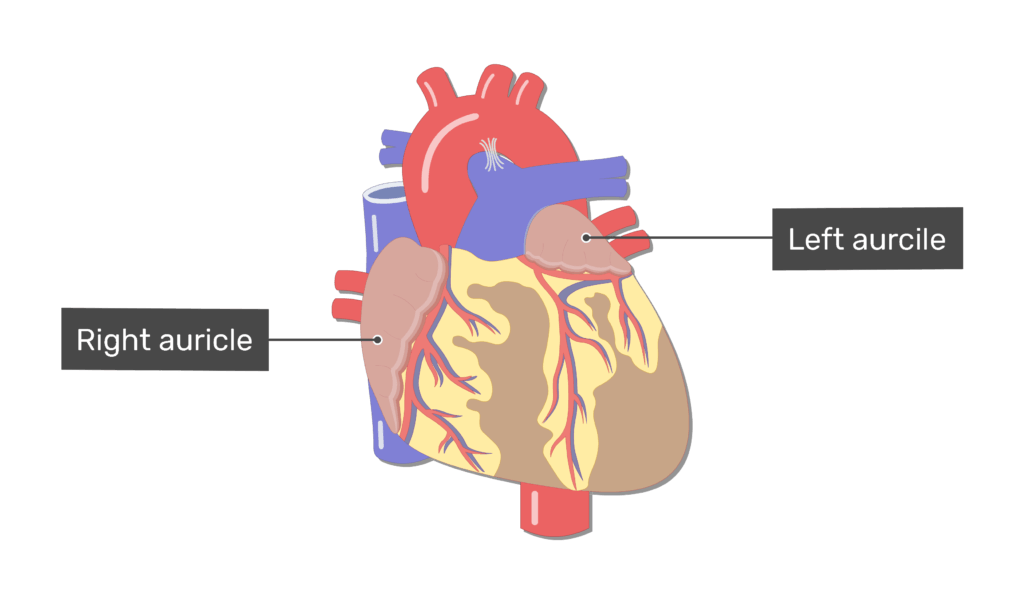
Auricles
ear-like appendages on each atrium that increase atrial volume
What is the main function of the right atrium?
It receives deoxygenated blood from the body.
What are the structural features of the right atrium?
Anterior wall is smooth
Posterior wall has ridges formed by pectinate muscles
Superior and inferior vena cava
drain into the right atrium
both carrying deoxygenated blood from the body.
What is the main function of the left atrium?
It receives oxygenated blood from the lungs.
Which vessels return blood to the left atrium?
The four pulmonary veins, carrying oxygenated blood from the lungs.
ventricles
They are the discharging chambers and the actual pumps of the heart.
have thicker walls than the atria.
Where does the right ventricle pump blood?
pumps blood into pulmonary trunk
Where does the left ventricle pump blood?
pumps blood into aorta
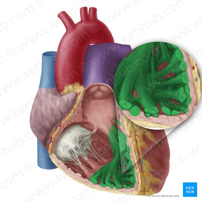
What are trabeculae carneae?
Irregular ridges of muscle on the inner walls of the ventricles.
What are papillary muscles and what do they do?
Muscles that project into the ventricular cavity
-anchor the chordae tendineae
What is the primary function of heart valves?
To ensure unidirectional blood flow through the heart.
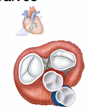
What are the two major types of heart valves?
Atrioventricular (AV) valves – between atria and ventricles
Semilunar valves – between ventricles and major arteries
What is the function of the atrioventricular (AV) valves?
To prevent backflow into the atria when the ventricles contract.
What is the tricuspid valve and where is it located?
The right AV valve, made of three cusps, located between the right atrium and right ventricle.
What is the bicuspid (mitral) valve and where is it located?
The left AV valve, made of two cusps, located between the left atrium and left ventricle.
What are chordae tendineae?
Tough, fibrous cords that anchor the cusps of AV valves to the papillary muscles.
What is the function of the chordae tendineae and papillary muscles?
Hold valve flaps in the closed position during ventricular contraction
Prevent valve flaps from everting (flipping backward) into the atria
What is the function of the semilunar (SL) valves?
prevent backflow from major arteries back into ventricles
Where is the pulmonary semilunar valve located?
located between right ventricle and pulmonary trunk
Where is the aortic semilunar valve located?
located between left ventricle and aorta
What is the function of the papillary muscles and chordae tendineae?
They anchor the AV valve flaps and prevent them from inverting (everting) into the atria during ventricular contraction, ensuring one-way blood flow.
Q: Is the left ventricle wall thicker than the right?
A: ✅ Yes — it pumps to the whole body, needing more force.
Q: Does the left ventricle pump at higher pressure?
A: ✅ Yes — systemic circulation has more resistance.
Q: Does the left ventricle pump more blood per beat?
A: ❌ No — both sides pump equal volumes each beat.
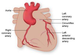
Where do the coronary arteries originate?
From the base of the aorta.
What are anastomoses in coronary arteries?
Junctions
•Provide additional routes for blood delivery
•Cannot compensate for coronary artery occlusion
What is the function of coronary veins?
To collect blood from the capillary beds of the heart muscle.
Where does the coronary sinus empty?
Into the right atrium
What veins form the coronary sinus?
The great, middle, and small cardiac veins.
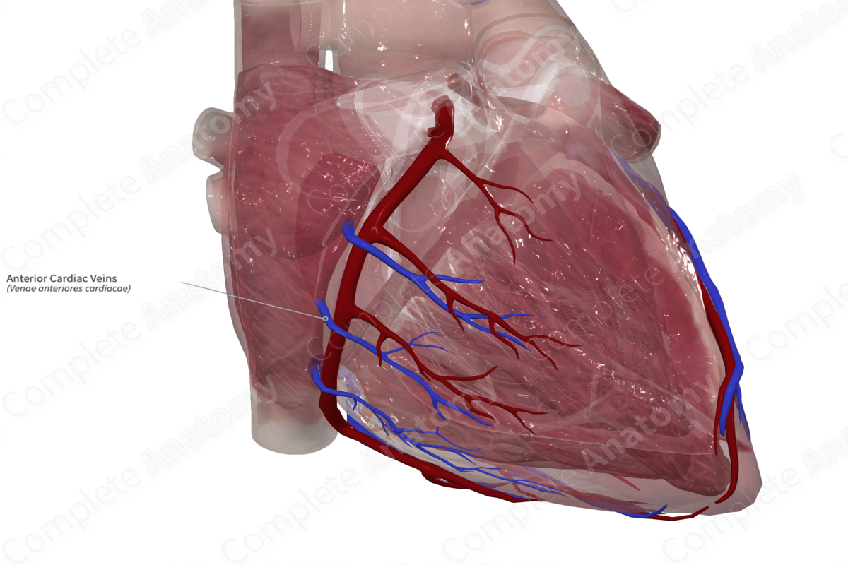
What do the anterior cardiac veins do?
They empty directly into the right atrium from the front of the heart.
What are the structural features of cardiac muscle cells?
They are striated, short, branched, fat, and interconnected.
Why are cardiac muscle cells resistant to fatigue?
They contain numerous large mitochondria.
How do cardiac muscle T tubules and sarcoplasmic reticulum (SR) compare to skeletal muscle?
T tubules: Wider but less numerous
SR: Simpler, and no triads
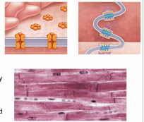
What are intercalated discs?
Connecting junctions between cardiac muscle cells.
Allows heart to be a functional syncytium, a single coordinated unit
What is the function of desmosomes in intercalated discs?
They hold cells together and prevent separation during contraction.
What is the function of gap junctions in intercalated discs?
They allow ions to pass between cells, enabling electrical coupling
What are the differences between skeletal and cardiac muscle cells? What are the functional reasons for these differences?
Cardiac muscle is built for endurance and coordination, while skeletal muscle is designed for voluntary, forceful contractions.