The Back - OTH 504 WVU
1/80
There's no tags or description
Looks like no tags are added yet.
Name | Mastery | Learn | Test | Matching | Spaced |
|---|
No study sessions yet.
81 Terms
1. Superficial Group associated with the shoulder girdle, Intermediate muscles involved with respiration, and deep muscles with the vertebral column
What are the 3 Main Groups of back muscles?
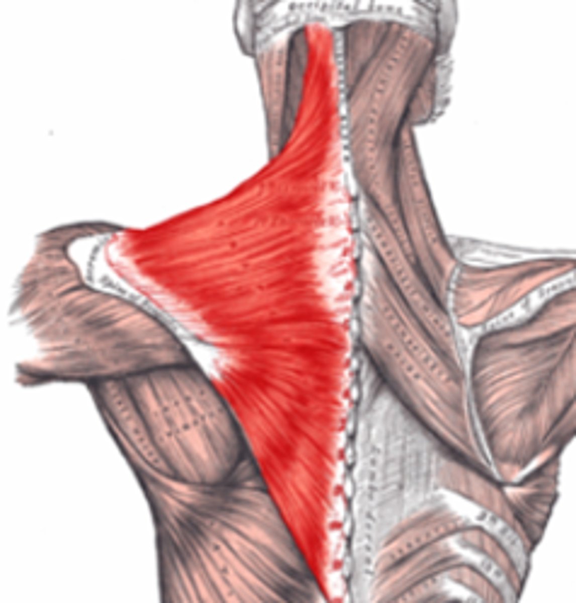
Trapezius
There is an upper, middle and lower part to this muscle
Origin: nuchal line, occipital protuberance, ligament niche, spinous process C7-t12
Insertion: lateral clavicle, acromion, spine of scapula
Nerve: spinal accessory nerve 11
Action: elevate and depress scapula, retraction
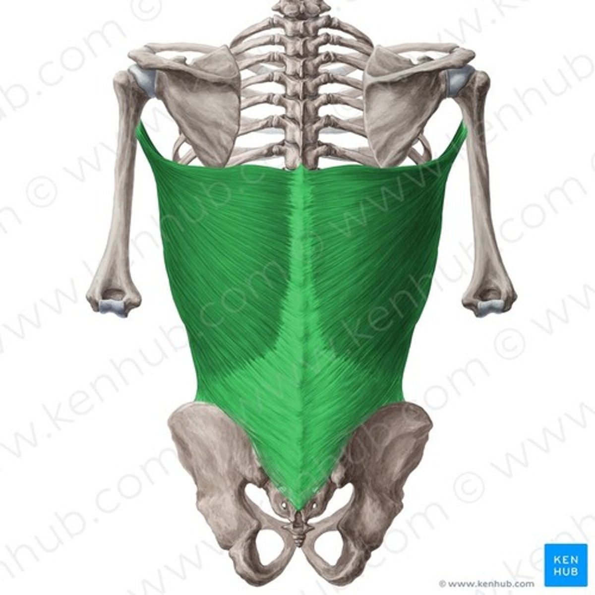
Larissimus Dorsi
Origin: spines from T7 to sacrum, iliac crest, lower ribs
Inserton: intertubecular groove humerus
Nerve Supply: thoracodorsal nerve, posterior cord of brachial plexus
Acton: extends, adducts, and medially rotates arm
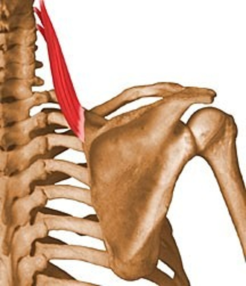
Levator Scapulae
Origin: transverse
processes C1-4
Inserton: medial border of scapula
Nerve supply: 3rd and 4th cervical nerves and dorsal scapular nerve (C5)
Acton: elevates the scapula
Rhomboid Minor
Origin: ligamentum nuchae; C7-T1 spines
Inserton: medial border of scapula
Nerve supply: dorsal scapular nerve (C5)
Acton: with rhomboid major and levator scapulae, elevates, retracts and rotates the scapula
Rhomboid Major
Origin: T2-T5 spines
Inserton: medial border of scapula
Nerve supply: dorsal scapular nerve (C5)
Acton: with rhomboid minor and levator scapulae, elevates, retracts and rotates the scapula
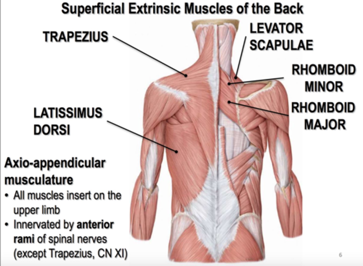
Superficial muscles of the back
trapezius, latissimus dorsi, rhomboid major, rhomboid minor, levator scapulae
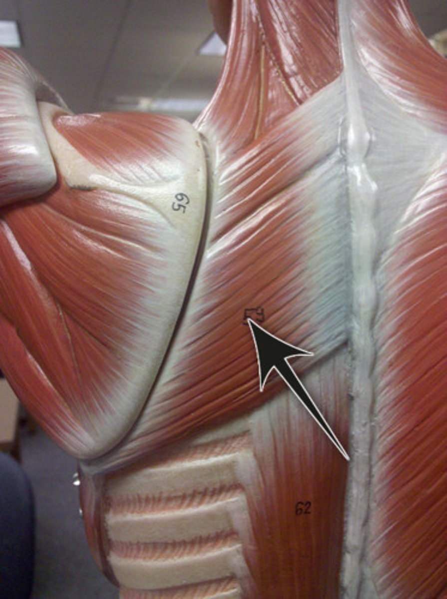
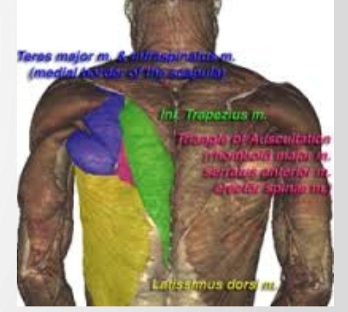
Triangle of Auscultaton
Medially= trapezius
Superiorly= rhomboid major Inferiorly= latssimus dorsi
Floor= thoracic wall
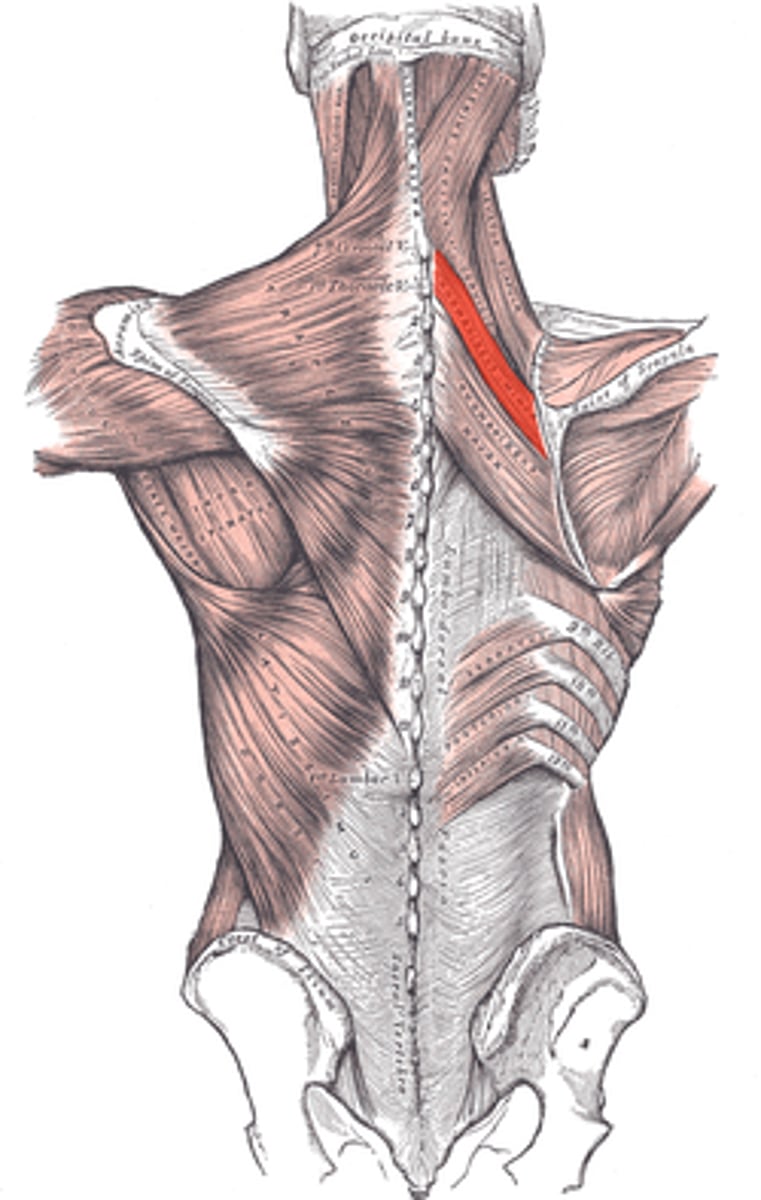
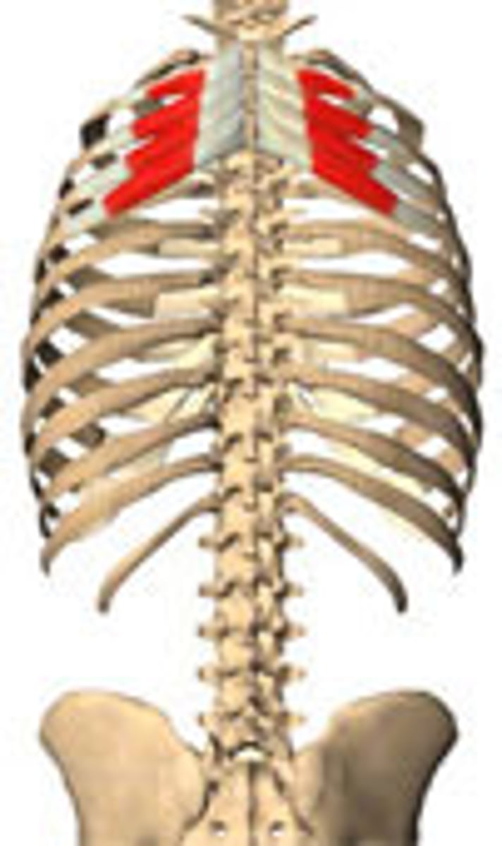
Serratus Posterior Superior
Origin: ligamentum nuchae, C7, T1-3 spines
Inserton: ribs 2-5
Acton: elevates ribs (i.e. an inspiratory muscle)
Nerve supply: intercostal nerves
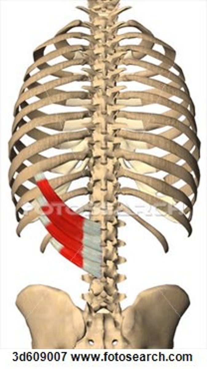
Serratus Posterior Inferior
Origin: thoracolumbar fascia, T11-12, L1-2 spines
Inserton: ribs 9-12
Acton: depresses ribs (i.e. an expiratory muscle)
Nerve supply: intercostal nerves
Deep Muscles of the Back
postural tone
responsible for maintenance of normal vertebral curves
form broad, thick column of muscle tssue, which occupies each side of spinous processes
extend from sacrum to skull and lie beneath thoracolumbar fascia
vertebral spines and transverse processes serve as levers that facilitate muscle actons
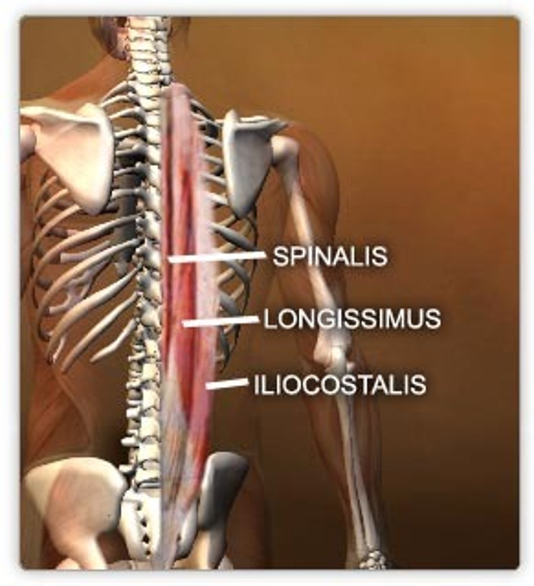
erector spinae
Deep, vertically running back muscles: iliocostalis, longissimus, spinalis
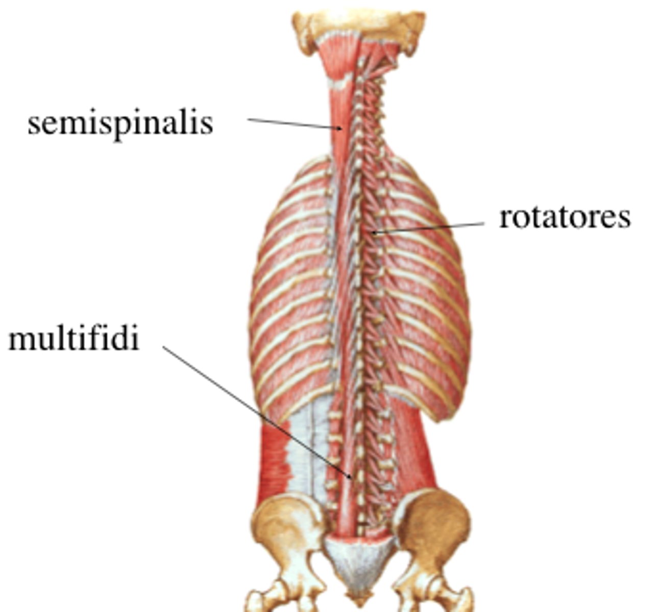
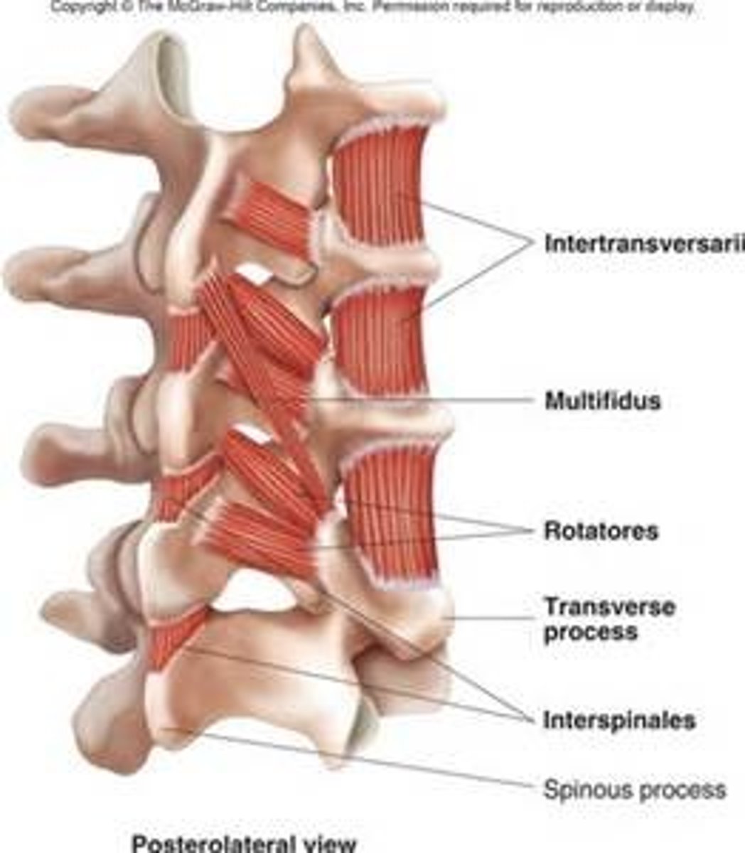
Transversospinalis
Oblique running back muscles: semispinalis, multifidus, rotatores
Interspinales, intertransversarii
deepest back muscles
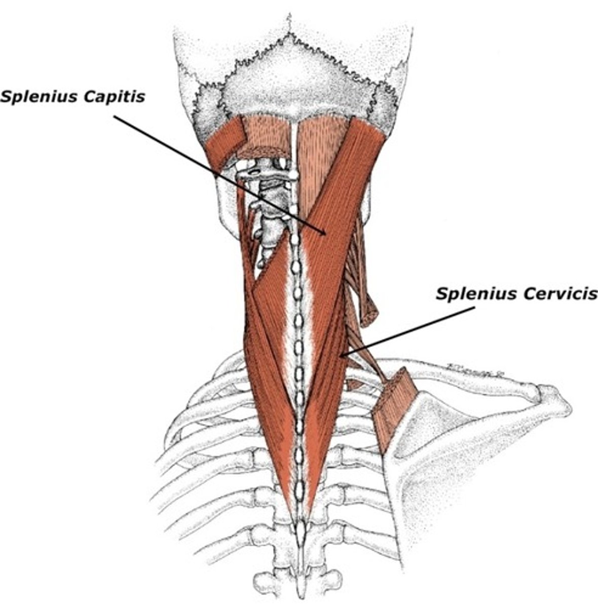
Splenius capitis and Cervicis Muscles
Other Muscles
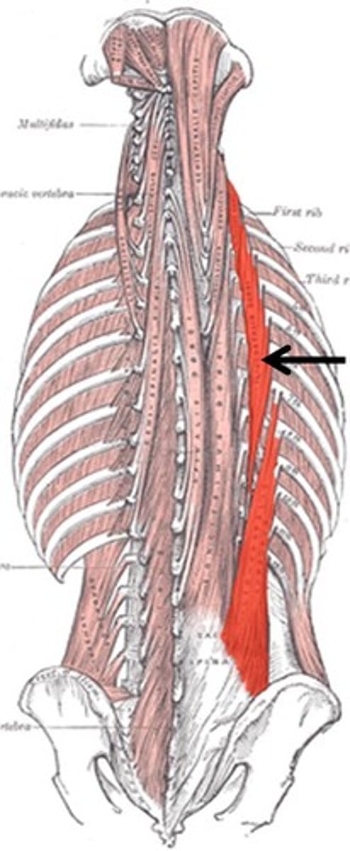
Iliocostalis (erector spinae)
•• Inserton: angles of ribs
Origin: iliac crest, sacrum
• Innervaton: spinal nerves C4-S5
•
Acton: extends and laterally bends trunk and neck
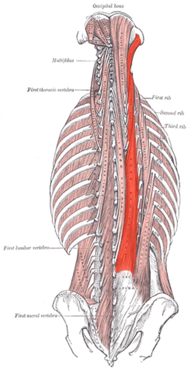
Longissimus (erector Spinae)
• Inserton: transverse processes of superior vertebrae; mastoid
Origin: transverse processes of inferior vertebrae
process•
Innervaton: spinal nerves C1-S1•
Acton: extends, laterally bends trunk, neck and head
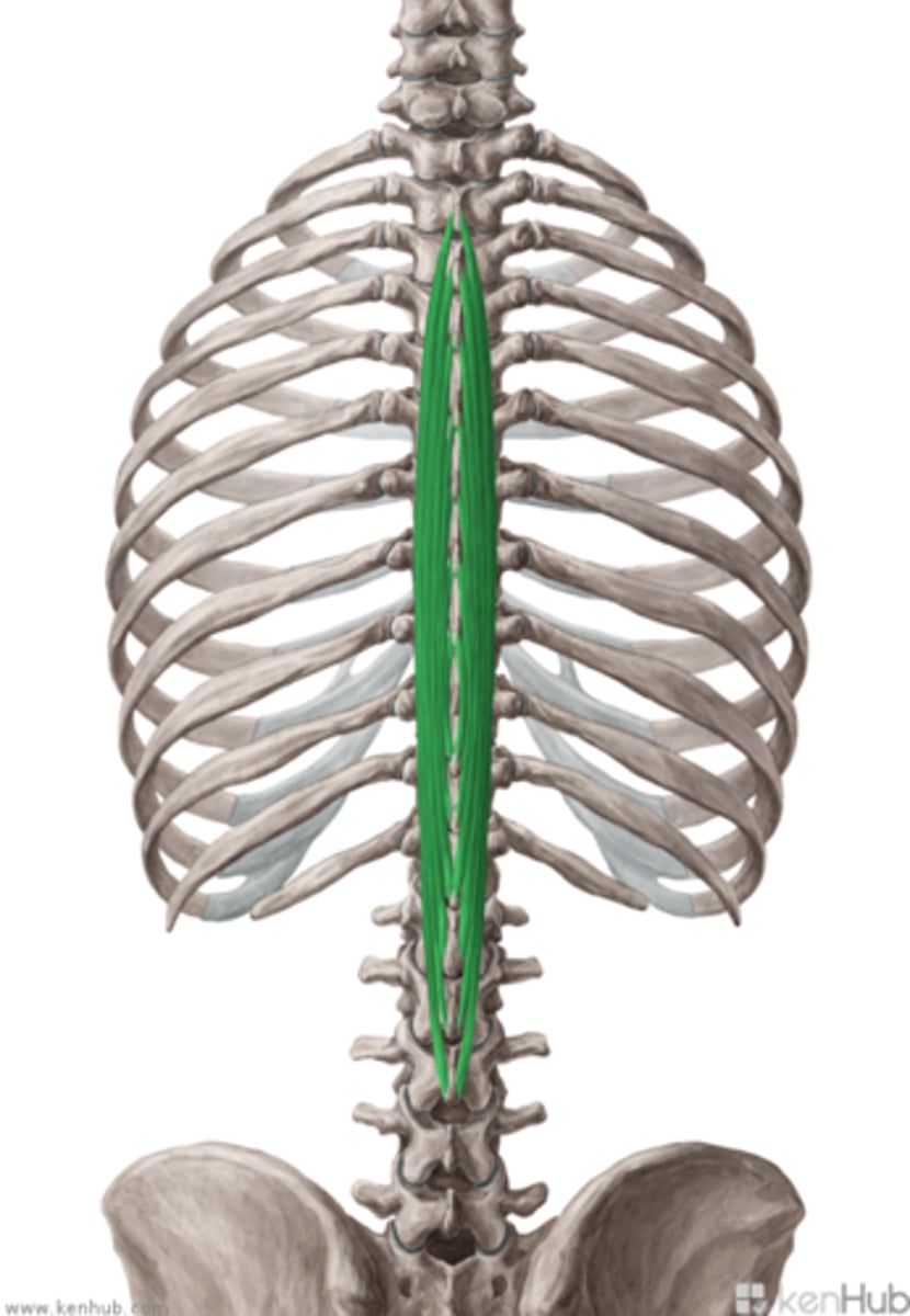
Spinalis (erector Spinae)
• Inserton: spinous processes of superior vertebrae, base of
Origin: spinous processes of inferior vertebrae
skull• Innervaton: spinal nerves C2-L3• Acton: extends and laterally bends trunk and neck
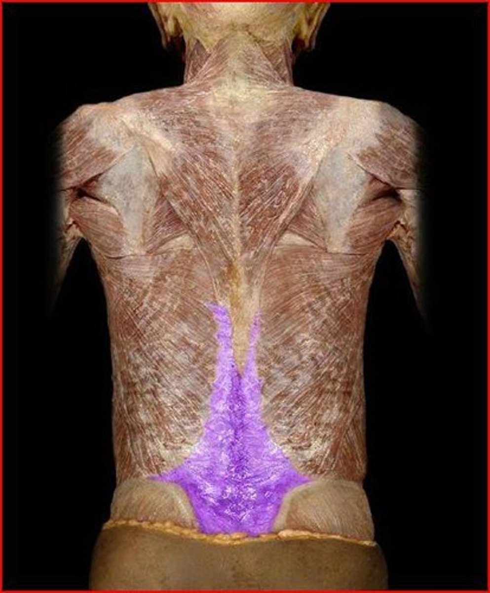
thoracolumbar fascia
sheet of connective tissue on lower portion of the back
lumbar part of deep fascia situated in interval between iliac crest and twelfh rib
forms strong aponeurosis
covers posterior surface of deep muscles of back
Anterior
These spinal Ligaments are wide and strong, and is atached to the front and sides of the margins of vertebral bodies and intervertebral discs
Posterior
These spinal ligaments are weak and narrow, attracted to the posterior border of discs
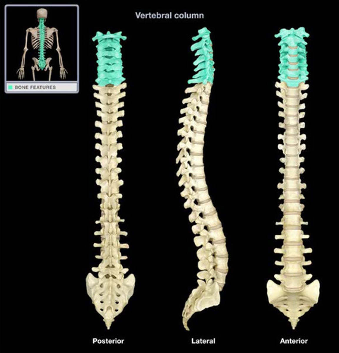
Two primary and two secondary
fully developed individuals how many curves?
Primary Curve
Infant curves?
Primary and secondary where the child begins to stand upright
End of first year curves?
lordosis
abnormal anterior curvature of the lumbar spine (sway-back condition)
kyphosis
excessive outward curvature of the spine, causing hunching of the back.
Babies are born with a primary curve a " ) ", as they gain head control they develop their first secondary curve a "(" in the cervical spine region.
Which of the following developmental milestones leads to an infant developing the first secondary curve of the spine?
Short head of the Biceps femorius which originates on the linea Aspera of the femur. Now, you might be thinking "But wait, the Vastus Lateralis also does not originate on the Ischial Tuberosity!" This is correct however the question asks which Hamstring muscle does not originate on the tuberosity and the Vastus Lateralis is not a hamstring muscle. This might seem tricky but it is checking both your comprehension of which muscles are hamstrings and the origins of the hamstrings.
This structure is the only muscle of the Hamstrings that does not originate on the ischial tuberosity
Cervical, Thoracic, Lumbar, Sacrum, Coccyx
5 regions of the vertebral column
cervical vertebrae
C1-C7
thoracic vertebrae
T1-T12
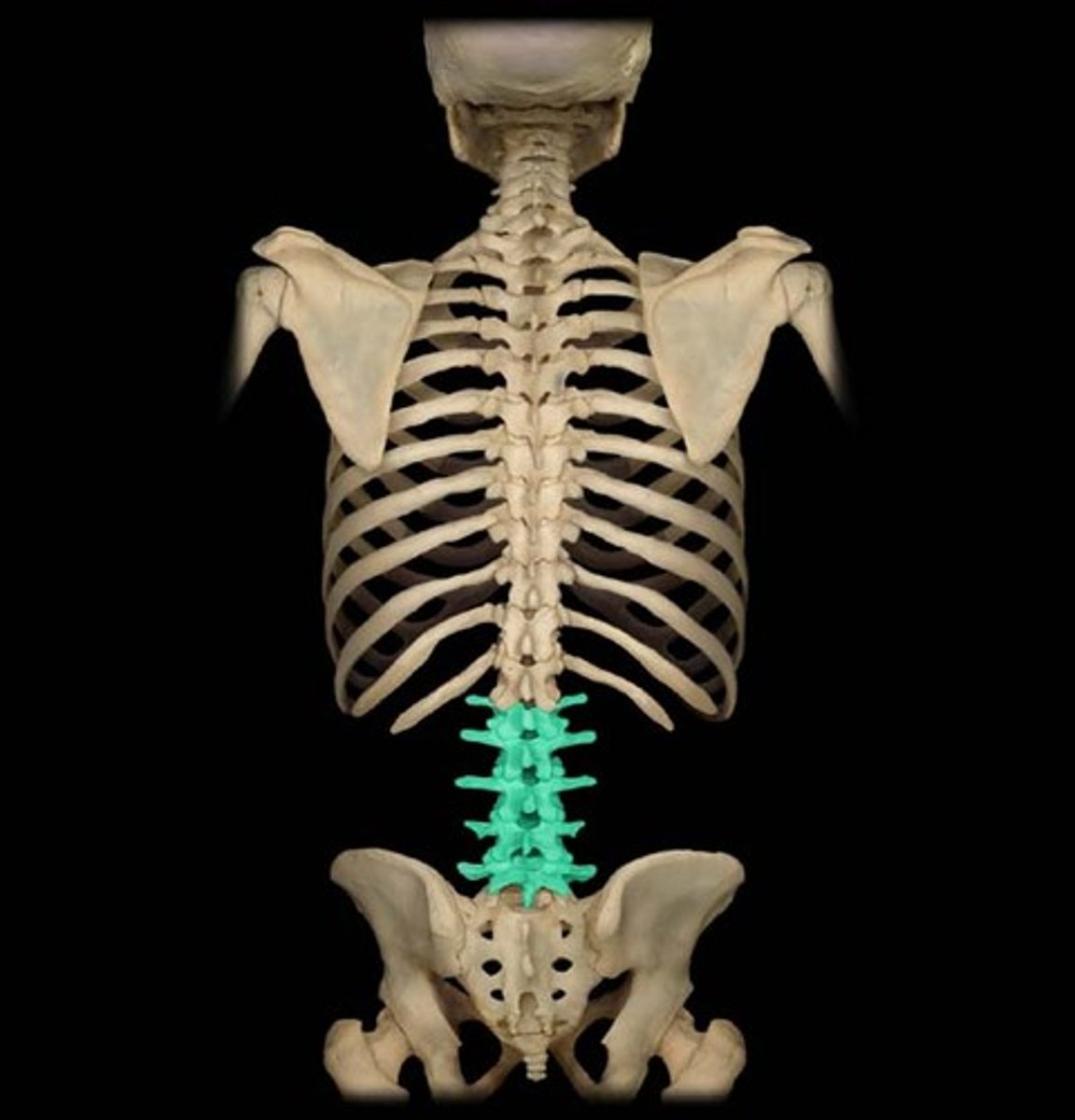
lumbar vertebrae
L1-L5
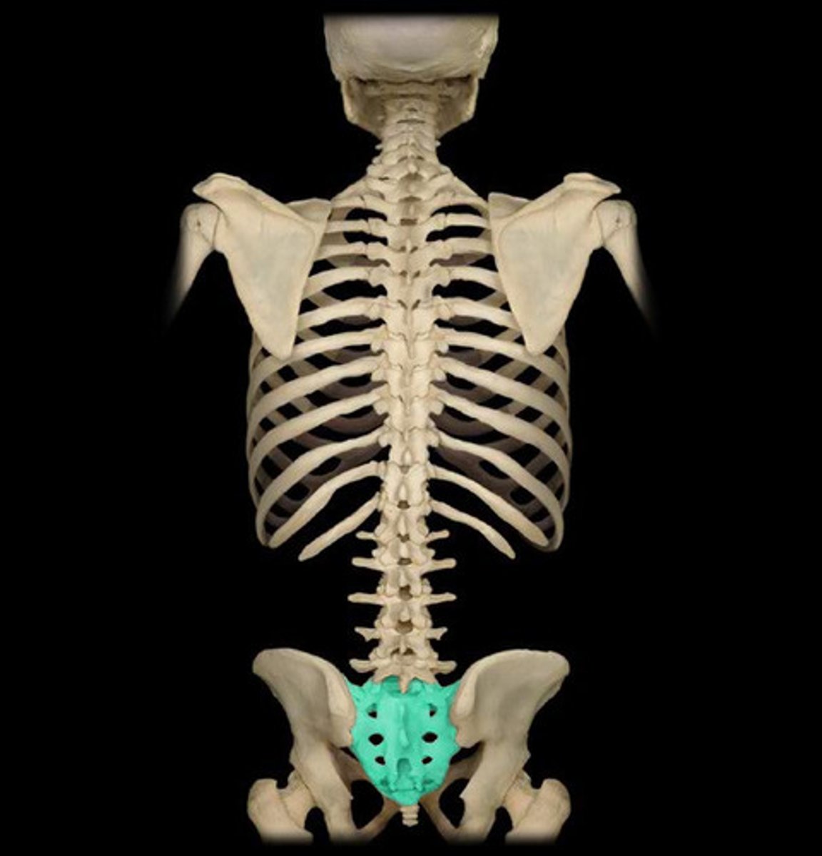
Sacral
Fused 5 vertebrae
Coccyx
Fused 4 vertebrae
Pedicles of the vertebral arch
walls of the vertebral arch
laminae
roof of the vertebral arch
vertebral body
main portion of the vertebra, separate from the arches of the vertebra, supports the bodyweight
4 (2 superior and 2 inferior)
How many and what are the vertebra articulations to make the foramen?
Cervical
Most unprotected part of the vertebrae?
vertebral foramen
canal through which spinal cord passes formed by the two superior processes articulating with the inferior processes
articular process
this causes restriction of movement in the vertebrae
vertebral arch
composed of pedicles, laminae, and sometimes spinous process- it represents the junction of all posterior extensions from the vertebral body and protects the spinal cord
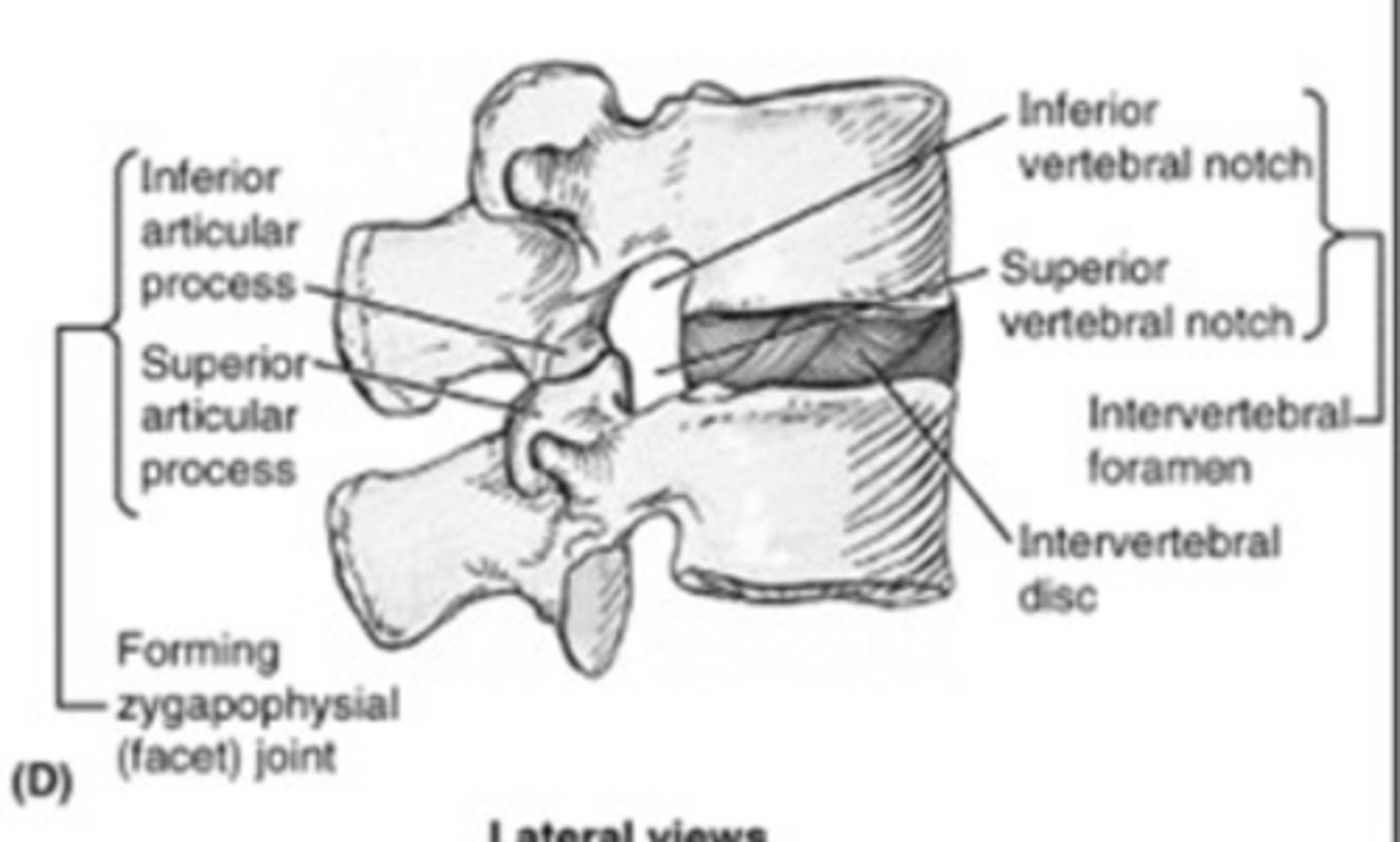
intervertebral foramen
superiornotchofone vertebra and inferior notch of adjacent vertebra form
ormingsuperiorandinferior vertebral notches
two superior articular processes of one vertebral arch articulate with two inferior articular processes of arch above
BLANK; on each side
Cervical Vertebrae (C1-C7)
first set of seven bones, forming the neck,. Transverse processes have a foramen
Spinous processes are small and bifid
Body is small and rectangular
Vertebral foramen are large and triangular
C3-C6
What part of the cervical have BIFOIDS?
C7
has a longer spinous process (not bifid) and smaller transverse foramen - also the bump on the back of the neck
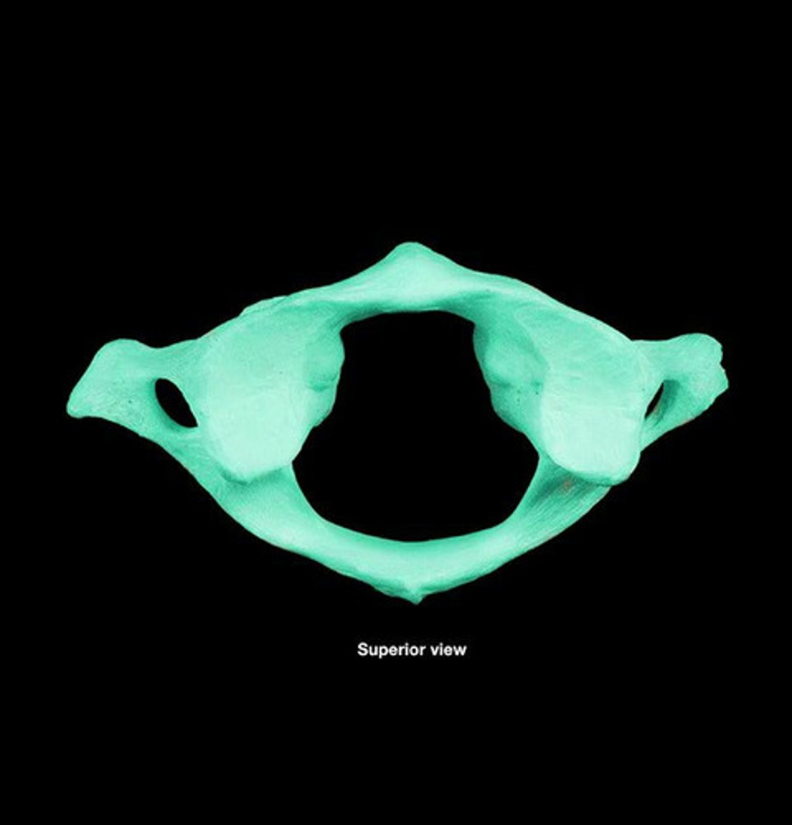
Atlas
C1• Ring shaped• Widest of the cervical vertebrae • No spinous process or body
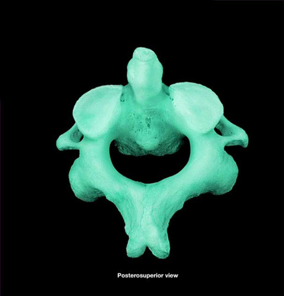
Axis
C2• Strongest cervical vertebrae• Carries the skull, allows for rotation• Has a dens-odontoid process-projects superiorly from the body
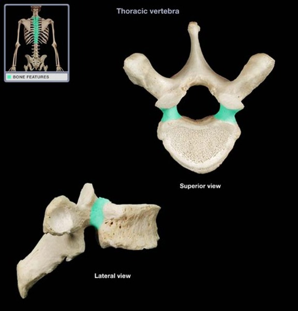
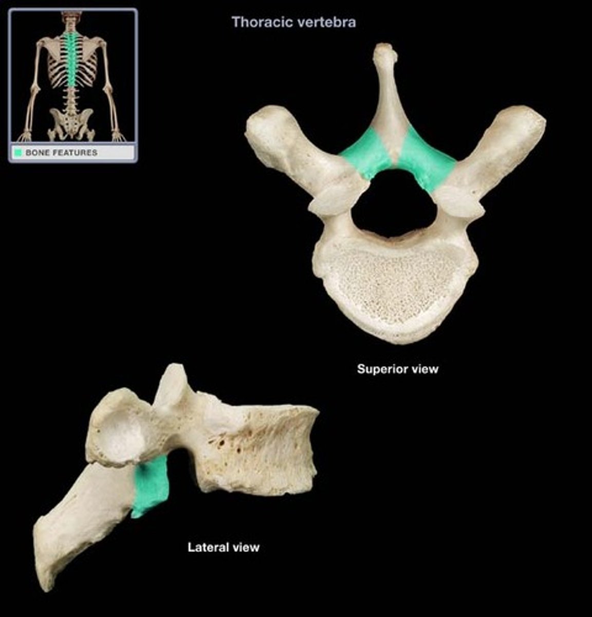
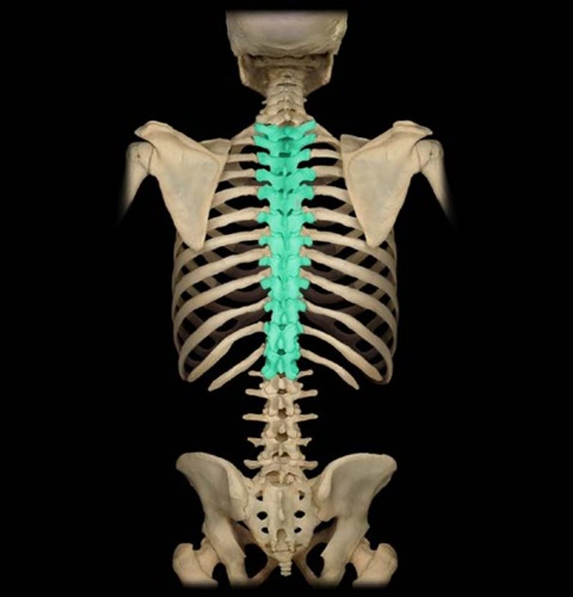
Thoracic Vertebrae
Heart-shaped, medium-sized body
Bodies increase in size from superior to inferior
Smaller,circularvertebralforamen
Spines are long and downward
Coastal facets are lateral to the body articulates with the head of the ribs
Transverse process for articulation with the tubercles of the ribs
Lumbar Vertebrae
Body is large and kidney shaped
Thick laminae
Triangular foramen
Long and slender transverse processes
Spinous processes are short, flat, and quadrangular
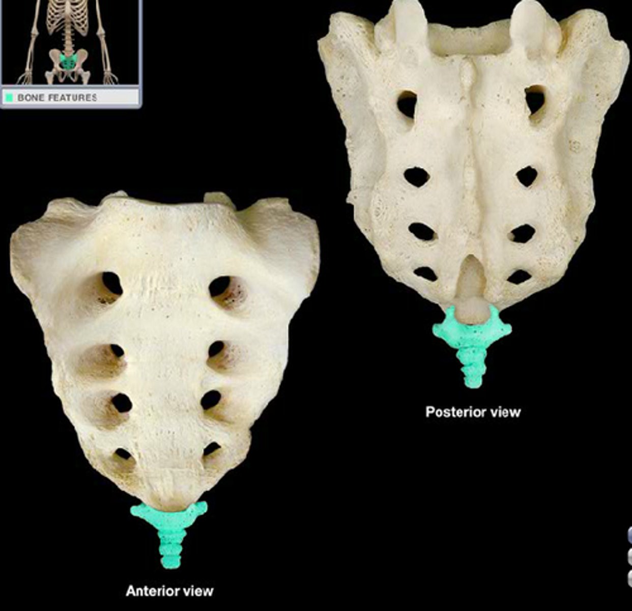
Sacral Vertebrae
5 fused vertebrae
Forms a wedge shaped
bone
Superiorly connects with the 5th lumbar vertebra
Inferiorly connects with the coccyx
Vertebral foramina formed together form the sacral canal
Coccyx Vertebrae
4 fused vertebrae
Forms a single small
triangular bone
Articulates with the end of the sacrum
Co.1 has 2 transverse processes
Remnant of the skeleton of the tail
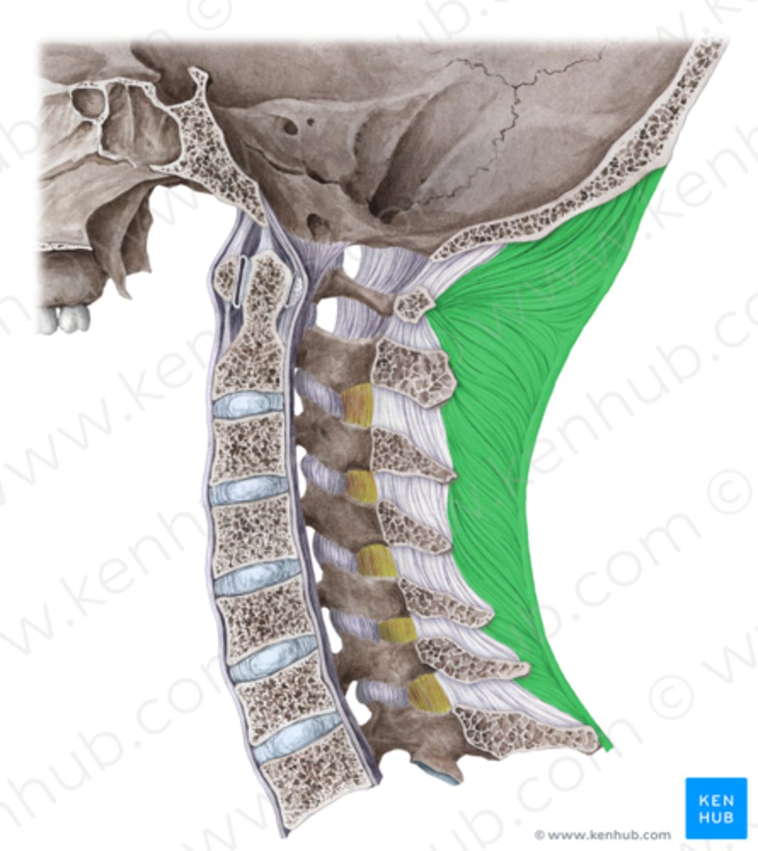
Ligamentum nuchae
Occipital protuberance,
spine of C7,
Formed from aponeurosis of cervical muscles
Attaches to dura • Whiplash
(Synovial) atlanto-occipital joint
What kind of joints are the vertebral column? Synovial joints formed between occipital condyles and superior
facets on the atlas
flexion, extension and lateral flexion
three movements of the the vertebral column
Anterior atlanto-occipital membrane
Continuation of the anterior longitudinal ligament
Connects the anterior arch of the atlas to the anterior margin of foramen magnum
posterior ligament of back
weak and narrow ligament of back
anterior ligament of back
wide and strong ligament of back
Ventral
Afferent Root?
Dorsal
Efferent Root
Efferent, afferent, and autonomic signals
spinal nerve signals
C7-T1
Hand and Finger Movement
C5
Wrist and Elbow
L2
Hip Motion
L3
knee extension
L4-S1
Foot Motion
L5
Knee Flexion
T2-T12 myotome
Trunk stability
abdominal muscles
c5-c8
Dermatome of Arm and hands
C5-C8
Myotome Movement of ARM, SHOULDER, HAND
L1-S2
Myotome of Legs
T9-L1
ABS MYOTOMES
trapezius (superior fibers), rhomboid minor, serratus posterior superior, splenius capitis and spinalis.
Muscle Attachments of Ligamentum Nuchae
L1-S2
Lower Body Nerve Function Dermatome
T2-T12
Torso Nerve Function
8
Cervical Nerve Numbers
1
Coccyx Nerves Number
5
Sacral Nerve Number
5
Lumbar Nerve Number
12
Thoracic Nerve Number