Posterior Leg + Ankle
1/23
There's no tags or description
Looks like no tags are added yet.
Name | Mastery | Learn | Test | Matching | Spaced | Call with Kai |
|---|
No analytics yet
Send a link to your students to track their progress
24 Terms
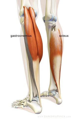
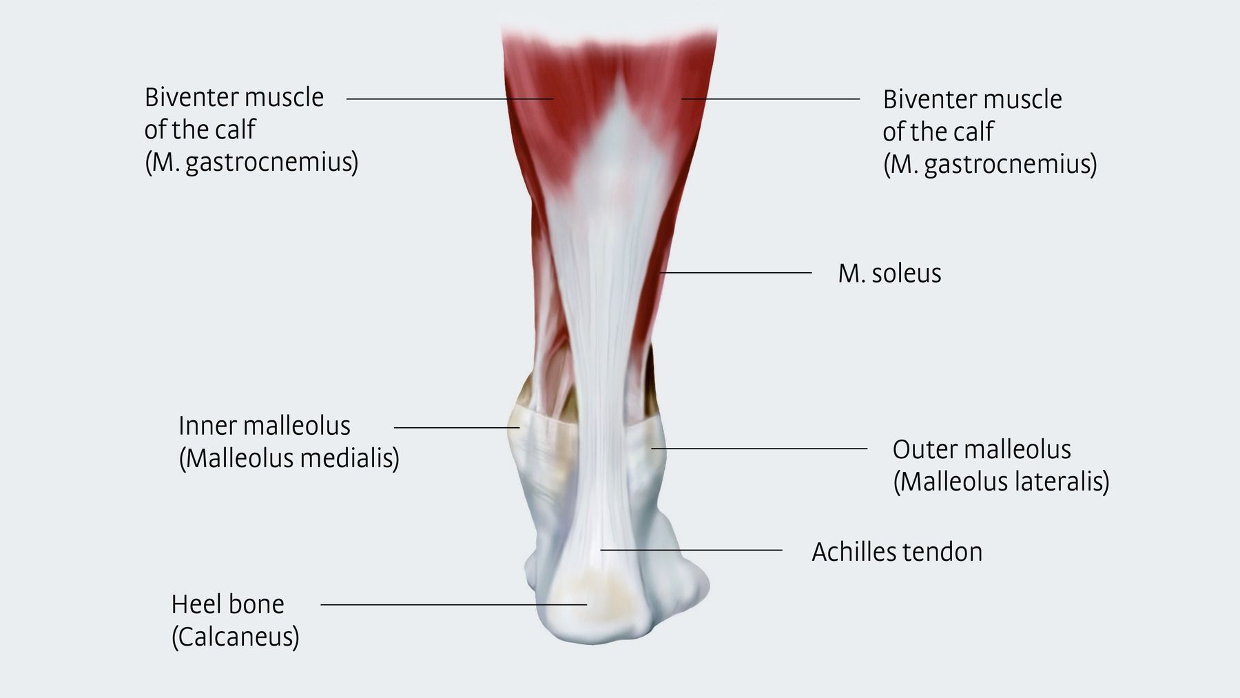
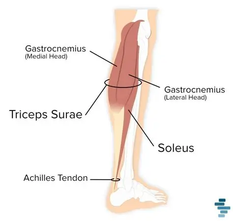
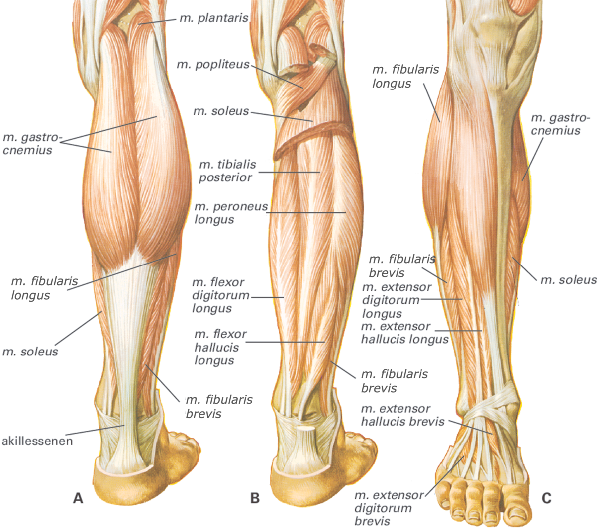
M. gastrocnemius
Large superficial calf muscle with two heads plantar-flexes ankle and flexes knee. (N. tibialis S1-S2). 🔎 Visible on surface—two big red bellies forming the calf bulge. 💡 "Gas gives you push."

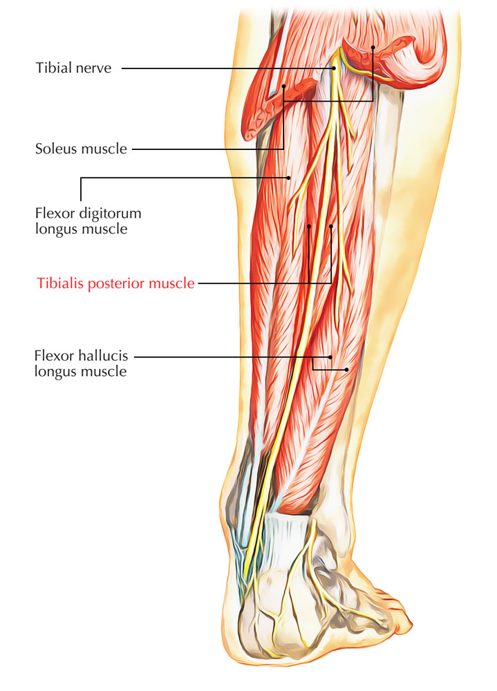
M. soleus
Broad flat muscle deep to gastrocnemius strong plantar flexor for standing. (N. tibialis). 🔎 Slice under gastrocnemius—thick red sheet. 💡 "Soleus = solid support."
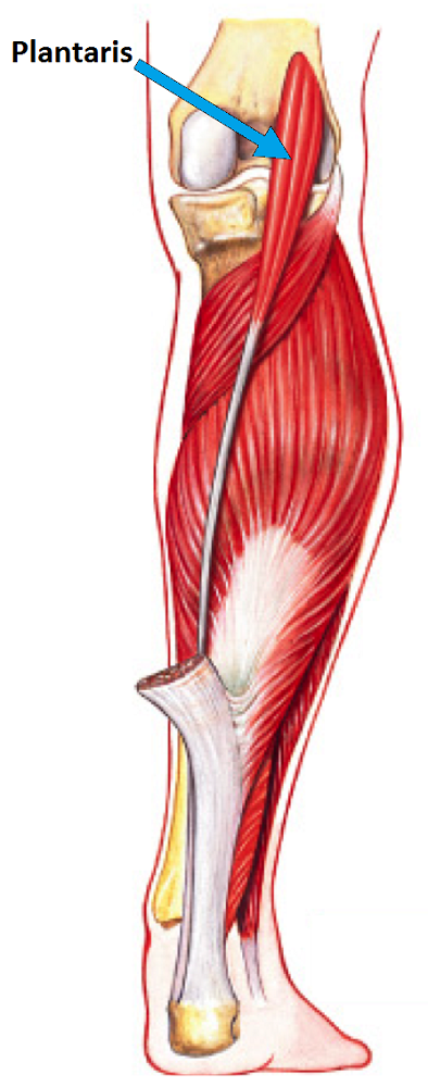
M. plantaris
Small muscle with long thin tendon between gastroc & soleus weak plantar flexor. (N. tibialis). 🔎 Slender line running to Achilles tendon. 💡 "Plantaris = tiny helper."
Tendo calcaneus (Achilles tendon)
Common tendon of gastrocnemius + soleus inserting on calcaneus strongest tendon in body. 🔎 White cord at heel. 💡 "Achilles = power cord."
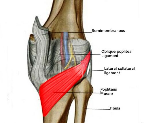
M. popliteus
Small diagonal muscle behind knee unlocking the joint (flexion + rotation). (N. tibialis). 🔎 Short red triangle in popliteal fossa. 💡 "Pop = unlock."
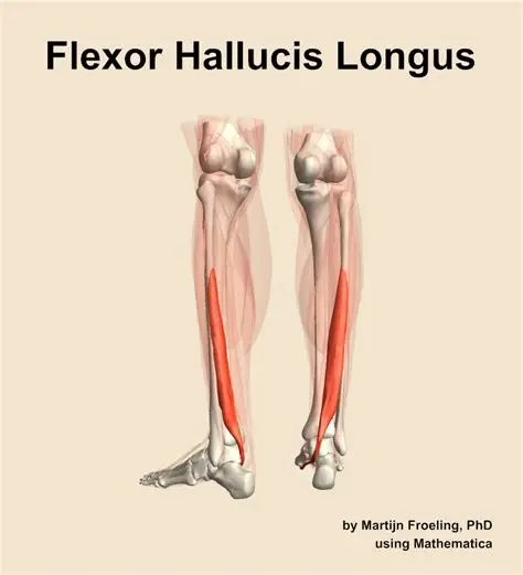
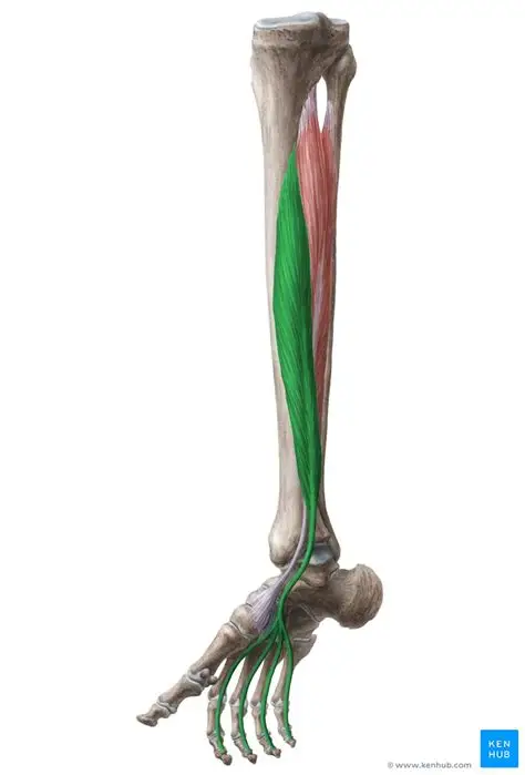
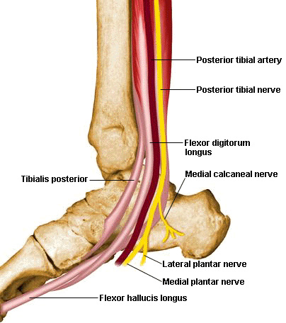
M. flexor hallucis longus
Deep muscle on fibular side flexes big toe & assists plantar flexion. (N. tibialis). 🔎 See tendon crossing posterior ankle to hallux. 💡 "Hallucis = hello big toe."
M. flexor digitorum longus
Medial deep muscle flexes toes 2-5 and plantar flexes foot. (N. tibialis). 🔎 Medial to tibialis posterior tendons divide to digits. 💡 "Digits = deep flex."
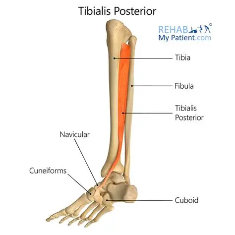
M. tibialis posterior
Deepest central posterior muscle inverts + plantar flexes foot maintains arch. (N. tibialis). 🔎 Between FDL & FHL tendon behind medial malleolus. 💡 "Tibialis posterior = tendon police of arch."
Triceps surae
Collective term for gastrocnemius + soleus (± plantaris). Powerful plantar-flexor. 🔎 Identify both layers together on Anatomage. 💡 "Tri = three calves."
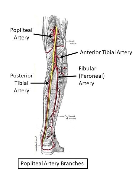
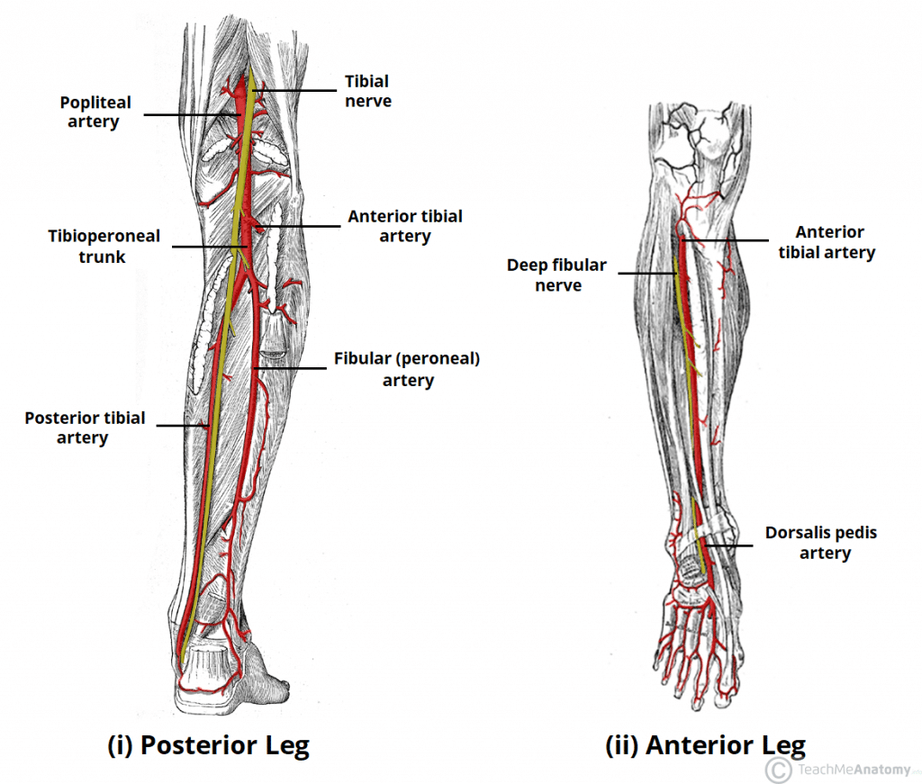
A. tibialis posterior
Main posterior-leg artery continuation of popliteal supplies deep flexors. 🔎 Runs with tibial nerve behind medial malleolus. 💡 "Posterior = behind malleolus."
A. fibularis (peronea)
Branch of A. tibialis posterior supplying lateral leg & fibular muscles. Lateral red vessel. 💡 "Fibular = side flow."
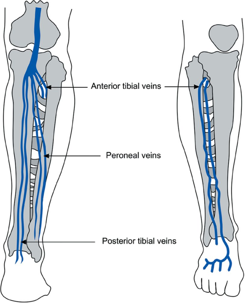
V. tibialis posterior
Vein 💙 accompanying artery → drains into V. poplitea. 🔎 Blue partner vessel posteriorly. 💡 "Vein = very close posterior."
N. tibialis
Continuation of sciatic ⚡ supplies posterior-leg muscles & foot sole. 🔎 Yellow cord central in calf → behind medial malleolus. 💡 "Tibial = to toe plantar."
N. cutaneus surae medialis
Branch of N. tibialis providing skin sensation of posterior leg joins fibular branch → N. suralis. ⚡ 🔎 Small yellow strand mid-calf. 💡 "Sura = skin strip."
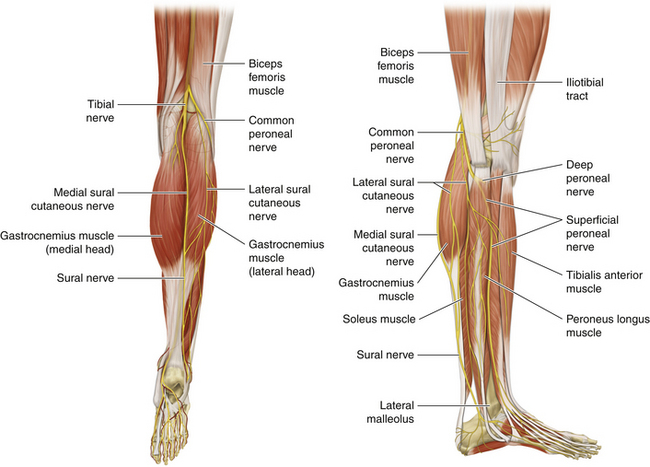
N. suralis
Union of medial & lateral sural cutaneous nerves sensory to lateral foot. ⚡ 🔎 Yellow line with small saphenous vein. 💡 "Sural = side sense."

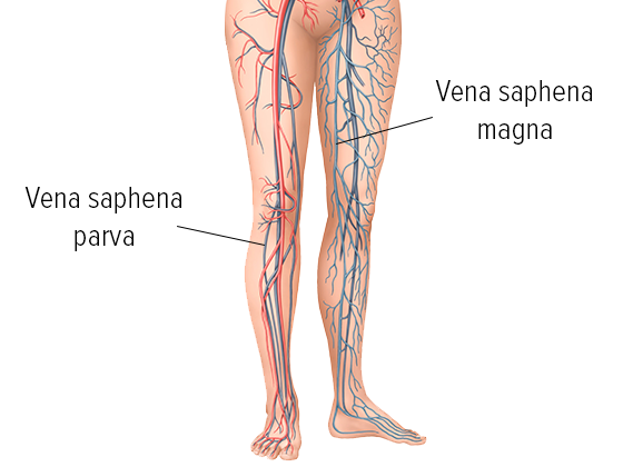
V. saphena parva
Small saphenous vein 💙 superficial posterior calf → drains to V. poplitea. 🔎 Blue surface line behind calf. 💡 "Small = short posterior."
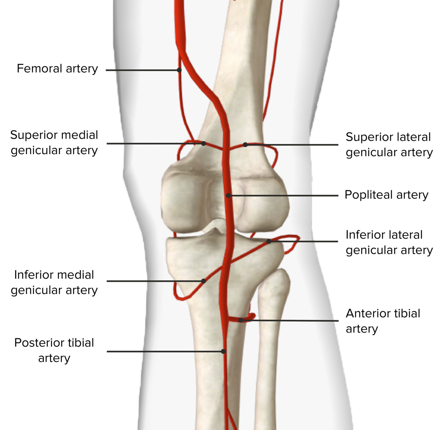
A. poplitea
Continuation of femoral → posterior leg arteries. Deep in fossa behind knee. 💡 "Pop = posterior pivot."
Canalis cruralis posterior (posterior leg compartment)
Contains tibial nerve + posterior tibial & fibular vessels + deep flexor muscles. 🔎 Cross-section view → posterior compartment. 💡 "Posterior pack."
Retinaculum flexorum (flexor retinaculum)
Fibrous band forming tarsal tunnel holds tibial nerve + tendons (FDL, TP, FHL). 🔎 At medial ankle. 💡 "Tom Dick And Nervous Harry."
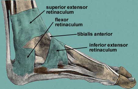
Tarsal tunnel
Space behind medial malleolus containing TP, FDL, A./V./N. tibialis posterior, FHL. 🔎 Locate by "Tom Dick ANd Harry" order. 💡 "Tunnel = tight passage."

A. plantaris medialis et lateralis
Branches of posterior tibial artery forming plantar arch . 🔎 Visible in foot sole. 💡 "Plantaris = plant supply."
M. fibularis longus
Superficial lateral-leg muscle everts foot + supports arch. (N. fibularis superficialis). 🔎 Lateral red belly to 1st metatarsal. 💡 "Longus = lateral lift."

M. fibularis brevis
Shorter lateral-leg muscle inserting on 5th metatarsal everts foot. (N. fibularis superficialis). 🔎 Deep to longus, tendon to base of 5th. 💡 "Brevis = base of 5th."
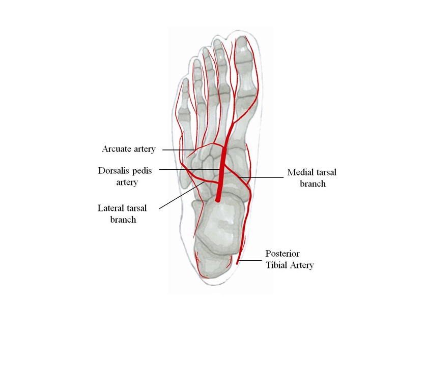
A. dorsalis pedis
Continuation of A. tibialis anterior on dorsum foot → 1st interdigital space. 🔎 Red line over navicular. 💡 "Pedis = pulse point."
