[CYTOGENETICS] Karyotyping
1/91
There's no tags or description
Looks like no tags are added yet.
Name | Mastery | Learn | Test | Matching | Spaced |
|---|
No study sessions yet.
92 Terms
Karyotyping
is the number and appearance of chromosome in the nucleus of a eukaryotic cell
Karyotyping
The process of Pairing and Ordering of chromosomes
Metaphase
Phase of cell division where chromosomes are studied
bright field microscope or fluorescent microscope
two microscopes used in karyotyping
Size of chromosome
> Compare sizes of Chromosome 1 and Y chromosome
Position of centromere
> Acrocentric, Metacentric etc
Presence of secondary constrictions
Size of satellites
What are the 4 things we can get from Karyotyping
Karyotype
comes from the Greek word "Karyon," which means nucleus.
Karyology
The study of whole sets of chromosomes.
Idiogram or Karyogram
The standard format of representing chromosomes as a diagram is when the haploid set of chromosomes of an organism is ordered in a series of decreasing size.
Idiogram or Karyogram
Chromosomes are arranged from largest to smallest
Manually take pictures using a camera first
Asymmetric Karyotype
show larger differences between smaller and larger chromosome in a set.
Have more acrocentric chromosomes and relatively advanced feature.
Symmetric Karyotype
show lesser difference between smaller and larger chromosome in a set. Have more metaphase chromosomes and no advanced feature.
Asymmetrical
more complex; more asymmetrical
Describe the karyotype of humans and animals
GA Levitzky
a Russian scientist suggested that in flowering plants there is a predominant trend towards karyotype asymmetry.
This trend has been carefully studied in the genus Crepis of the family compositae
Crepis
Compositae
Species of flowering plants used by GA levitzky
Green-top vacutube (heparin)
note: EDTA citrate can interfere with chromosomes
What type of tube is used for the patients blood after harvesting
1. SHORT TERM LYMPHOCYTE CULTURE
2. HARVESTING OF LYMPHOCYTES
3. FIXING THE CELLS
4. MAKING THE CHROMOSOME SLIDES
5. SLIDE ANALYSIS
5 major steps of Karyptyping
SHORT TERM LYMPHOCYTE CULTURE
The collected blood will be grown in vitro by adding cell culture growth medium, fetal bovine serum, antibiotics, and phytohemagglutinin (PHA) - the reagent that induces mitotic activity
The cultured blood cells will be grown at 37 °C incubator for 3 days
Note that the cells must be in logarithmic phase because splitting of a cell line 2 days before harvesting, and changing the medium 1 day before harvesting, stimulates cell proliferation significantly.
phytohemagglutinin (PHA)
fetal bovine serum
antibiotics or antifungals
1) SHORT TERM LYMPHOCYTE CULTURE
the reagents that induces mitotic activity
3 components of the growth medium
HARVESTING OF LYMPHOCYTES
Addition of pre-warmed colcemid (also known as colchicine), the reagent that arrests the cell cycle at metaphase stage, into the culture and incubate for 15 mins.
Optimal exposure time to colcemid requires a balance between proliferative activity index of cells and concentration of colcemid
Centrifuge the tube at 1000 RPM for 10 mins and the cell pellet was resuspended in warm hypotonic solution (can be KCl or sodium citrate) and the solution was mixed.
Incubate at room temperature for 15 mins.
Colcemid or Colchicine
(2) HARVESTING OF LYMPHOCYTES
Used to arrest the lymphocyte cells in metaphase
1000RPm for 10mins
(2) HARVESTING OF LYMPHOCYTES
Centrifuge the tube at ______________ and the cell pellet was resuspended in warm hypotonic solution (can be KCl or sodium citrate) and the solution was mixed. Incubate at room temperature for 15 mins.
HARVESTING OF LYMPHOCYTES
additional modifications:
allow for enrichment of long (prometaphase) chromosomes by using Actinomycin D or ethidium bromide (added before harvesting), or bromodeoxyuridine (BrdU), added before colcemid treatment.
Cell synchronization can significantly increase the total yield of metaphase chromosomes. Cells are arrested at S phase by adding an excess amount of BrdU overnight (16 h). After this, the block is released by washing the cells and adding thymidine for 5.5 h before colcemid treatment (Hirai et al., 1994).
Cell synchronization
can significantly increase the total yield of metaphase chromosomes. Cells are arrested at S phase by adding an excess amount of BrdU overnight (16 h).
FIXING THE CELLS
The cell suspension in hypotonic state will be centrifuged for 1200 RPM for 5 mins.
The cell pellet will be treated with fixative solution (absolute methanol:glacial acetic acid; 3:1) or Carnoy’s fixative and will be centrifuged at 1200 RPM for 5 mins
The process will be repeated 3x and the final addition of fixative solution will require incubation at 4 °C for 10 mins.
methanol:glacial acetic acid; 3:1
or Carnoy’s fixative
Fixative used
Carboy's Fixative
Solution made up of methanol and glacial acetic acid
Methanol (1)
Preferred fixative for proteins (histones)
Glacial Acetic Acid (3)
Fixative of nucleic acids (DNA)
MAKING THE CHROMOSOMES SLIDE
5 or 6 cold slides will be layered next to each other in a paper towel.
2 or 3 drops of the samples will be dropped onto each slide and dry them spontaneously
The slide will be stained by GTG-banding (G-bands by Trypsin using Giemsa), the most common method of staining chromosomes for differentiation which uses trypsin that digests the chromosomes at regions rich in basic amino acids (Arg and Lys).
Trypsin
digests the chromosomes at regions rich in basic amino acids (Arg and Lys).
Giemsa Stain with Trypsin
Stain used for GTG banding
SLIDE ANALYSIS
Slides that will be chosen for analysis and visualization must be:
> Properly trypsinized chromosomes
> Clearly defined metaphase spreading
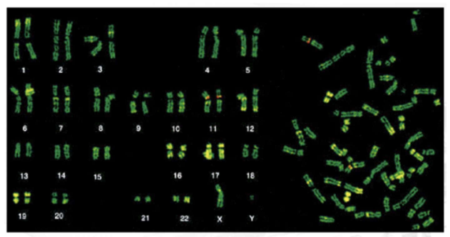
Requires a microscope with automated computer software program primarily, CytovisionTM by Applied Imaging Inc. which follows the International System of Human Cytogenetic Nomenclature (ISCN) that arrange chromosomes according to size and banding patterns
10-20mL
How much peripheral blood is drawn
Because they have the greatest nucleus to cytoplasm ratio (malaki nucleus)
Why are lymphocytes used and not RBC?
Fish and amphibians
We cannot culture RBCs because they are anucleated. However, _______ and ________ have a nucleus in the RBC
Antibiotics
Antifungal
Added to the nutritious culture medium so we can ensure ONLY the growth of Lymphocytes
Phytohemagglutinin
Reagents that stimulate the growth of lymphocytes and Removal of RBCs
Hypotonic Solution
Solution where we need to put the cultured media so cell membrane are broken and the chromosomes are exposed
Band
A ______ is a part of a chromosome that is clearly distinguishable from its adjacent segments by appearing darker or lighter with various banding methods (Paris conference, 1971)
Light Staining Area
Contains the euchromatin
Where gene expression happens
Dark Staining Areas
Contains the heterochromatins
Epigenetics
A branch of genetics that studies gene expression
"Does an organism express or use this gene?"
1958. Caspersson et al
published there first paper describing the use of quinacrine mustard to stain chromosome there by ushered in a new era of chromosome banding
The Paris Report (1971)
was the first attempt to provide nomenclature for chromosome banding in any species and thus its recommendations have been adopted to nonhuman species as well.
Banding Patterns
allows you to see smaller pieces of the chromosome, so that you could identify smaller structural chromosome abnormalities not visible on a routine analysis.
GC and At rich regions
Contitutive Heterochromatin Region
What are the basis of classification of banding techniques?
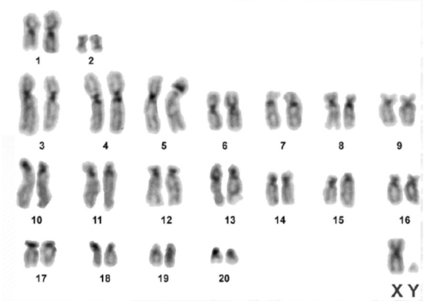
Metaphase Chromosomes
chromosomes whose size has condensed and whose diameter is increased are used for chromosome banding studies after fixing the stage
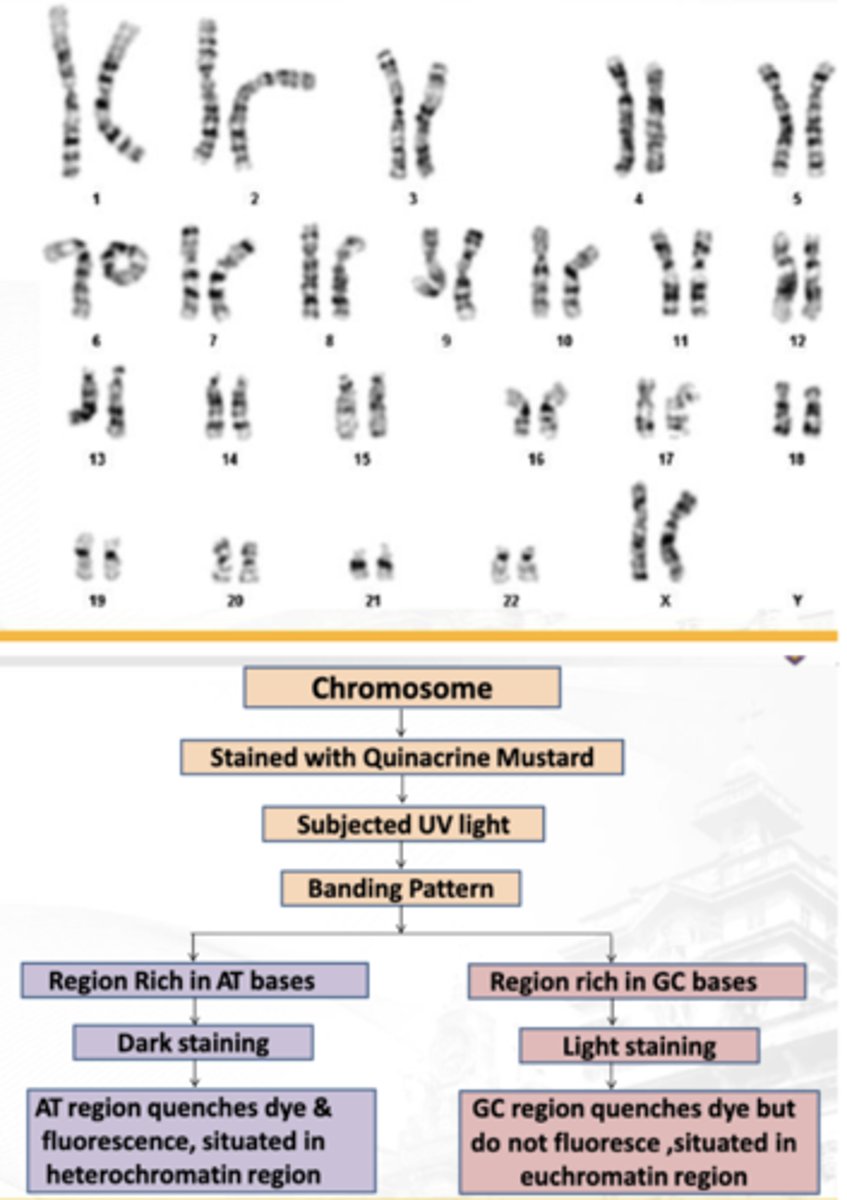
Q-BANDING
Banding technique by: CASPERSON 1958
G-BANDING
Banding technique by: SUMMER 1971
Nucleolar organization regions (N-BANDING)
Banding technique by: MATSUI AND SASAKI 1973
Centromeric (C-banding)
Banding technique by: LINDE AND LAURSEN 1978
Q-BANDING
We use UV light to visualize it
> Shorter wavelength
> Higher frequency and energy
Light staining areas are green
> G and C-rich regions
Dark staining areas are the apple green ones or the yellow-green
> Contains adenine and thymine-rich regions
Q-BANDING
Note: GC rich regions do not fluorescence
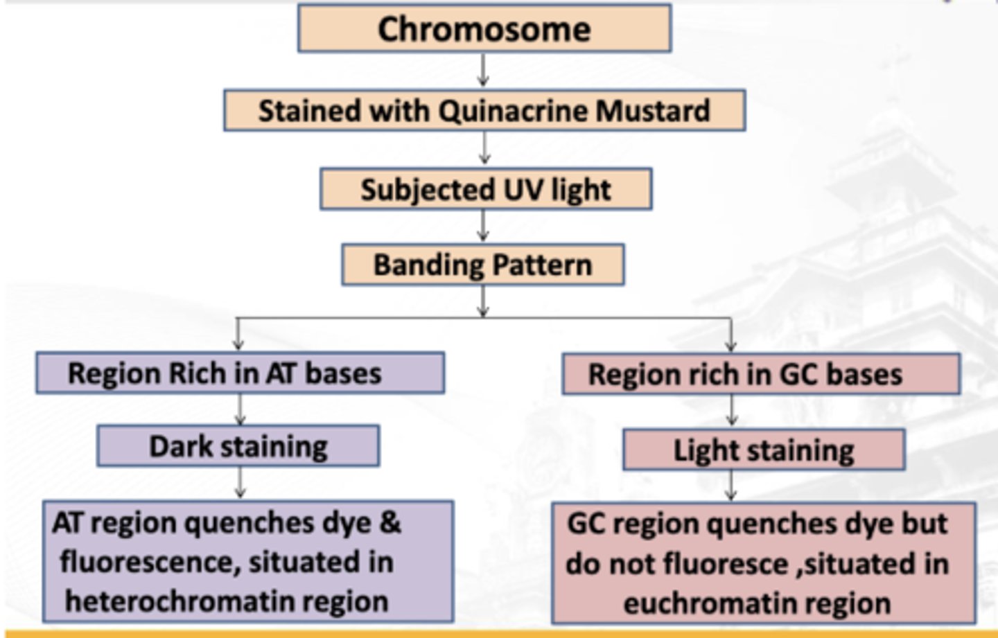
Q-BANDING
Banding technique used for revealing polymorphisms in Chromosomes 3,4,13,14,15,21,22
Quinarcine or mustard
The first ever discovered banding technique in Karyotyping (ito lang daw itatanong)
Color: Apple green
Light base or alkaline
Ideally, there is NO denaturation in Q banding, but we use this for denaturation of Q banding when needed
Apple green
Color of Quinarcine or mustard (Q-BANDING)
AT rich regions
What regions do Quinacrine fluorescent dye stains (light staining regions - green)
Fluorophores (fluorescent stains)
undergo photobleaching or quenching when exposed to light
High frequency
Higher Energy
Shorter wavelength
Describe the UV light (frequency wavelength and energy)
Visible light
Light to which the fluorescent light will be converted to
has:
longer wavelength
Lower energy and frequency
Q-BANDING
Banding used in study of chromosome heteromorphism
Nucleolar organization regions (N-BANDING)
The ability of specific chromosomes to generate ribosomal RNA
What part of the human chromosomes produces ribosomal RNA?
Nucleolar organization regions (N-BANDING)
Banding pattern is:
Dark bands
> Nucleolar Organizer Regions (NORs) – rDNA sites
Rest of the chromosome (usually not stained)
Nucleolar organization regions (N-BANDING)
Banding pattern used for PLANTS
Nucleolar organization regions (N-BANDING)
Uses: Trichloroacetic Acid and HCl
Stains: Structural non-histone proteins
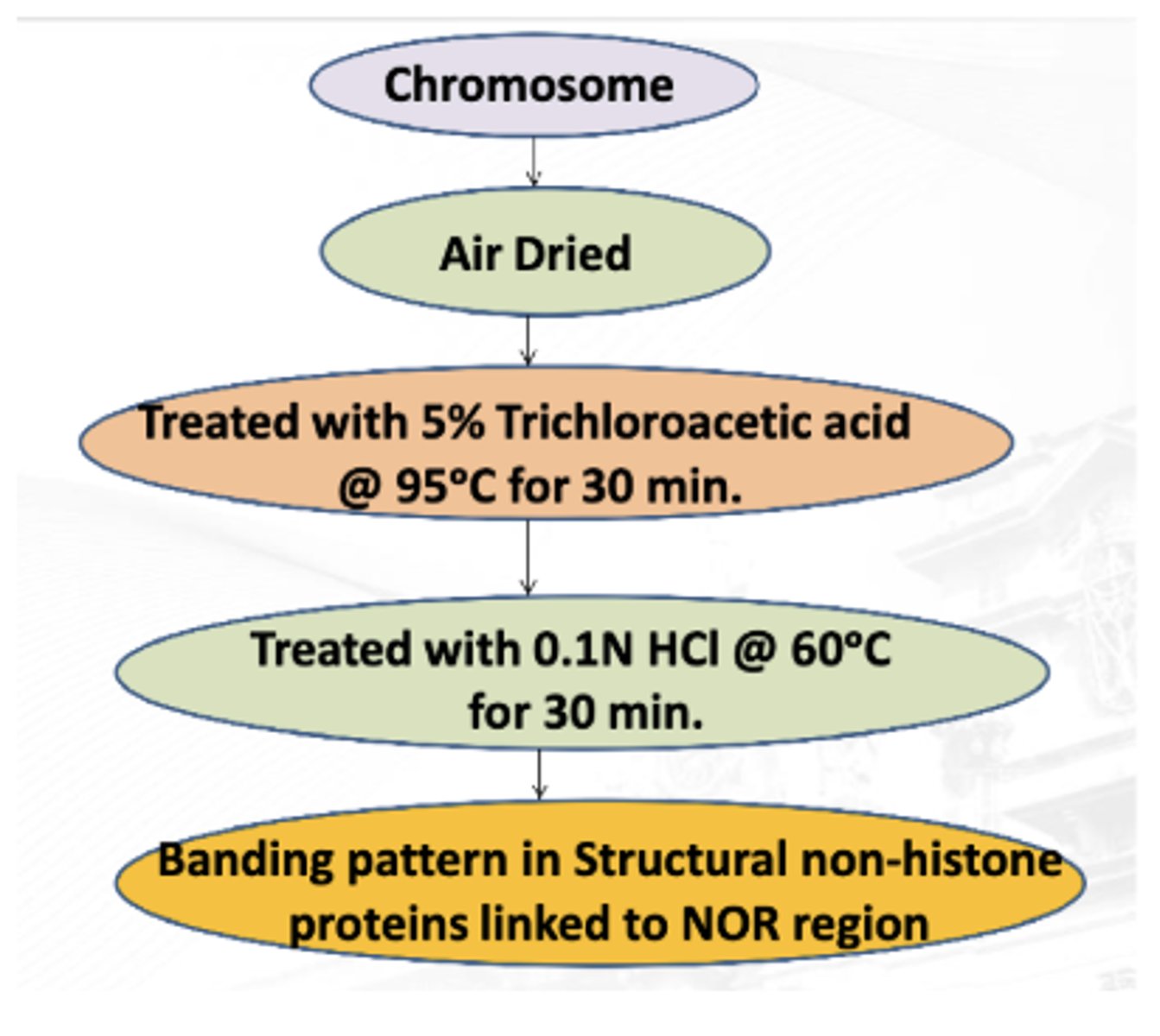
AgNO3
Stain technique in Nucleolar organization regions (N-BANDING)
Nucleolar organization regions (N-BANDING)
Reveals polymorphisms and rearrangements of acrocentric chromosomes
Centromeric (C-banding)
Dark-staining areas are only the centromeres.
There is denaturation of the DNA.
> Destroys the hydrogen bonds
> The denatured parts are lightly staining.
Tendency of repetitive DNA (like centromere, if we denature it, it can naturally renature
We use a brightfield microscope
Centromeric (C-banding)
Detection of the centromere
Is there a centromere or no?
Centromeric (C-banding)
Stains the heterochromatin regions close to the centromeres
Note: The euchromatin are usually unstained
Centromeric (C-banding)
Usually stains the entire long arm of the Y chromosome
BaOH
Denaturating agent for the C-banding technique
Centromeric (C-banding)
Treated with alkali solution
Washed with Sodium Citrate
Stained with Giemsa
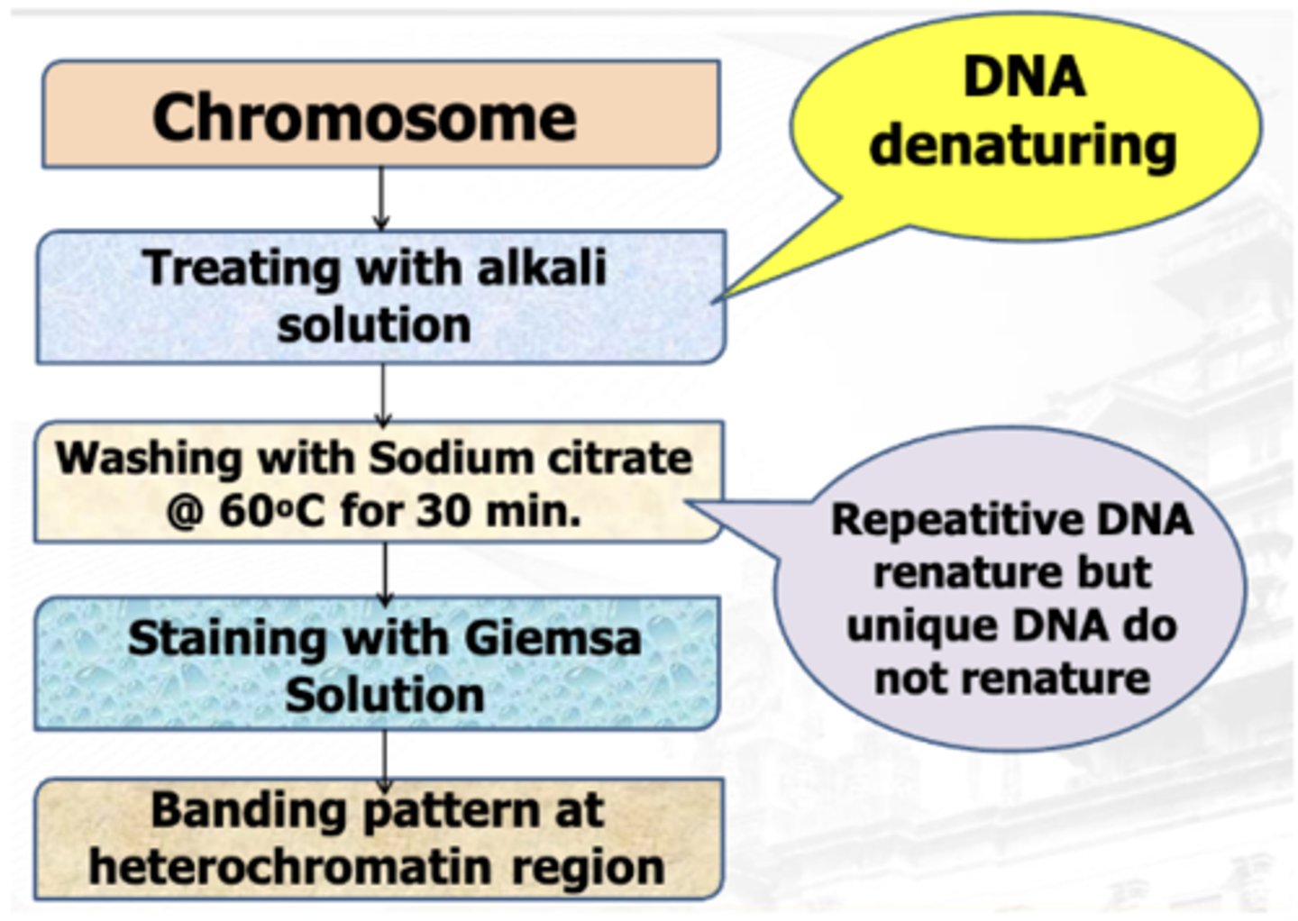
Sodium Citrate
Used in C banding to ensure that repetitive DNA centromeres so not renature
Centromeric (C-banding)
Identification of chromosomes, particularly in insects and plants.
Identification of bivalents at diakinesis using both centromere positions.
Paternity testing.
Gene mapping
Centromeric (C-banding)
Reveals polypormisms including heterochromatin inversions; evaluation of ring and dicentric chromosomes
GIEMSA (G-BANDING)
Required partial denaturation process using Trypsin
Loosen the bonding between the DNA and the histone
GIEMSA (G-BANDING)
Dark staining areas are AT-rich.
Light staining areas are CG-rich regions.
Targeted by acetyltransferases (CG) :: There is acetylation → increased gene expression
Wrights (uses bright field as well)
GIEMSA (G-BANDING) alternative staining technique
GIEMSA (G-BANDING)
Banding technique for simple chromosome photography
Q-BANDING
> Tendency to fade during examination; UV light breaks the chemical bond.
> Photo-degradation
> Chromopore- absorb light of a particular wavelength due to a chemical bond formed between dye and light.
Trypsin
The denaturing agent used in Giemsa or G STAINING
> Proteolytic enzyme by cracking open histones
We use a brightfield microscope
Boiling
Alternative to Trypsin if we want to denature the DNA
G Banding Techniques
Advantages
▪ Used in identification of bands rich in Sulfur content.
▪ Used in the identification of chromosomal abnormalities
▪ Gene Mapping
G Banding Techniques
Disadvantages
▪ Not used in plants.
R-BANDING
Makes use of heat to denature the proteins
The banding area is reversed
Dark: CG
Light: AT
R-BANDING
Used for visualization of ends of chromosomes and small positive R-bands
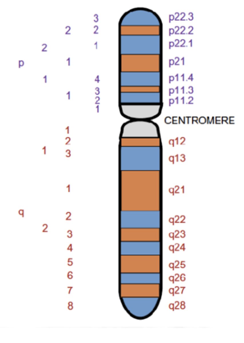
7q31.2
This indicates that the gene is on chromosome 7, q arm, band 3, sub-band 1, and sub-sub-band 2.
The ends of the chromosomes are labeled ptel and qtel
The notation 7qtel refers to the end of the long arm of chromosome 7
International System for Human Cytogenetic Nomenclature
ISCN
Closest or Proximal to the centromere
As per ISCN, Each area of chromosome given the lowest number
____________ of closest (proximal) to centromere

Highest number
As per ISCN, Each area of chromosome at tips (distal) to centromere