EKG review: Axis, BBB, MI rules
1/28
There's no tags or description
Looks like no tags are added yet.
Name | Mastery | Learn | Test | Matching | Spaced |
|---|
No study sessions yet.
29 Terms
Normal axis
both leads I and aVF are positively deflected
LAD
Lead I is positively deflected, but aVF is negatively deflected
RAD
Lead I is negatively deflected, but aVF is positively deflected
eRAD
both leads I and aVF are negatively deflected
subendocardial ischemia
represented by ST segment depression
transmural ischemia (injury)
represented by ST segment elevation
pathological Q waves
longer than 0.4 sec or deeper than 2 small boxes or deeper than 25% of the R wave (if present)
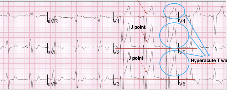
STEMI
1+ mm ST elevation in 2 or more contiguous leads w/ reciprocal changes
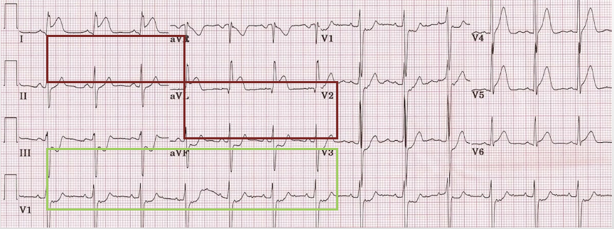
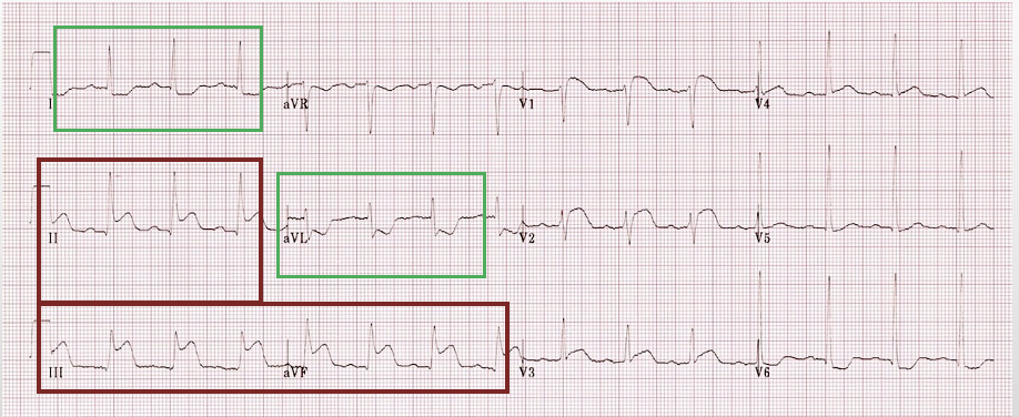
Lateral wall STEMI
ST elevations in 2+ lateral leads (I, aVL, V5, V6) + reciprocal changes in the inferior leads (II, III, aVF)
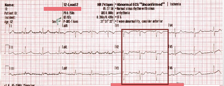
Anteroseptal STEMI
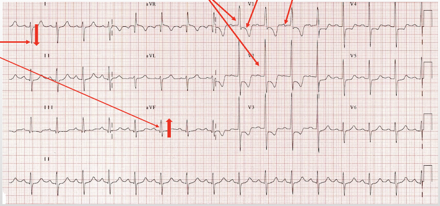
ST elevation in leads V1-4
Posterior MI (12 lead)
ST depression in leads V1-4
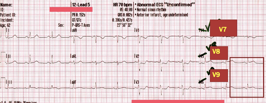
Posterior MI (15 lead)
ST depression in leads V1-4 w/ ST elevation in leads V7-V9
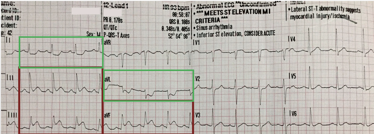
Inferior MI
ST elevation in inferior leads (II, III, aVF) w/ reciprocal changes in leads I and aVL
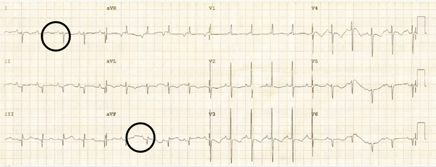
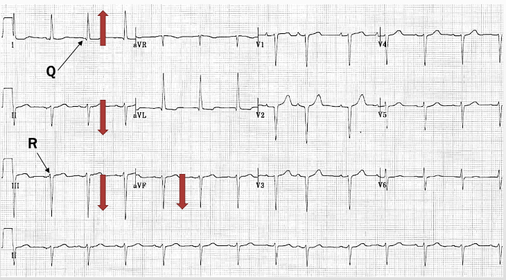
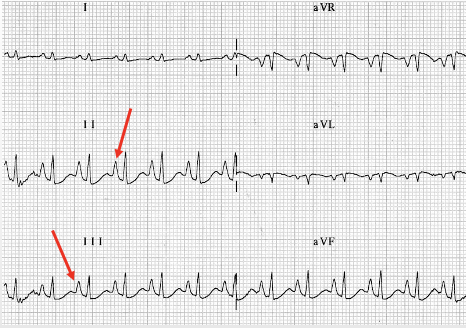
left anterior fascicular block (LAFB)
LAD, Q wave in lead I + R wave in lead III, mostly - lead II & III
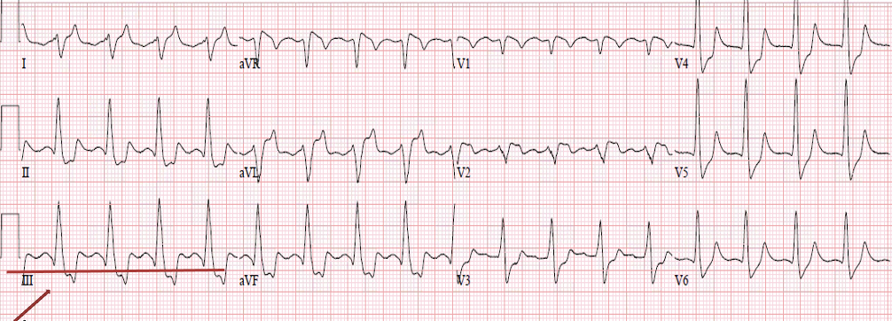
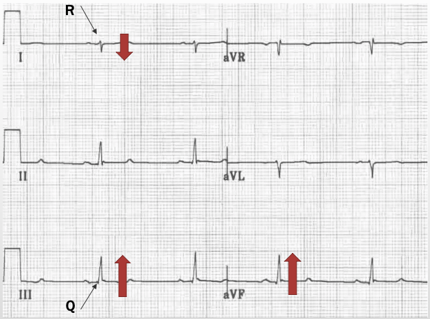
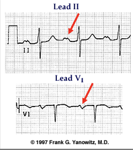
left posterior fascicular block (LPFB)
RAD, R wave in lead I and Q wave in lead III, mostly + lead III
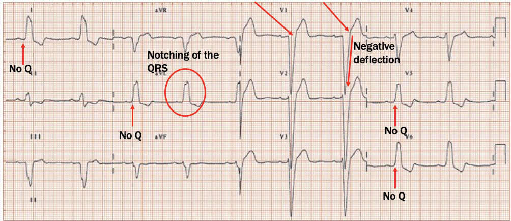
LBBB
V1: dominant S wave (W)
Lead I: wide QRS, RS pattern
Lateral leads: no Q waves, notched QRS (double QRS) =broad notched R (M)
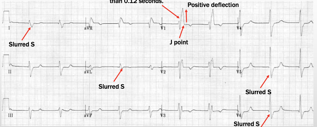
RBBB
Lead V1: wide QRS, rSR pattern (M) (usually V1-3)
Lateral: slurred S (sometimes W seen)
RAE (P-pulmonale)
Leads I, II, III: upright P taller than 2.5 mm
Lead V1: biphasic P w/ bigger + initial deflection & smaller - terminal deflection
LAE (P-mitrale)
Leads I, II, II: upright humped P wave
Lead V1: biphasic P wave w/ small + initial deflection and larger - terminal deflection
RVH
R:S ratio of 1+ mm in leads V1 & V2
supported by: RAE, RAD, strain pattern (concave ST segment turning into an inverted T wave)
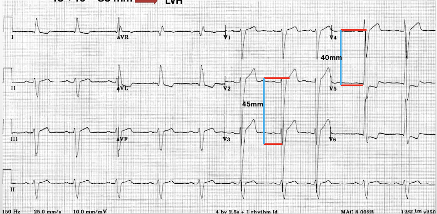
LVH
deep S waves in leads V1 & V2, tall R waves in lateral leads, LAD
-add the deeper S to the taller R; must be + or = to 35 mm
-if R wave is > 12 mm or if any chest lead is > 45 mm
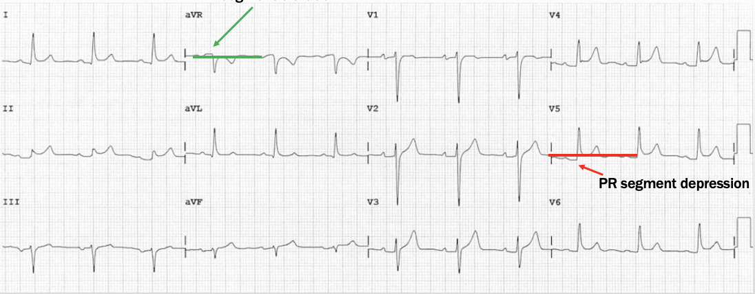
Pericarditis
PR depression & global concave ST elevation w/ no reciprocal changes
PR segment elevation in aVR (if there is ST depression in any lead other than aVR and V1 or any Q is not pericarditis)
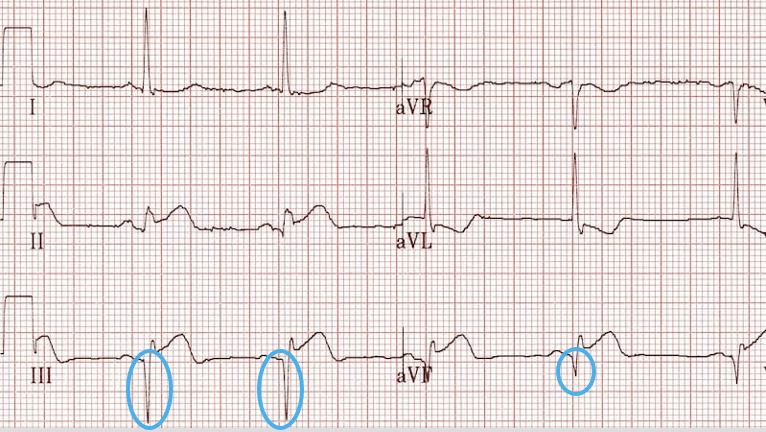
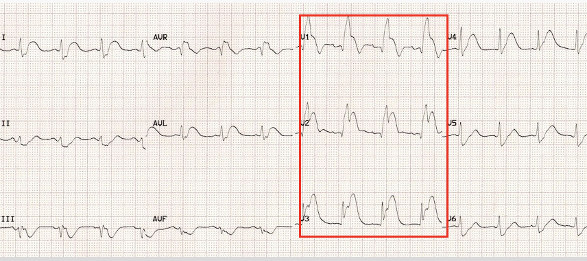
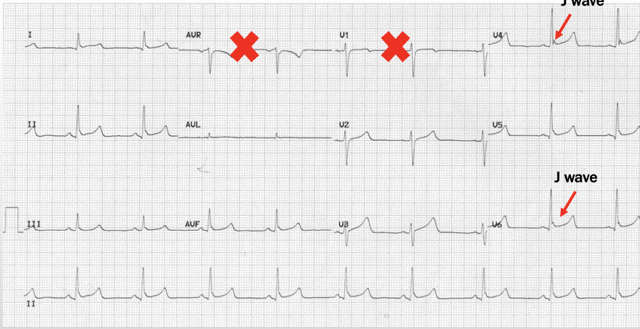
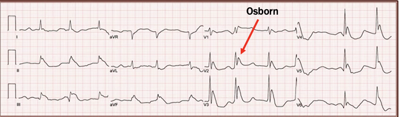
Early repolarization (BER)
global concave ST elevation, terminal QRS notching (J wave, Osborn wave), large T waves, no reciprocal ST depression (outside of aVR and V1)
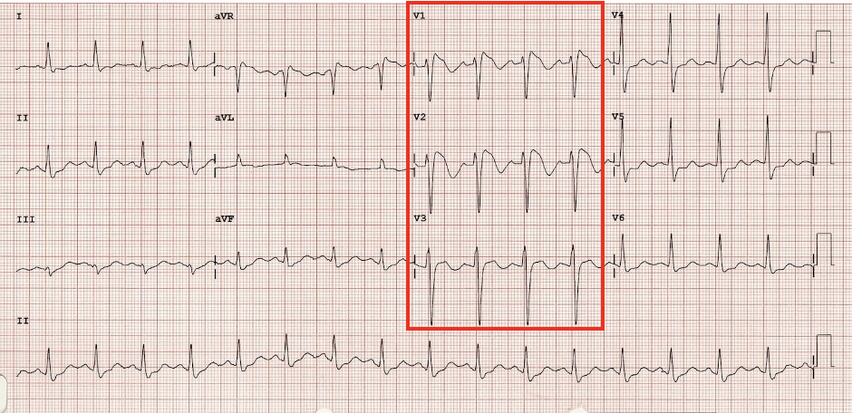
Brugada Type 1
convex ST elevation in V1-3 (coved shaped)
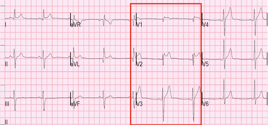
Brugada Type 2
saddle shape ST elevation in V1-3 (carousel horses sign)
Hypothermia
J waves; prolonged PR, QRS, QT; various bradyarrythmias
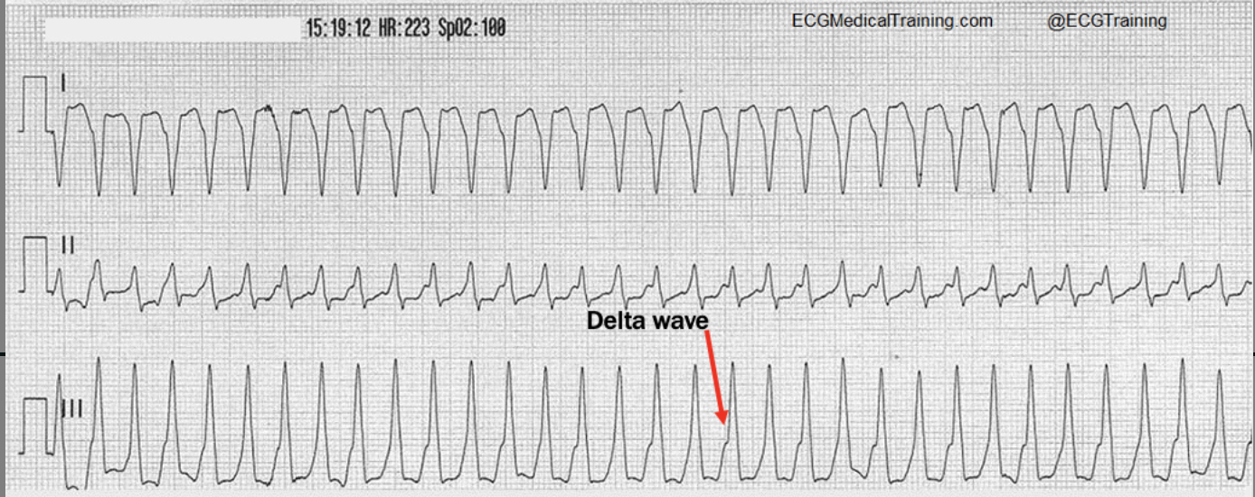
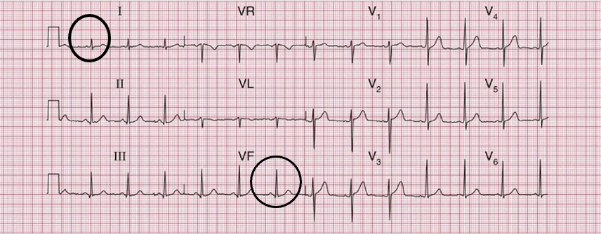
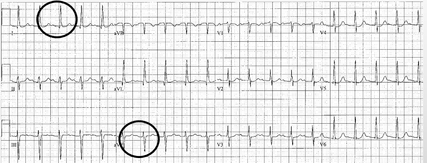
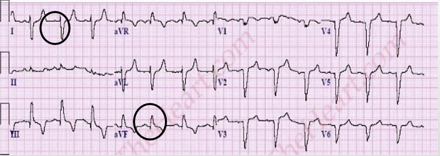
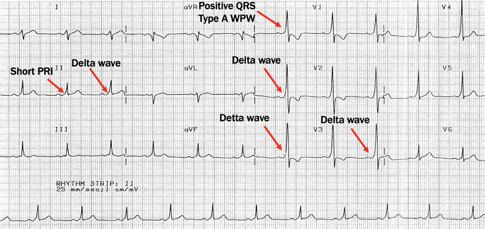
WPW
short PR, wide QRS, delta wave
Orthodromic narrow tachycardia
narrow QRS, no P wave
Antidromic wide tachycardia
wide QRS, no P waves