Cochlea, Inner Ear, and Vestibular System Concepts
1/103
Earn XP
Description and Tags
A set of vocabulary flashcards summarizing key concepts from the cochlea, inner ear, vestibular system, and associated pathways.
Name | Mastery | Learn | Test | Matching | Spaced | Call with Kai |
|---|
No analytics yet
Send a link to your students to track their progress
104 Terms
What are the three scalae of the cochlea?
The Scala Vestibuli, scala media, and scala tympani
Scala Vestibuli
The upper chamber of the cochlea that contains perilymph.
Scala Media
The middle chamber of the cochlea that contains endolymph and houses the Organ of Corti.
Scala Tympani
The lower chamber of the cochlea also containing perilymph and ending at the round window.
Endolymph
The fluid found in the scala media of the cochlea.
Perilymph
The fluid in the scala tympani
Basilar Membrane
Becomes wider and more flexible toward the apex, coding for low frequencies
What is the strong positive charge that’s drives K+ into hair cells?
Endocochlear potential
What does the tectorial membrane make direct contact with?
Outer hair cells
What hair cell sends 95% of auditory information to the brain?
Inner ear cells
Helicotrema
The apex of the cochlea that connects the scala vestibuli and scala tympani.
Spiral Ganglion Cells
Nerve cells that form the cochlear or auditory nerve.
Frequency encoding on Basilar Membrane: Base
HIGH frequency sound (narrow, stiff)
Frequency encoding on Basilar Membrane: Apex
LOW frequency sounds (wide, flexible)
tonotopic organization
The arrangement of auditory neurons in the cochlea that corresponds to different frequencies of sound, where specific areas are tuned to specific frequencies, creating a map of sound frequency along the membrane.
How are traveling wave peaks happen?
based on the point of most resistance
Scala Vestibuli (superior-chamber)
Contains perilymph, begins at oval window, separated from scala media by Reisner’s membrane
Scala Media (middle/cochlear duct chamber)
contains endolymph, houses organ of Corti, maintained by stria vascularis
Scala Tympani (inferior chamber)
contains perilymph, ends at round window, round widow ensures sound energy doesn’t reflect back
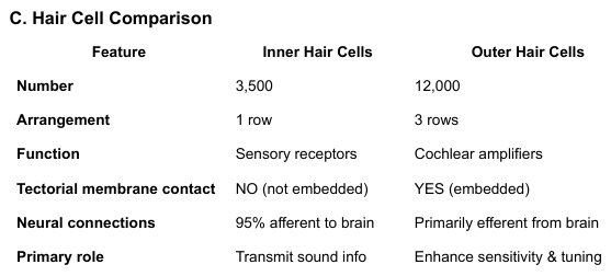
Hair cell comparison chart
what hair cells have motor function?
Outer hair cells have motor function.
Hiar cell Transduction Process
Sound>BM Movement> Stereocilia bend> Mechanotransduction channels open> K+ enters hair cell (driven by Endocochlear potential)> depolarization occurs> Neurotransmitter (glutamate) release>auditory nerve fired
What is depolarization caused by?
K+ Influx
Stria Vascularis
Contains capillary loops and blood vessels, vascular tissue with epithelial cells, produces endolymph, creates endococlear potential (+80 mV)
Tectorial Membrane
Semi-transparent structure over hair cells, indented by OHC stereocilia, purely mechanical role
extra important supporting cells
Deiter’s cells, Hensen’s cells, Claudius cells, Pillar cells (NOT epley’s cells)
Inner hair cell depolarization occurs due to an influx of what?
K+
Structure responsible for maintaining endolymphatic volume
stria vascularis; a specialized tissue in the cochlea that regulates the production and absorption of endolymph, maintaining ionic balance.
frequency tuning in the cochlea results from?
Basilar membrane mechanical properties
Outer hair cells?
They sharpen frequency tuning
Auditory nerve fibers for high frequencies are located?
along the base of the cochlea
structure ensuring sound energy doesn’t reflect back?
round window
What is the osseous?
Bone
Afferent
toward the brain (sensory input)
Efferent
away from the brain (motor output)
tonotopic organization
frequency mapping preserved throughout system
The semicircular canals detect what acceleration?
angular/rotational
The otolith organs detect what acceleration and head position/tilt?
linear
The sensory structure in the otolith organs is the?
macula
The gelatinous structure containing otoconia is called?
the otholithic membrane.
Endolymph movement bends the hair cells?
Stereocillia/kinocillium
What does VOR do?
stabilizes vision/gaze during head movement
What does the push-pull dynamic refer to?
one canal being excited while the paired canal is inhibited
The horizontal canals are activated most by?
turning the head to the left or right (yaw)
How is the saccule oriented?
in the vertical/sagittal plane
How is the utricle oriented?
on the horizontal plane
The Five Mechanoreceptors (per ear): Utricle
horizontal linear acceleration
The Five Mechanoreceptors (per ear): Saccule
vertical linear acceleration
The Five Mechanoreceptors (per ): Superior semicircular canal
pitch (nodding "yes")
The Five Mechanoreceptors (per ear):Posterior semicircular canal
roll (tilting side-to-side)
The Five Mechanoreceptors (per ): Horizontal semicircular canal
yaw (shaking "no")
Utricle details
Larger of two membranous sacs, Elliptical, lies most laterally, Hair cells oriented with kinocilium TOWARD striola, Responds best to side-to-side/horizontal movement
Saccule details
Smaller membranous sac, Primary sensor for vertical movement (jumping, elevator), Oriented in vertical plane
Macula Structure details (both organs)
1. Otoconia - calcium carbonate crystals (top layer)
2. Otolithic membrane - gelatinous layer
3. Stereocilia - hair bundles
4. Kinocilium - tallest cilium
5. Type II Hair Cells - sensory receptors
Otoconia facts
Composed of calcium carbonate crystals
● HIGH density (increase specific gravity above endolymph)
● Provide inertia to detect linear acceleration
● When they break loose → cause BPPV (Benign Paroxysmal Positional Vertigo)
● Do NOT cause Meniere's disease
Three canals per ear: Superior
Superior (anterior) - pitch movements
Three canals per ear: Posterior
Posterior - roll movements
Three canals per ear: Horizontal
Horizontal (lateral) - yaw movements
Key canal pairings:
● Left horizontal ↔ Right horizontal
● Right superior ↔ Left posterior (contralateral)
● Left superior ↔ Right posterior (contralateral)
Each ampulla contains what?
crista
Why is endolymph flow opposite of head movement?
due to inertia
what does exciting one canal do to its paired canal?
inhibitsthe activity of the paired canal.
Vestibular hair cell properties
● Have continuous activity with directional sensitivity
● Always firing—just change firing rate
● Found in cristae (canals) and maculae (otoliths)
● NOT located in Organ of Corti
Directional responses: toward kinocilium
Toward kinocilium → Depolarizes → Increases firing rate
Directional response: Away from kinocilium
Away from kinocilium → Hyperpolarizes → Decreases firing rate
Vestibular activation sequence
1. Head rotates
2. Endolymph lags behind (inertia)
3. Cupula deflects
4. Hair cells bend
5. Depolarization occurs
6. Signal travels through vestibular nerve (CN VIII)
In unilateral vestibular loss, the brain interprets “resting asymmetry” as?
constant rotation/spinning
In BPPV what breaks loose and falls into the semicircular canal?
Otoconia (ear crystals)
Cupula deflects because of movement of
endolymph in the semicircular canals.
Reflex stabilizing body during sudden shifts
called the vestibulo-ocular reflex (VOR). It helps maintain stable vision while the head moves.
Utricle responds best to?
side-to-side movements
Hair cells within the ampulla are oriented towards?
the kinocilium, the tallest stereocilia.
The superior canal detects?
pitching/nodding yes
Three primary functions of the vestibular function
1. Image stabilization (VOR)
2. Balance control (VSR)
3. Spatial orientation
Thinf the vestibular function does not do
provide hearing, aid in sound localization, drain mucus from the middle ear
What three systems are required to balance information sources?
1. Vestibular system
2. Visual system
3. Proprioception (joint/muscle sense)
correction response
automatic responses to unexpected changes in the center of gravity
Other types of balance responses
maintenance responses, stabilization response, preservation response
What is the purpose of the vestibulo-oculomotor reflex
keeps visual target in view when the head moves by stabilizing the eyes during head movement.
Vestibulo-oculomotor reflex (VOR) Neural pathways
Input: CN VIII (vestibular nerve)
Processing: Vestibular nuclei
Output: CN III, IV, VI (oculomotor, trochlear, abducens)
Result: Eye muscles contract/relax appropriately
Nystagmus (VOR)
Eye movement response to vestibular stimulation, seen during acceleration and deceleration, NOT seen during constant velocity
Medial Vestibulospinal Tract
Controls head and neck movements only, NOT full body (common misconception!)
Lateral Vestibulospinal Tract
Controls head and neck movements only, NOT full body (common misconception!)
Lateral Vestibulospinal Tract
Controls trunk and limb movements, Maintains posture and balance
ECOLI-MA
E = Eighth nerve (CN VIII/cochlear nerve)
C = Cochlear nucleus
O = Olivary complex (superior)
L = Lateral lemniscus
I = Inferior colliculus
M = Medial geniculate body (MGB)
A = Auditory cortex
Cochlear Nucleus (medulla/pons)
First central processing station, all auditory fibers synapse here, Wave III of ABR
Superior Olivary Complex
Critical for sound localization
ITD (Interaural Time Differences) = horizontal/azimuth sound clues ILD (Interaural Level Differences) = intensity/level sound clues Initiates acoustic reflex
ITD (Interaural Time Differences)
horizontal/azimuth sound clues
ILD (Interaural Level Differences)
intensity/level sound clues Initiates acoustic reflex
Lateral Lemniscus
Pathway connecting lower to upper brainstem, projects from cochlear nucleus to inferior colliculus
Inferior Colliculus (midbrain)
Integrates multisensory attention to sound
Contributes to reflexive orienting to sound
Nearly all auditory fibers synapse here
Medial Geniculate Body (MGB - thalamus)
Thalamic relay station
Lesion affects auditory attention and relay to cortex
Sends projections to auditory cortex
Auditory
Brodmann areas 41 & 42
High-level interpretation
Pitch discrimination, complex sound processing
Lesion does NOT cause complete deafness (bilateral input)
Tonotopy Preservation
Maintained by basilar membrane organization
Preserved through cochlear nucleus, inferior colliculus, MGB, auditory cortex
NOT preserved in: utricle, vestibular structures
Binaural Integration
Corpus callosum connects left and right hemispheres
Enables integration of information from both ears
Why cortical lesions don't cause complete deafness in one ear
Structure sending projections through lateral lemniscus
Cochlear nucleus
Structure integrating multisensory attention to sound
Medial geniculate body (also accept inferior colliculus)
Wave III of ABR associated with
Cochlear nucleus
Lesion in MGB would most affect
Auditory attention and relay to cortex
Everything that contains perilymph
Scala tympani, scala vestibuli