Normal Cardiac Mechanical Function
1/10
There's no tags or description
Looks like no tags are added yet.
Name | Mastery | Learn | Test | Matching | Spaced | Call with Kai |
|---|
No analytics yet
Send a link to your students to track their progress
11 Terms
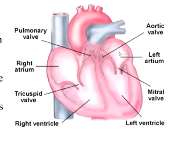
Describe the two pumps and direction of blood flow occurring within the heart.
• The heart is a muscular pump
• Blood comes into the atrium and flows into the ventricle. Atrial contraction helps it along (can be important), then the ventricle contracts, closes mitral valve, opens aortic valve and blood flows into the aorta.
• The heart is in fact two pumps acting in synchrony
Describe the two components of the cardiac cycle.
• (a) Ventricular systole: period of ventricular contraction
• (b) Ventricular diastole: ventricular relaxation during which ventricle fills with blood
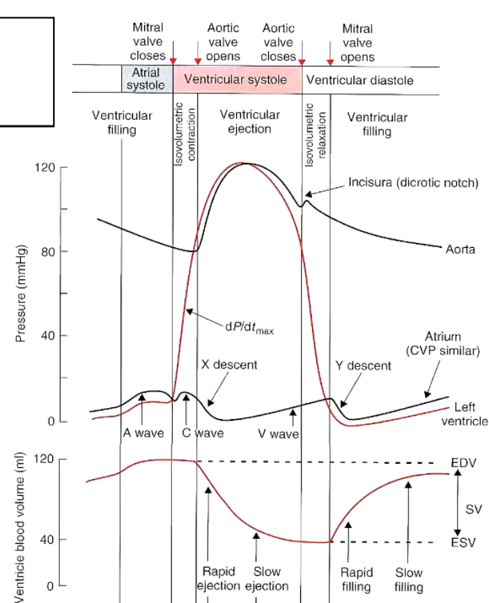
Explain what is occurring in the following graph.
Left Ventricular Pressure
• LVP is low at start of systole
• Contraction of muscles causes a rapid increase in pressure momentarily causing a back flow of blood from left ventricle to left atrium and this closes the mitral (left AV) valve.
• There is no immediate ejection of blood from the ventricle, however as the aortic valve remains closed until left ventricular pressure exceeds aortic pressure
No change in volume, but contraction increases pressure which means aortic v. opens
Iso-Volumetric Contraction
• Note that there is no change in ventricular volume (or aortic blood flow) in early systole despite an increase in ventricular pressure. This is called iso-volumetric contraction
• Once ventricular pressure goes above aortic pressure the aortic valve is opened and blood is rapidly ejected (see volume graph).
• At the ventricular pressure peak, blood ejection starts to slow and as pressure begins tc decrease the blood ejection ends
Relaxation
• As pressure in ventricle drops just after peak the pressure in ventricle is slightly below aortic but blood flowing out (momentum) keeps aortic valve open.
• As this slows there is a momentary back flow of blood from the aorta which closes the aortic valve. This begins ventricular diastole.
• Muscles relax, pressure continues to decline although no refilling occurs at this point since pressure inside the ventricle is greater than atrial pressure. Once it drops below atrial pressure the mitral valve opens
Ventricular Filling
• Once the mitral valve opens there is a phase of rapid ventricular filling followed by a period of slow filling until atrial contraction occurs (atrial systole).
Atrial Systole
• The atrial systole doesn' t have a marked effect at rest most of the blood (~90%) is already in the ventricle - atrial contraction just tops it up - so you can survive atrial dysfunction (c.g. atrial fibrillation).
• Atrial contraction becomes more important during exercise because the increased HR leaves less time for ventricular filling so you find exercise intolerance in atrial dysfunction.
Do the ventricles completely empty in dystole?
• Note that the ventricles don' t empty in systole.
• Just before ventricular systole the ventricles contain 40-60 ml (dog) this the end-diastolic volume. At rest a dog ejects 50-65% (ejection fraction) and we are left with end-systolic volume
• The volume ejected is the stroke volume. In heart failure you tend to get a lower stroke volume. (CO = SV X HR)
What is the end-systolic volume?
End-systolic volume (ESV) is the amount of blood left in a ventricle at the end of systole, after the ventricle has finished contracting and ejected blood.
What is end-diastolic volume?
End-diastolic volume (EDV) is the amount of blood in a ventricle at the end of diastole, just before ventricular contraction begins.
What is diastole?
What is systole?
Diastole is the phase of the cardiac cycle when the heart muscle relaxes and the chambers fill with blood.
Systole is the phase of the cardiac cycle when the heart muscle contracts and pumps blood out.
Do the same pressure + volume changes occur in the left side and right side of heart?
Yes, but the pressure on the right hand side is lower, only about 20 mmhg
What is blood pressure a measure of?
Blood pressure readings denote the peak arterial pressure during systole over the lowest arterial pressure during diastole.
Top number — Systolic pressure
Measured during ventricular systole
This is the maximum pressure in the arteries
Happens when the left ventricle contracts and ejects blood into the aorta
👉 Example: 120 mmHg
Bottom number — Diastolic pressure
Measured during ventricular diastole
This is the minimum pressure in the arteries
Happens when the left ventricle relaxes and fills
👉 Example: 80 mmHg
What is a heart sound?
The turbulence caused by blood crashing around due to a closed valve
Describe the four heart sounds in a single cardiac cycle.
1st - “Lub”, occurring when the mitral valve closes
When ventricular pressure exceeds that of the atria and closes the valves
2nd - “Dub”, occurs when the aortic valve closes (Dicrotic notch)
3rd - rapid filling of the ventricle
4th - when the atria contract
**Lub / dub is usually always heard