Lecture 6: Haploinsufficiency, Imprinting, and PWS/AS
1/33
There's no tags or description
Looks like no tags are added yet.
Name | Mastery | Learn | Test | Matching | Spaced | Call with Kai |
|---|
No analytics yet
Send a link to your students to track their progress
34 Terms
What is haploinsufficiency?
One gene copy is inactivated/deleted and the remaining copy is NOT adequate for normal function.
Caused by deletion or loss-of-function mutation.
Contrast: haplosufficiency = one copy IS adequate.
How common are autosomal deletions?
1/7000 live births.
Usually part of a chromosome or a few genes.
Most not clinically recognized.
Often small.
Remove non–dosage-sensitive genes.
The delete genes do not require two copies to function normally.
One copy is sufficient (no haploinsufficiency).
Cause mild or no phenotype → no testing.
What is a contiguous gene syndrome?
Multiple neighboring genes from same chromosome are lost together.
Caused by deletions at low copy repeat sequences.
Low copy repeats = similar DNA sequences that can misalign during meiosis → unequal crossover → deletion of neighboring genes.
Results in variable clinical phenotypes.
What causes genomic disorders?
Centromeric and telomeric breakpoints cluster.
Homologous recombination between low copy repeat sequences.
Altered gene dosage causes phenotype.
Phenotypic variability even with same dosage imbalance.
What is epigenetics?
Study of gene expression and phenotype.
Heritable gene expression changes that do NOT depend on DNA sequence.
Mechanisms: DNA methylation and histone modification.
DNA methylation: addition of methyl groups to DNA that usually silences gene expression.
Histone modification: chemical changes to histone proteins that loosen or tighten chromatin to regulate transcription.
What is genomic imprinting?
Monoallelic gene expression from only ONE parental chromosome.
Affects ~1% of mammalian genes (several dozen to few hundred).
Normal, reversible process using DNA methylation.
DNA methylation turns one parental allele on or off. This mark is not permanent and is reset in eggs/sperm.
Marks parental origin of chromosome/subregion.
When does genomic imprinting occur?
The cell is erased and reset during gametogenesis (before fertilization).
Marks genes as paternal or maternal using DNA methylation.
Imprinted genes retain memory even during paternal genome demethylation at fertilization.
Persists postnatally through hundreds of cell divisions.
Why must imprinting be reversible?
Methylation at CpG dinucleotides (differentially methylated regions - DMRs).
A paternal allele inherited by a female must have its male-specific methylation removed and be re-marked as maternal in her eggs.
Similarly, a maternal allele inherited by a male must be re-marked as paternal in his sperm.
Governed by imprinting centers.
Ensures proper parent-of-origin gene expression in the next generation.
What is the parental gene growth rule?
Paternal genes generally ENHANCE growth.
Maternal genes generally SUPPRESS growth.
Expression depends on whether mutant allele inherited from father or mother.
Describe germ cell methylation during development.
Primordial germ cells: start highly methylated.
Demethylation occurs during development.
After gonadal differentiation: de novo methylation occurs.
Gonadal differentiation: the stage in development when the primitive gonads in the embryo develop into testes or ovaries.
What happens during embryo reprogramming?
Fertilization: sperm & egg highly methylated (maternal/paternal differences).
Early embryo (morula/blastula): most DNA demethylated.
Pregastrulation: de novo methylation begins.
Pregastrulation = the stage just before gastrulation, which is when the embryo begins forming the three primary germ layers: ectoderm, mesoderm, and endoderm.
Somatic cells: heavily methylated.
Placenta/trophoblast: less methylated.
Primordial germ cells: mostly unmethylated → will get sex-specific methylation at gonadal differentiation.
What are the three causes of PWS and AS?
Cytogenetic deletion (70%): information from only one parent.
Can do array CGH to see the deletion!
Uniparental disomy (30%): both chromosomes from one parent.
Isodisomy = two copies of the same chromosome from one parent.
Heterodisomy = both homologs from one parent.
Imprinting center defect (5%): gametogenesis switches do not occur.
What is Prader-Willi syndrome?
First human genomic imprinting disorder
Loss of PATERNAL 15q11-q13 → only maternal genes expressed.
Long arm of chromosome 15; contiguous gene syndrome.
1/10,000-15,000; both sexes and all ethnicities.
What are infant PWS phenotypes?
Hypotonia.
Feeding difficulties.
Almond-shaped eyes.
Small hands and feet.
Hypogonadism and genital hypoplasia.
Respiratory problems.
What are childhood PWS phenotypes (2-4 years)?
Obesity (leading genetic cause).
Excessive and indiscriminate eating.
Food-seeking behavior and increased appetite.
Reduced metabolic rate.
Abnormal body composition.
Moderate cognitive impairment.
What are PWS obesity complications?
Cardiorespiratory insufficiency.
Diabetes.
Obstructive sleep apnea.
How is PWS diagnosed prenatally?
Prenatal onset hypotonia
Decreased fetal movement
Abnormal fetal heart rhythm
Children born full term and normal size.
The other phenotypes do not affect overall growth or cause early delivery.
Hypotonia also occurs in Down syndrome, Fragile X, other chromosomal disorders
What are the genetic causes of PWS?
~70% paternal deletion:
Missing part of chromosome 15 (15q11–q13) from dad
Region is ~4 Mb with ~50 genes
~25% maternal uniparental disomy (UPD):
Both chromosome 15s come from mom, none from dad
Often due to an embryonic trisomy that “loses” the paternal chromosome
Risk increases with maternal age
2–3% chromosome rearrangements:
Translocations or other structural changes
2–3% imprinting center defects:
Tiny deletion or mutation that prevents normal imprinting
How do deletion vs UPD differ in PWS?
Maternal UPD less likely:
Almond-shaped eyes.
Thin upper lip.
Hypopigmentation.
Maternal UPD more likely:
Higher IQ.
Milder behavioral problems.
Psychosis and ASD.
What is SNRPN and its role in PWS?
Small nuclear ribonucleoprotein N.
Controls gene splicing.
Expressed from paternal chromosome.
Highest expression in heart and brain.
What are MAGEL2, MRKN3, and IPW in PWS?
MAGEL2: Highest expression in hypothalamus and brain (late development).
MRKN3: Zinc finger gene, function unknown.
Zinc finger = DNA-binding protein that can control gene activity.
IPW: Non-coding gene, function unknown.
What is Necdin (NDN) in PWS?
Maternally imprinted gene.
Involved in cell cycle control and apoptosis.
Highest expression in hypothalamus.
What are P gene and GABRB3 in PWS?
P gene (nonimprinted): Tyrosinase positive albinism; causes hypopigmentation in 1/3-1/2 patients.
Not fully albino.
GABRB3: Implicated in seizure susceptibility.
How is PWS molecularly diagnosed?
DNA methylation test: looks at the 5′ CpG island of the SNURF-SNRPN gene.
Detects >99% of PWS cases.
Shows if the paternal copy is missing or silenced.
Cannot tell whether it’s due to deletion, UPD, or imprinting defect.
Other tests to clarify the cause:
Cytogenetics (chromosome analysis).
FISH using SNRPN probe.
Array CGH (aCGH).
What is Angelman syndrome?
Loss of MATERNAL 15q11-q13 → only paternal genes expressed.
1/12,000-20,000.
“Flat heads, jerky movements, protruding tongues, bouts of laughter.”
What is the early presentation of AS?
Normal at birth.
Feeding problems in first months.
Developmental delays by 6-12 months.
Seizures begin 2-3 years.
Severe speech impairment (little to no words).
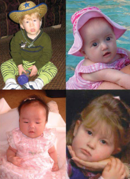
What are characteristic AS features?
Microcephaly (decreased brain growth).
Severe cognitive impairment.
“Puppet-like” ataxic gait with jerky movements.
Seizures.
Distinct facial appearance.
Short stature.
What are additional AS features?
EEG abnormalities.
Hyperactivity.
Bouts of laughter (happy demeanor).
Severe speech and language limitations.
What are the AS genetic classes and severities?
Class 1 (70%): 15q11-q13 deletion → most severe.
Class 2 (5%): Uniparental disomy → less severe.
Class 3 (5%): Imprinting defect → less severe.
Class 4 (10%): UBE3A mutation → intermediate, displays obesity.
Class 5 (10%): Unknown → most severe.
Classes 4 & 5 have normal methylation and PWS-imprinting center (PWS-IC).
Classes 4 & 5 can’t be diagnosed by standard methylation testing; they need gene sequencing or other approaches.
What is the AS imprinting center?
880 bp exon.
Mediates switch from paternal to maternal imprint during oogenesis.
AS-IC methylates PWS-IC and silences expression.
Most class 3 AS cases have NO defect in AS-IC → suggests epimutations.
Epimutations: abnormal changes in gene expression caused by faulty epigenetic marks (like DNA methylation or histone modification) without changing the DNA sequence itself.
What is UBE3A and its function?
A protein that tags other proteins with ubiquitin → marks them for degradation or regulation.
The E3 ligase part decides which proteins to tag.
The HECT domain at the end is needed to actually transfer ubiquitin.
Can also interact with nuclear receptors and help turn certain genes on.
How do UBE3A mutations cause AS?
Mutations cause premature termination and HECT domain disruption.
Loss causes substrate protein accumulation → neurological abnormalities.
Inherited through MATERNAL line (maternally expressed).
Expressed in hippocampal neurons and cerebellar Purkinje cells.
Purkinje cells = large neurons in the cerebellum (the brain region that controls movement and coordination).
What are neurological effects of UBE3A loss?
Microcephaly = abnormally small head size, usually due to reduced brain growth.
Impaired motor function and long-term potentiation.
Defects in contextual and spatial learning.
May regulate estrogen receptor stability (decrease linked to cancer).
What’s the key PWS vs AS difference?
PWS: Loss of PATERNAL 15q11-q13.
AS: Loss of MATERNAL 15q11-q13.
Same region, opposite inheritance, completely different phenotypes!