Primary Visual Pathway
1/20
There's no tags or description
Looks like no tags are added yet.
Name | Mastery | Learn | Test | Matching | Spaced |
|---|
No study sessions yet.
21 Terms
What are the main components of the primary visual pathway?
The main components include the optic nerve, optic chiasm, lateral geniculate nucleus (LGN), and the primary visual cortex (V1).
The primary visual pathway from eyes to primary visual cortex in occipital lobe
- Each eye has a visual field from which visual information (light) can enter the retina and excite component cells.
- Central area of field of view (sharpest vision + colour vision) excites the fovea.
- From the retina via the optic nerve crossing into the optic chiasm, visual information is transmitted further up to the primary visual cortex via the lateral geniculate nucleus (thalamic relay station).
- Left field of view ends up in right visual cortex (vice versa).
Information processing stages in the Primary visual pathway
Visual information processing is hierarchal; it happens sequentially across several stages.
What are photoreceptors?
- Photoreceptors (rod s and cones) in retina detect presence or absence of light.
- Bipolar cells mediate between photoreceptors and ganglion cells.
- Photoreceptors and bipolar cells vary their voltage as they are stimulated (analogue signal) whereas all subsequent cells vary spike rate (all-or-nothing, digital signal).
- Photoreceptor detection of light is translated into excitation or inhibition of retinal ganglion cells via bipolar cells.
What type of signal do photoreceptors and bipolar cells use?
Photoreceptors and bipolar cells vary their voltage as they are stimulated, which is an analogue signal.
What types of cells make up the primary visual pathway?
The primary visual pathway is composed of photoreceptors, bipolar cells, and retinal ganglion cells.
What is the function of centre-surround receptive fields?
Centre-surround receptive fields help in detecting contrasts and edges in visual stimuli.
Area of the retina/visual field in which visual stimulation will evoke a change in the firing rate of a given visual neuron.
What are the different types of photoreceptors?
The two types of photoreceptors are rods and cones.
What are the main features of rods?
Rods are more numerous than cones, about 120 million in the human retina, sensitive to low light levels, do not mediate color vision, and have low spatial acuity. They are more abundant too.
What are the main features of cones?
Cones are less numerous than rods, about 6 million in the human retina, require bright light for activation, mediate color vision, and have high spatial acuity.
Retinal ganglion neurons
Receive input from multiple photoreceptors (via bipolar cells).
ON-OFF centre-surround receptive fields.
Light presented in 'ON' regions excites cells.
Light in 'OFF' regions inhibits cells.
ON and OFF regions are organised in 'centre-surround' fashion.
Response rate of cell is based on the sum of stimulation in ON region - stimulation in OFF region.
Neurons in the lateral geniculate body respond to visual stimuli in similar ways to retinal ganglion cells
Neurons in the lateral geniculate body respond to visual stimuli in similar ways to retinal ganglion cell
What is the significance of colour-oponency in retinal ganglion cells?
Colour-oponency allows retinal ganglion and LGN neurons to process color information and is associated with effects like negative afterimages
- We only need to respond to changes and boundaries (edges) → efficient.
- Luminance of features represented relative to their surround: helps preserve appearance of objects regardless of light levels in the environment → can also result in illusions..
Colour sensitivity of retinal ganglion and LGN neurons
Receive input from cones (differentially sensitive to different wavelengths) and are sensitive to colour.
- Colour-sensitive retinal ganglion and LGN neurons have receptive fields that show centre-surround colour opponency.
- Colour opponents either between yellow and blue, or red and green (e.g. when blue presented in periphery of receptive field, firing right is inhibited and where yellow presented in centre, firing rate increased → opposite effects when other way around: same for red and green).
- Colour opponency along with firing-rate adaptation (rebound effects) can explain negative afterimages.
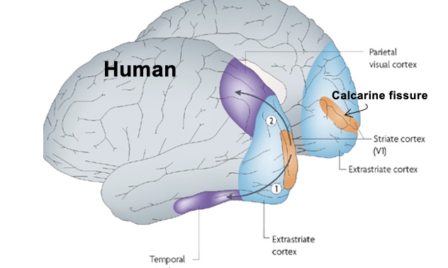
Primary visual cortex (striate cortex, V1)
- Sits around calcarine fissure, between the two hemispheres.
- Striate cortex that receives input from the lateral geniculate nucleus.
Orientation-selective cells in V1
- Respond to elongated stimuli of a particular orientation.
- Two main types of orientation V1 neurons: simple and complex cells.
- Simple cells: make up receptive fields of complex cells: have inhibitory and excitatory regions and ON and OFF regions.
- Complex cells: do not have discrete ON and OFF regions; wherever presented in receptive field, leads to excitation of the cell as long as stimulus presented in correct orientation, if not no response.
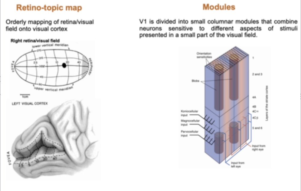
Maps and modules in V1
- Retino-topic map: orderly mapping of visual field onto visual cortex.
- Modules: V1 divided into small columnar modules that combine neurons sensitive to different aspects of stimuli presented in a small part of the visual field.
What is blindsight?
Blindsight refers to the ability of subjects who are 'blind' due to lesions in the primary visual cortex to respond to visual stimuli without conscious awareness.
Subjects with lesions to primary visual cortex and apparent 'blindness' can show appropriate response to visual stimuli of which they are not 'conscious'.
Examples include looking (i.e. moving the eyes) or pointing towards visual stimuli; detection of movement etc.
Blindsight highlights that there are additional visual pathways.
Also highlights that brain can perform visual information processing which can guide subjects' behaviour without their conscious awareness.
What roles do simple and complex cells in V1 serve?
Simple cells respond to specific orientations of stimuli, while complex cells are more responsive to moving stimuli and have no discrete on/off regions.
How does the brain process visual information for perception and memory?
Visual information from V1 is combined and further processed in the visual association cortices (V2-V5, inferior temporal cortex, posterior parietal cortex) to perceive holistic visual properties.
What happens to visual perception without rods or cones?
Without rods, sight would only be in shades of black and white, and without cones, one would struggle to see in low light.