2. Arachnoentomology
1/22
Earn XP
Description and Tags
Oral part - Study of Arachnids - spiders, mites etc. and insects. (VS, lecture - intro, insects, mites). pink = fant ikke i pres.. men er fra notes.
Name | Mastery | Learn | Test | Matching | Spaced | Call with Kai |
|---|
No analytics yet
Send a link to your students to track their progress
23 Terms
1.General Morphology and Classification of ARTHROPODA
Arthropods - 3 major classes are of medical importance:
Insecta (hexapoda) - includes mosquiotes, flies, bugs, lice and fleas
Arachnida (octopoda) - ticks, mites, spiders, scorpion
crustacea - organisms such as cyclops and crabs
Classification:
Phylum: Arthropoda (Arthropods)
subphyl:Trilobitomorpha
subphyl: Chelicerata
Class: Arachnida
subclass: Acari - mite & Tick (distribution according to localization of stigmata):
Order: Ixodida (metastigmata)
Order: Gamasida (mesostigmata)
Order: Trombidiformes (prostigmata)
Order: Sarcoptiformes (astigmata)
Order: Oribatida
Subphylum crustacea:
Class: Crustacea
Subphylum Tracheata:
Class: Insecta
Order: Heteroptera (true bugs)
Order: Phthiraptera (lice)
Order: Diptera (flies)
Order: Siphonaptera (fleas)
Morphology of Arthropods:
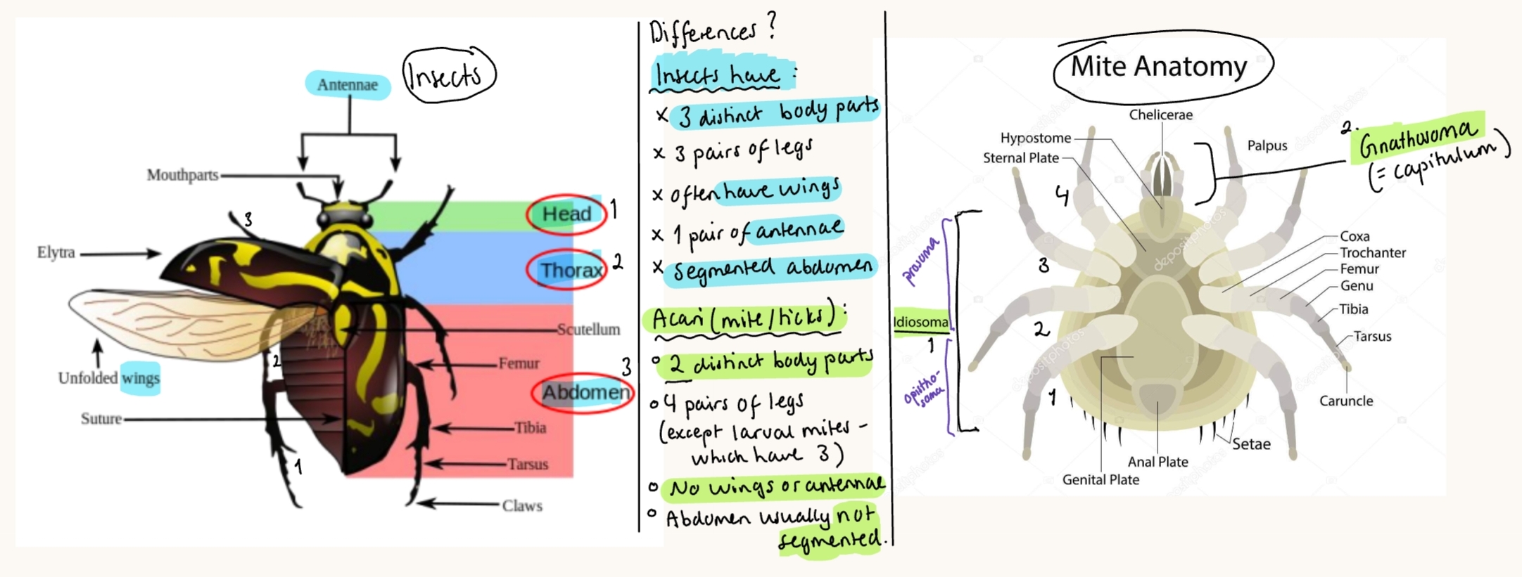
Body structure: segmented (tagmata)
Insects → 3parts = head, thorax + abdomen
Arachnids (spiders) → 2parts = cephalothorax/gnathosoma (head + chest fused) + idiosoma (abdomen)
Exoskeleton (outer): hard outer covering of chitin, prevents drying out, must moult to grow. Antenna is here.
Jointed limbs → can move easily, segmented (6), ends in claws. parts + segments are joined by flexible membranes.
Internal systems:
Open vascular system/circulatory - haemolymph (not true blood) flows through body (haemocoel)
NS: ventral nerve cord, sensory organs - anterior ganglia, paired nerve cords, antennae (in insects) - which may/not have receptors
Digestive system - complete: Mouth → intestine → anus.
Respiratory: resp. through skin, via openings called spiracles/stigmata in abdomen/thorax.
Reproduction: separate sexes - sexual or parthenogenesis (clones), most lay eggs.
Sensory system: compound eyes → many lenses (sees movement well) or simple eyes → one lense for detecting light
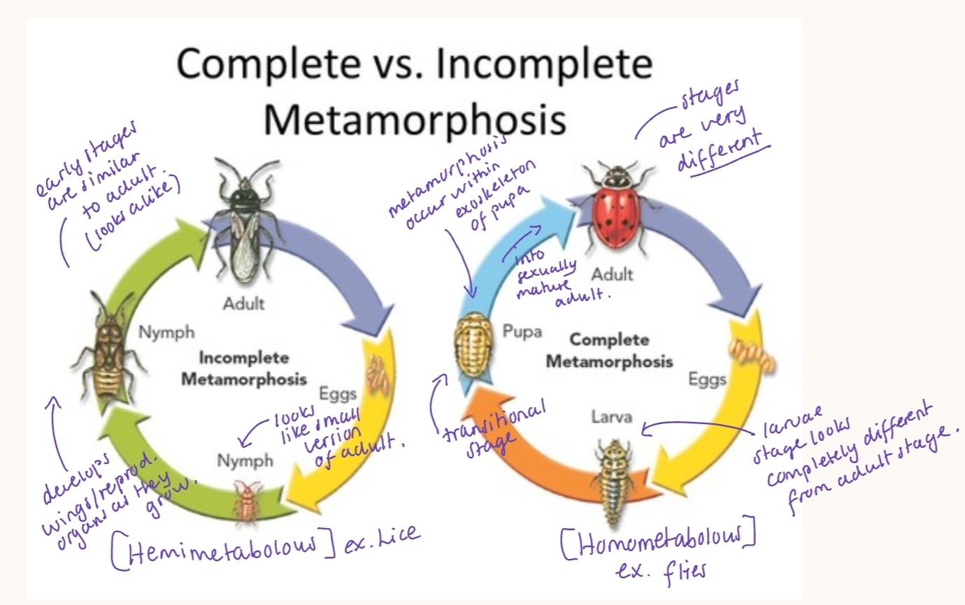
Development by Incomplete & Complete Metamorphosis:
2.Veterinary significance of arthropods as vectors of pathogens
Arthropods can be intermediate hosts for various parasites. They can be vectors of bacteria, virus, parasites and other pathogens. They are very adaptable organisms.
As ectoparasites, they can cause direct harmful effect: blood loss (as bloodsuckers), myasis, skin inflammation, pruritus (skin reactions), their saliva can lead to allergic reactions, and they may produce venom that can be toxic.
Or indirect harmful effect: Disturbance/social nuisance - large amounts can lead to stressed animals/affects behavior/interactions → immunosuppression over time, Self-wounding from trying to get rid of them → causing wounds that attract new flies.
Mode of transmission:
Mechanical vector (passive carrier - pathogen just gets a ride on the bug, no development of pathogen inside vector)
Viruses, bacteria
Biological vector - undergoes specific stages of their LC - development and/or reprod. of pathogen before it becomes infective. For protozoa, tapeworms (cestode), round worms (nematodes). Types of levels:
Propagative transmission: pathogen multiplies only, ex. yellow fever, eastern equine encephalitis (EEE)
Cyclodevelopmental: pathogen develops, but does not multiply, ex. in filaria - single microfilaria eaten by culex mosquito
Cyclopropagative: pathogen changes form + multiplies ex. malaria (plasmodium in mosquito, chagas disease)
Vector-disease concept: Disease occur when all connect - vector + host + pathogen + environment → transmission. These factors interacts between each other, as vector takes up the pathogen, pathogen develops, transmitted to host → causing disease.
Transmission methods:
Direct contact: Host contact with bedding, clothing contaminated with eggs/pupae. (simply treating the animal is not enough in this case → must disinfect the environment).
Adults: actively seek host to feed or lay eggs, mosquitoes (fly to host), fleas jump onto passing host, eggs (repellents + environmental barriers needed).
Eggs: contaminating shared environment (risk of whole herd/household)
Parasitic stages:
both larval and adult stages can be parasitic, depending on species.
Adult parasites - adult mosquitoes and flies (ex. tse tse & horse flies) act as primary vectors of blood-borne pathogens (malaria/Leishmania) because they need blood meals for reproduction. Adult stage = parasitic.
Larval parasites - in some, the larval stage cause disease. Larvae causing myiasis invade tissues/organs, like Hypoderma bovis → going into spinal cord, causing direct tissue damage.
Important groups:
Lice (order: phthiraptera): adults are ectoparasites of skin/hair/feathers. Eggs attach to hair/feathers, thus single treatments are often ineffective.
ex. sucking lice (haematopinus suis in pigs) and biting lice (bovicola/dalmalinia bovis in cattle)
Ticks (Ixodida): obligate blood-feeding ecto. Adults feed on blood.
Mites: feeds on skin debris, tissue fluids or lymph, adults and often nymphs + larvae can be parasitic.
ex. sarcoptes scabiei (mange) and demodex.
Fleas (siphonaptera): only adult stage is parasitic, feeding on blood. Larvae are free-living and feed on organic debris in the environment. Effective control is needed in treatment (both host + environment - bedding).
Ex. ctenocephalides felis (cat flea)
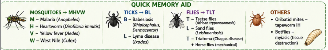
Common arthropod vectors - for pathogens:
mosquitoes (culicidae) transmits:
Malaria - by anopheles genus
Heartworm (dirofilaria immitis)
viruses: aedes & culex transmit yellow fever, west nile virus
Ticks (ixodida):
Babesiosis - by Rhiphicephalus sanguineus & dermacentor reticulatus
Lyme disease - by ixodes spp. (borrelia bacteria)
Flies (diptera):
Trypanosomosis - by tse tse flies (glossina), kissing bugs (triatoma) & horse flies (tabanidae)
Leishmaniosis - by sand flies (phlebotomus, lutzomyia)
Others:
Oribatid mites - IH for tapeworms - moniezia & anoplocephala spp.
Myiasis (tissue destruction) - direct harmful effect where larvae (maggots) like botflies (hypoderma bovis) develop in the living tissues of vertebrates.
3.Parasitic Nematocera
Nematocera = suborder. Order: Diptera (flies), Class: Insecta
Huskeregel: Cute Psychos Simulate Ceremony"
Families: Culicidae (mosquito), Psychodidae (drain flies), Simuliidae (blackflies), ceratopogonidae (biting midges, culicoides)
General - Nematocera:
Typical antennae (6 and more segments)
Slim body (except - simuliidae) + long limbs
Hematophagous LC (sucks blood). Only females feed on blood.
Found in aquatic/semiaquatic habitat, LC depend on moisture
Animals - protected by antiparasitic drugs, repellents, spot-on, collars
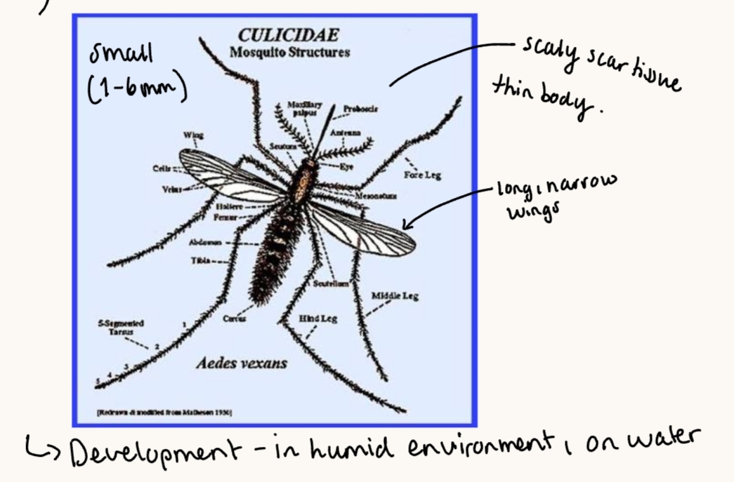
1) Culicidae (Mosquitoes)
3-6mm (small)
Subfam: anophelinae (genus: anopheles) & Culicinae (genus: aedes, culex - vectors for avian pox, filarial worms, equine encephalitis)
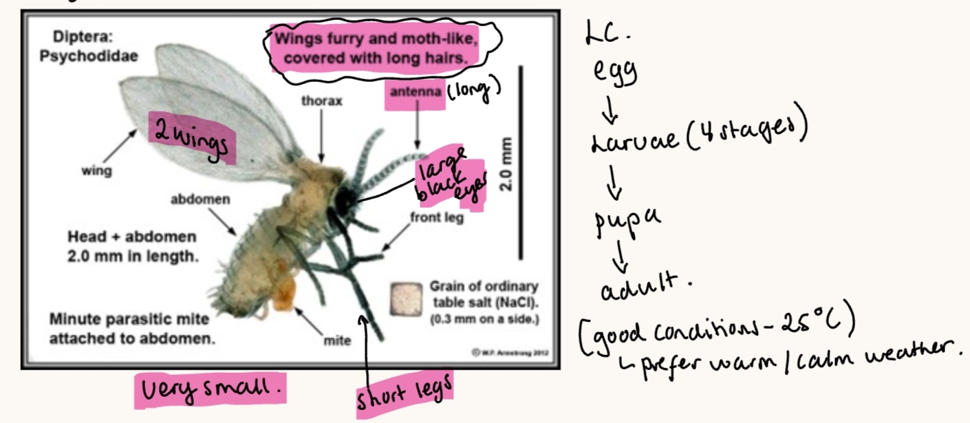
2) Psychodidae (Drain flies)
Genus: Phlebotomus, Lutzomyia - Vectors of Leishmania
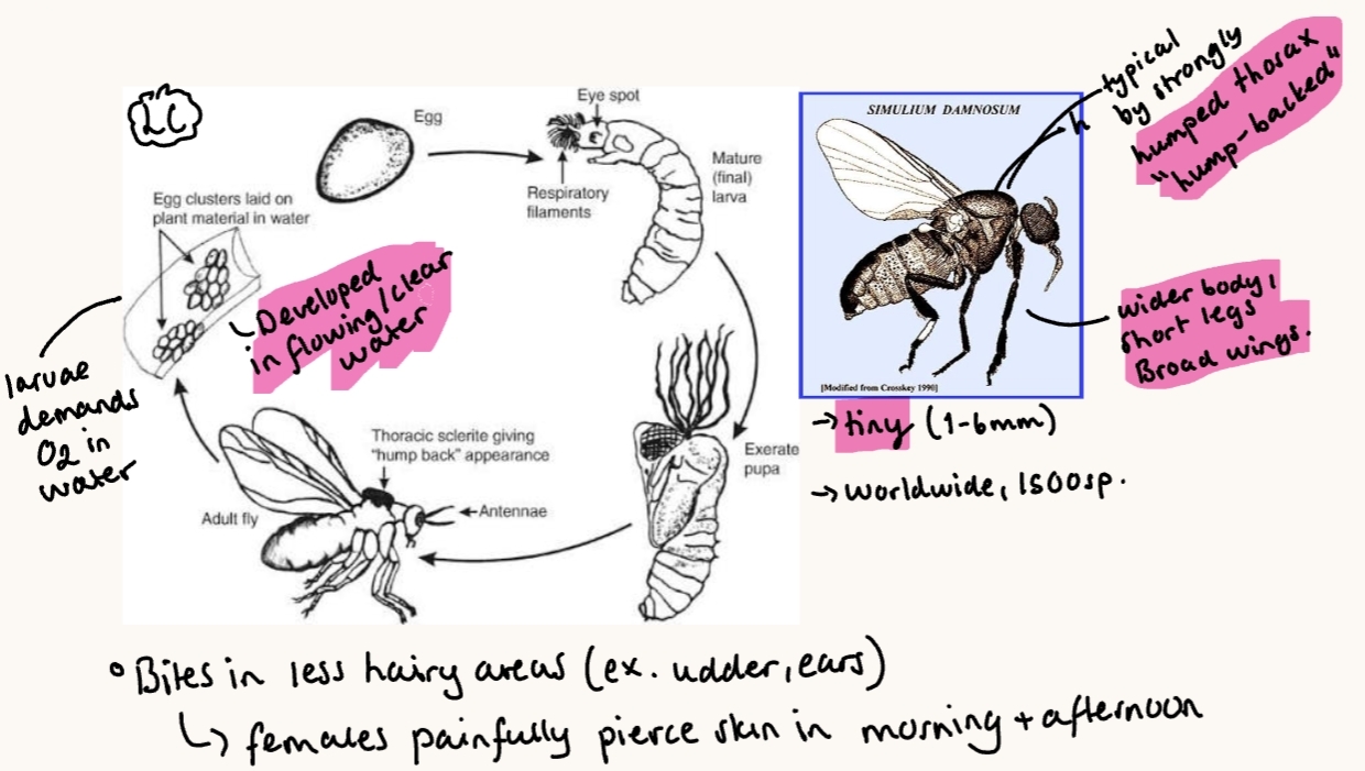
3) Simullidae (Black flies)
genus: Prosimulium, simulium
Clinical signs: Combined effect of anaphylactic reaction, blood loss, inhalation of flies + pulmonary edema → rapid death of animals.
Toxins in saliva → subcutaneous edema, pruritus, vascular damage, hypersensitivity reaction.
Simuliotoxicosis - vectors of: filarial nematodes (onchocerca species like O.gibsoni/O.cervicalis, Protozoa like leucocytozoon (L.simondi) & trypanosoma. Viruses of stomatitis like VSV (vesicular stomatitis virus).
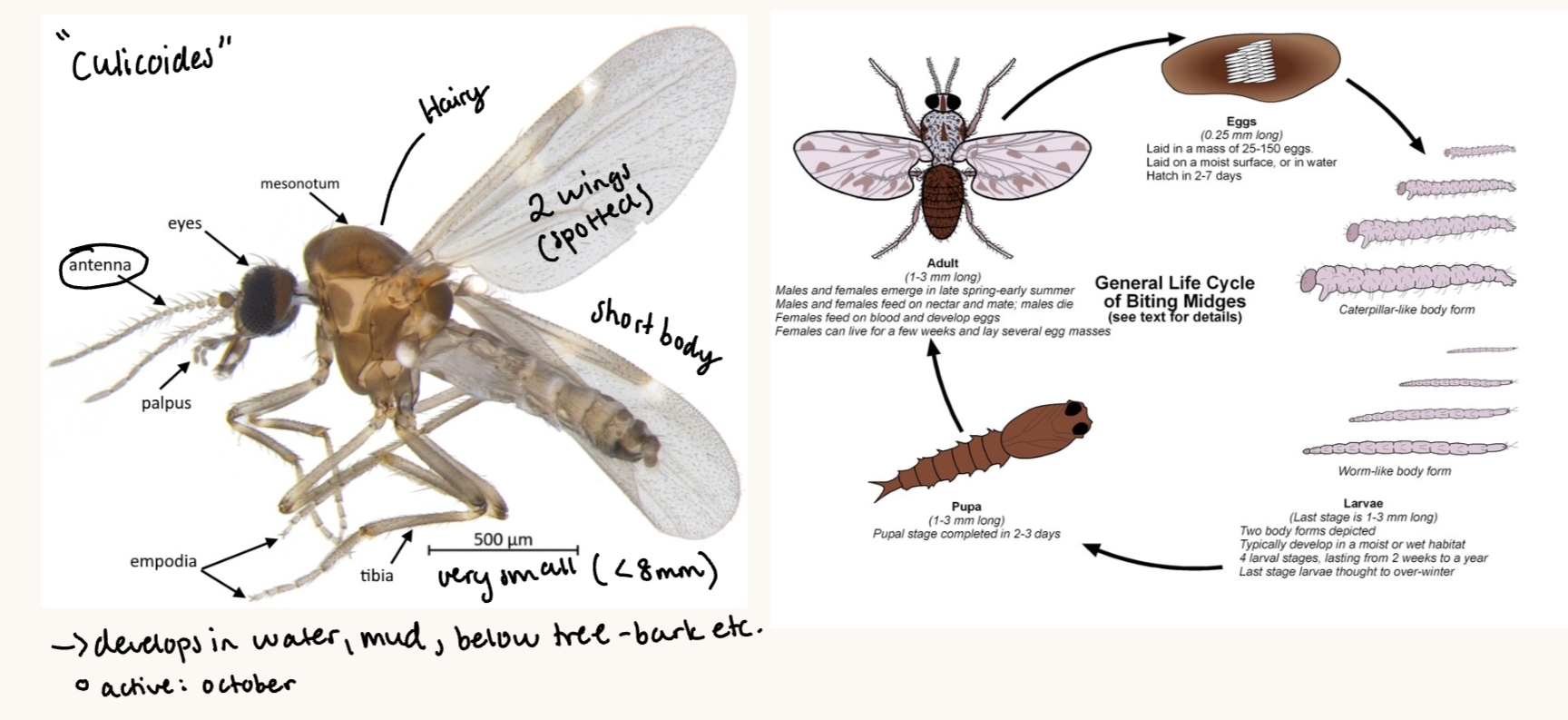
4) Ceratopogonidae (Biting midges)
Genus: Culicoides with species like: C. obsoletus, C. oulicaris, C. pulicaris
Vectors of protozoa like haemoproteus, heapatocystis, leucocytozoon, trypanosoma, Filarial nematodes like mansonella & onchocerca, blue tongue virus.
Clinical signs: cause chronic lesions of lichenification and alopecia in horses with Culicoides hypersensitivity. Use antihistamine and corticosteroids for treatment.
4. Parasitic Brachycera
Class: Insecta
Order: Diptera (Flies)
Suborder: Brachycera (Brachycera + Cyclorapha) - worldwide.
Family (w. ex. of genus)
Tabanidae - horse fly - “lower brachycera/more primitive” (tabanus, chrysops, Hamatopota)
Rest of families are “higher brachycera” - cyclorphapha
Glossinidae - tse tse (glossina)
Muscidae - House fly (muscus - vector for habronema muscae)
Calliphoridae - blood, bottle & screw worm fly (calliphora, lucilla)
Sarcophagidae - flesh fly (sarchophaga, wohlfartia)
Oestridae - botfly/warble (oestrus ovis, hypoderma bovis and lineatum, gastrophilus intestinalis)
Hippoboscidae - louse fly
Hippoboscha equina (with wings)
melophagus ovinus (wingless)
General Morphology/Brachycera:
3 segments of body, 3 pairs of legs, short antenna, compact, carnivorous larvae.
Mouth parts - important for these
oestridae have bad/missing mouth as an adult as it lives just 1 week as an adult, just for laying eggs.
Tabanus has painful bites, as it cuts up the skin to use a “sponge” in their mouth to suck the blood.
general on insecta: complete digestive system - mouth, intestine, anus. Mouth - 1 pair of jaws (mandibles), compound eyes (many lenses) or simple eyes, Reproduction by homometabolous (complete metamorphosis) for diptera; flies.
General LC:
egg (not oestrus ovis) → larva (long stage) → pupa → adult
Complete metamorphosis with min. 6 larval stages. Female lays eggs on underside of leaves/stones in muddy areas → eggs hatch → larvae drop into water → develops → pupa → develop for 1-3w. → adult.
Vector For: Active during hot, sunny weather. Worldwide.
many diseases such as surra, Q-fever, anthrax, hog cholera, equine infectious anemia, california encephalitis. - can give serious irritation to the livestock animals, may cause stress, self-harm.
Treatment: Insect repellent for tabanus, Musca. Ivermectin for Oestridae family, dipping for hippoboscidae, surgical removal for calliphoridae family.
Insecticides, repellent during summer - during warmer weather
5. Hypodermosis of cattle.
Class: Insecta
Order: Diptera
Family: Oestridae (myasis producing flies)
Cattle hypodermosis - Warble fly infestation.
Hypoderma Lineatum - common cattle grub
Hypoderma bovis - Northern cattle grub
Morphology:
Adult: Looks like a bee, abdomen is covered with yellow-orange hairs, moutparts does not work.
Adults do not eat, their only job is to mate & lay eggs
eggs: very small, 1mm. Laid on the hair of animals
Female: ovipositor - special tube-like organ for laying eggs. Long, extendable, helps fly place eggs precisely, then it can fold it inside like a telescope into abdomen when resting.
Mature larvae (L3): 28mm, Barrel-shaped (short, thick).
Spiracle plates (breathing openings) - looks like small ears
Dorsal (top side): slightly sunken, small narrow opening
Ventral (bottom): smooth (no spines)
White when newly emerged → Into dark-brown color
Pupa: almost black.
Geographical distribution: Worldwide.
Location: Through skin to either esophagus (Lineatus) or spinal cord (Bovis)
Life cycle: Long → 9-10m. Adults live only ish a week. In cold temperatures, females cannot lay eggs → fast deposit when temp. is high, thus most active during warm days (spring/summer)
Eggs are laid in hair of cattle (typ. distal hind limbs - “heel fly”) → larvae hatch and migrate into skin → continues into esophagus/spinal cord - dormant during winter, larvae migrates back to the skin again → matures (L1 → L3) and forms warble - L3 drop off host animal and pupate (1-3 month pupa stage) → pupa falls onto ground, emerge around may/july.
H.bovis - eggs are laid single, larva migrate along nerves to cord
H.lineatus - eggs are laid in batches, migrate along muscle/CT
Pathology & clinical signs: Nodules are seen in the skin on the back, each with a small opening, containing a larva inside. These are called “warbles”. Adult flies buzz and chase cattle → cattle becomes stressed, may run (stampeding).
cause irritation, possible self-mutilation. Pressure of many on spinal cord → NS.
Larval migration is not usually noticed clinically, but heavy infestation can reduce growth & milk production (Economic loss). Sometimes, pressure of larvae on spinal cord can cause paralysis.
Diagnosis: Presence of larvae under skin of back.
Treatment/Control:
Ivermectin (pour-on, spot-on)
Avoid late fall treatment becuase bloat/hind end ataxia may result due to host reaction around killed larva in esophagus or spinal canal
Control strategies: Make sure they do not conflict with each other, if using ivermectin, dectomax, or cytedectin. Treat during sept - oct. as they are active in may-august.
Dont treat during nov. 1 to feb. 1. - as they may die in spinal cord → release proteolysis → paraplegia (unable to feel, move legs)
6. Oestrosis of sheep and rhinoestrosis of horses.
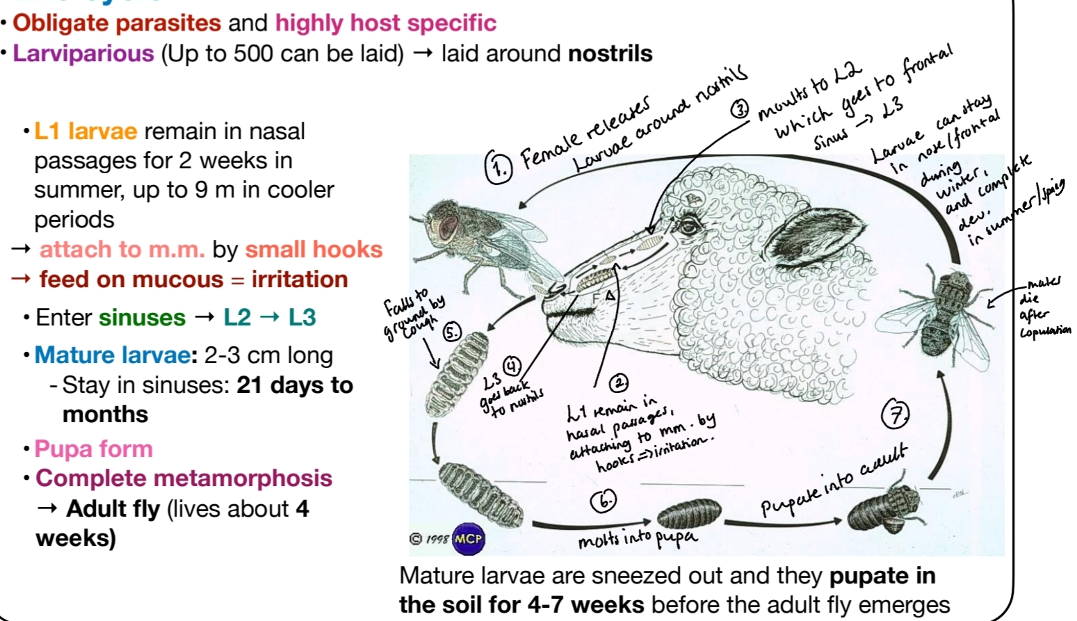
Oestrosis = Nasal Myiasis.
Class: Insecta, Order: Diptera, Family: Oestridae - these are nasal pharyngeal Myasises.
In sheep:
Caused by Oestrus Ovis (Sheep nasal bot fly). - “head maggot” - worldwide
Morphology:
Young larva: white-yellow in color.
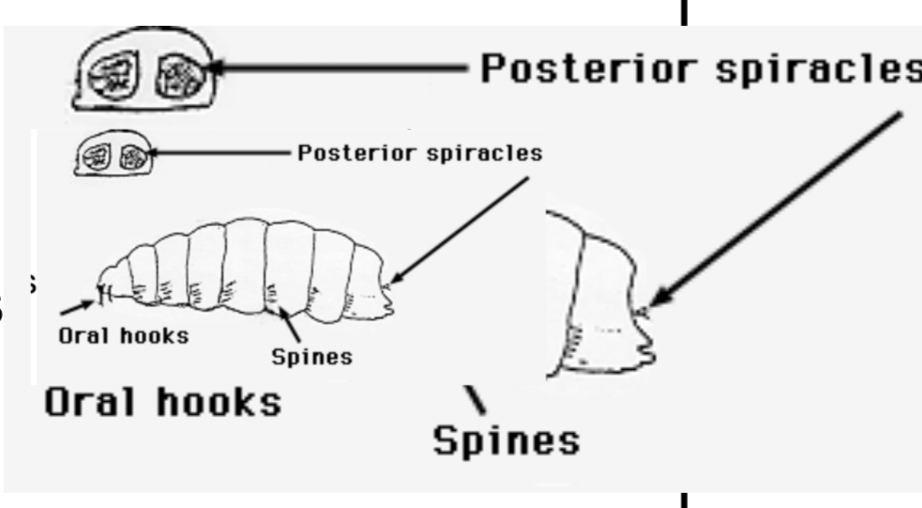
Mature Larva: Yellow/brown, matures - develops brown/dark bands on dorsal surface of segments. Full grown: 3cm. Has large black oral hooks, posterior spiracles, and spines.
Adult: 1cm, grey, small black spots on the body, short lived. Moutparts vestigial, non-feeding.
Life cycle: female is Larviparous (Lays live larvae).
Pathology and clinical signs:
Irritation & Inflammation due to larvae → impaired respiration, damage.
Nasal discharge, sneezing, nose rubbing, head shaking.
Weight loss, circling behavior, lack of coordination.
Sec. bacterial infections, abnormal migration → pneumonia & death.
Diagnosis: Look at the behavior of sheep, look for signs of the fly, CS. But it may be needed to differentiate the conditions from others with similar signs. Confirm with endoscopy, ELISA, necropsy.
Treatment: Ivermectin in heavy infections - not economical in low infections in humans - sometimes larva can make subcutaneous myiasis in young children - ocular myiasis (blindness) and spinal cord (paralysis) - oestrus ovis & rhinoestrus is considered zoonotic potential.
Closantel, Nitroxynil, Doramectin, Moxidectin
Rhinoestrosis of horses
Horse nasal myiasis - Seen worldwide
By Rhinoestrus Purpureus & R. usbekistanicus (Larvae of flies, belonging to genus Rhinoestrus)
Host: Horse, donkey, zebra.
Morphology: Adults are yellowish-gray-brown, hairy, mouthparts are rudimentary (absent). Glossy black stripes, whole body in wart-like dots. Larvae: like O. ovis, but recurved mouth hooks, single row of hooklets.
Life cycle: Similar to sheep. (Complete metamorphosis). larvae goes to turbinates/pharynx, moult to L2-L3, expelled and pupate on ground.
CS, dg, & treatment: same as in sheep.
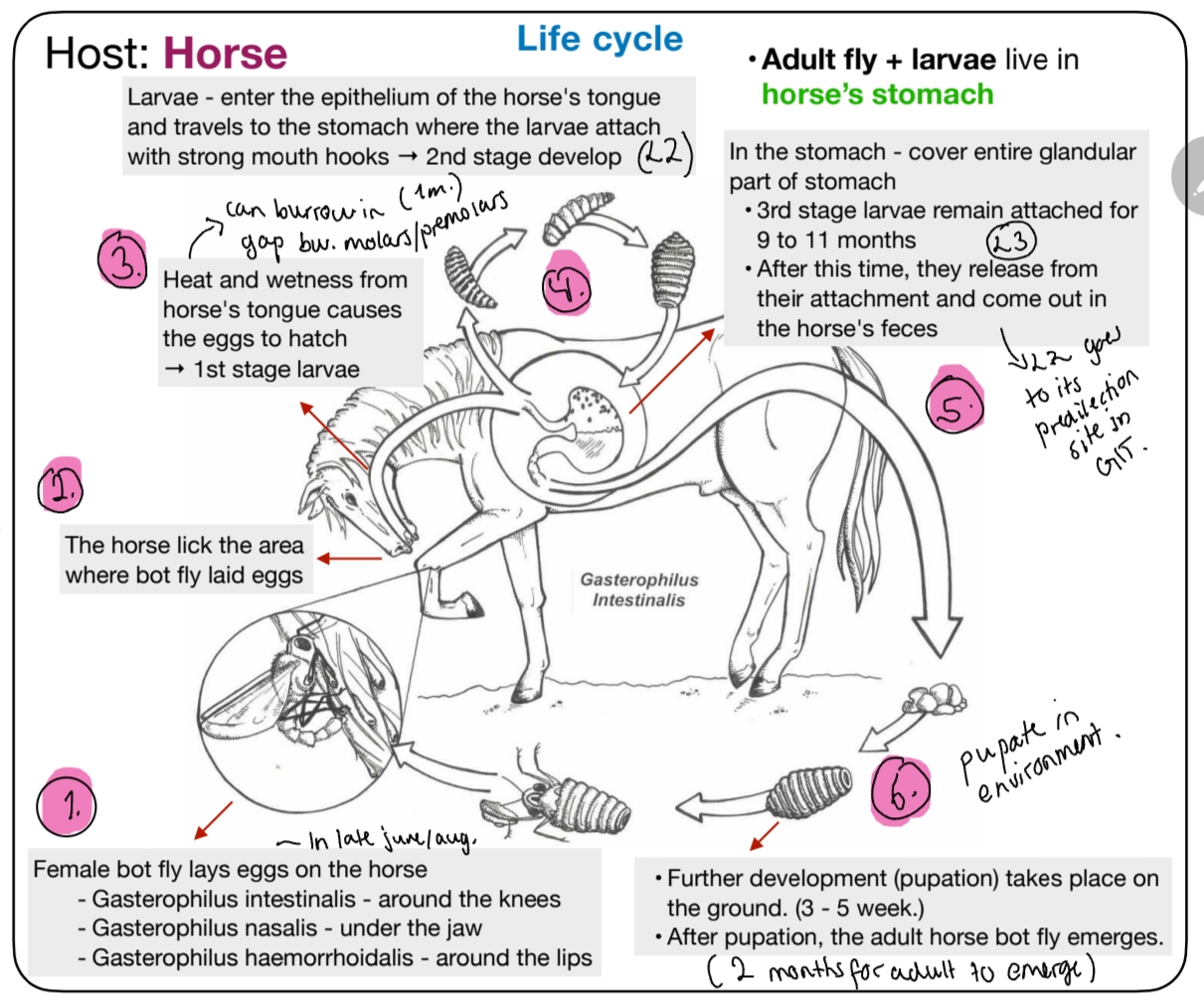
7.Gasterophilosis in horses.
Family: Oesteridae, Subfam: Gasterophilinae
Gasterophilosis - caused by:
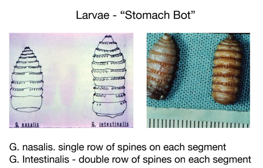
Gasterophilus intestinalis (botfly) – most common type, found in duodenum. Favors cardiac reg. of stomach.
G. haemorrhoidalis – Stomach and rectum
G. pecorum – stomach and rectum
G. inermis – found in esophagus and stomach
G. nigiricornis – duodenum
Morphology:
Adults: Robust dark flies, yellow hairs, transverse bands on wings. “honey bee - alike”. Female - protuberance for laying eggs.
Larvae: yellow-brown, oral hooks, transverse bands, 2 rows of blunt spines on all segments except the last. narrowed on one end.
Non-functioning moutpart → just mating + deposit eggs.
Life cycle:
Pathology and Clinical signs:
Burrowing L1 + L2 in tongue + mouth → lesions, tunnels that become infection.
Inflammation + ulceration of stomach and intestine.
Ulcers → irritation, colic, anemia, inappatence, can lead to necrosis, severe.
Less number of bots: minor problems, or none.
Lesions: Rings of inflammatory thickening with eroded centers where the larvae were attached.
Diagnosis: Visible eggs on hair/mane. Endoscopy show clusters looking like many grapes. Necropsy.
Treatment: Ivermectin.
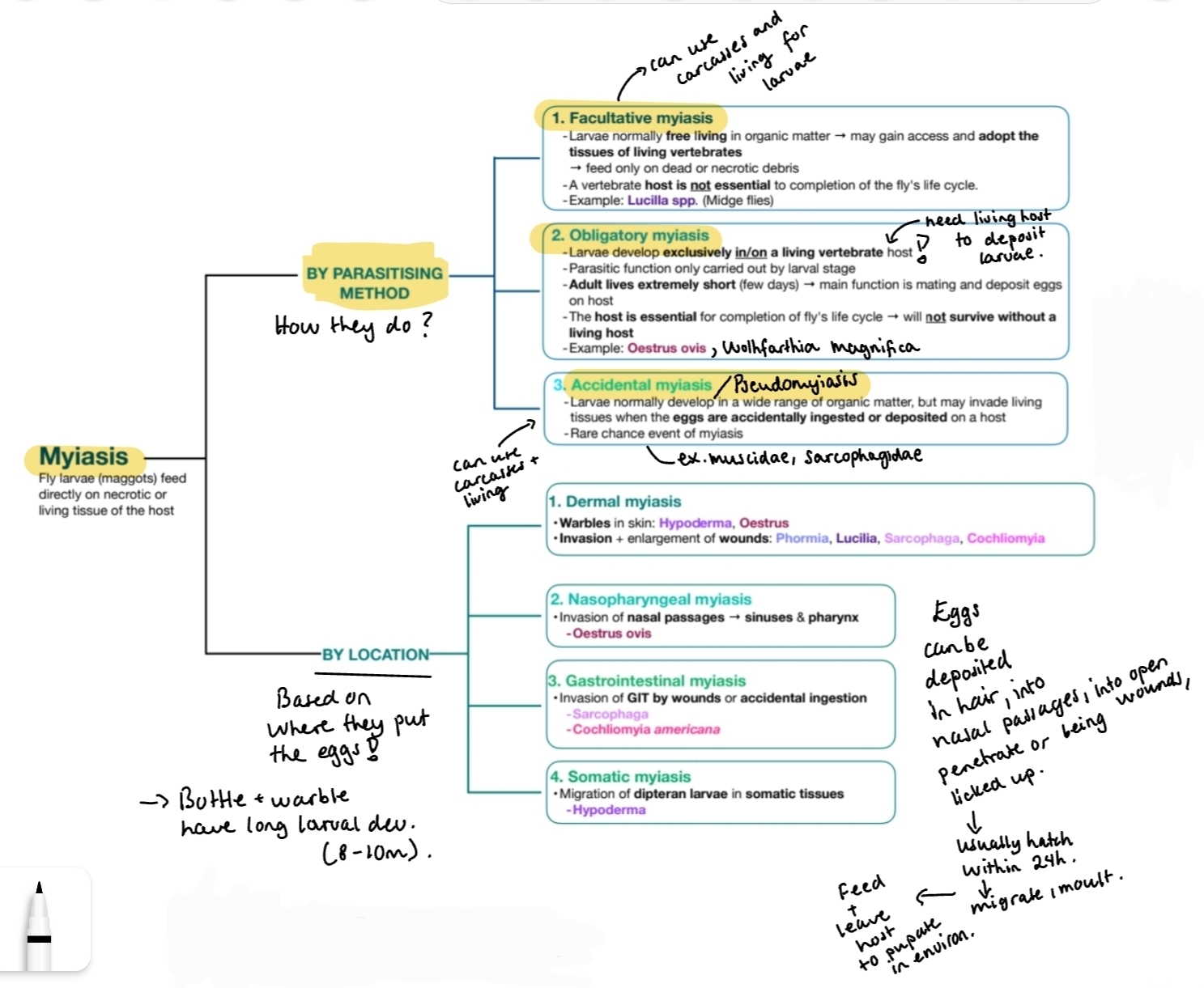
8. Myiasis
Myiasis: Parasitic infestation of organs & tissues by dipterous larva (maggots), which grow inside the host while feeding on living or dead tissue. Or in case of intestinal myiasis, living on hostˋs ingested food.
Hosts: mammals, humans, sometimes birds, reptiles & amphibians.
Order: Diptera
Three Main families causing Myiasis:
1) Oestridae (warble & bot flies). - Non-functional mouthparts. Bee-like, adults only live 1w. Host specific. Obligatory myiasis (not surviving without host, females deposit eggs/larvae onto host, living only for that). (not accidental → humans may be accidental host for oestridae).
Oestrus + Rhinoestrus (nasal cavity)
Gasterophilus (GIT)
Hypoderma & Dermatobia (under skin).
2) Sarcophigidae (permanent myiasis, flesh flies) - Facultative mostly. But some species of wolfharthia are obligatory. (also accidental).
Sarcophaga spp
Wolfarthia spp. (wound, meat, primarily infests humans).
3) calliphoridae (Metallic flies/Screw worm myiasis) - develop in animal flesh. Facultative & Obligatory.
Cochliomyia spp. - Screw-form. C. Hominivorax + Chrysoma Bezziana in humans invade nose, mouth & eyes → severe pain.
Phormia spp., Crymosoma spp.
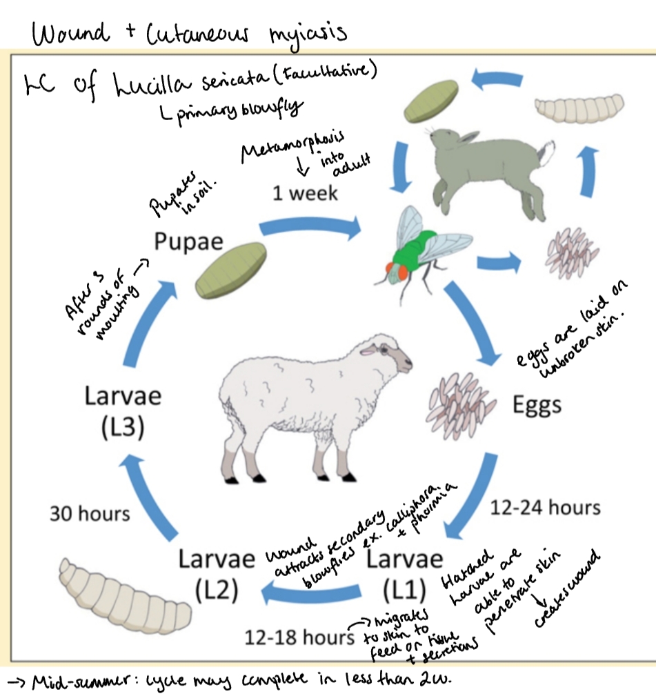
Lucilla spp. & Calliphora spp. - metallic flies. Lucilla sericata - blowfly-strike, eggs are layed in wounds, larvae move around → inflammation + skin damage, bacterial sec. infection, large economic loss.
Life cycle:
Picture: L. sericata (Facultative) - meaning it can use both carcasses & living hosts for completening LC as shown. It has 3 larval stages.
Flies - attracted to warm, wet skin, esp., urine + fecal stained areas, wounds.
Pathology and clinical signs:
Mechanical damage of tissues
Releases proteolytic enzymes - dissolving tissues, leading to deep, extensive damage & strong exudate formation
Bacterial infection, inflammation and animal may die.
CS: dull, lethargic, anemic, irritated, sec. bacterial infections. Often cause weight loss.
Diagnosis: Tunnelling lesions, Wounds with larvae, necropsy
Treatment:
Move animal to clean fly-free place. Treating predisposing factors!
Cutaneous myiasis larvae - Clip hair from lesions + clean area with antibacterial wash (ex. acetic acid, chlorhexidine). Should be dressed to prevent infection, avoid branding, dehorning + ear marking during fly season → as it makes wound.
Surgical debridement may be needed (subdermal warble forming larvae) - surgical removal
Insecticidal wash/spray can be used on rest of coat, ex. pyrethrin/pyrethroid based for dogs.
Systemic larvicidal drugs (unlicensed) may be beneficial: nitenpyram, ivermectin once, ATBs.
Nasophar, subdermal + GIT myiasis - ivermectin, moxidectin - particularly effective, but can also be for cutaneous.
Prognosis: varies depending on underlying cause, the level of tissue damage.
9.Melophagosis. (in sheep)
Order: Diptera, Suborder: Brachycera.
Family: Hippoboscidae (louse flies), Genus: Melophagus.
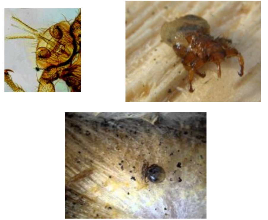
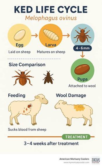
Melophagosis: in sheep. Caused by M. ovinus (Sheep ked).
Disease vector for bluetongue virus in sheep
Epizoo/Transmission: Permanent ectoparasite, host specific. Populations increase in winter due to longer wool. Spread by contact. Found worldwide.
Morphology:
Wingless, brown, hairy fly looking like a tick. Small head, flattened dorsoventrally, strong legs, tipped claws, 6 legs (true ticks have 8), small compound eyes. When the ked is released for its pupa they have wings, but break when they attach to the host (except Hipposca equina).
Female produce a single larvae at a time
Remaines inside the body for a week, then it pupates. Pupae is glued to the wool.
Female lives for 4months, makes only 10-13 puparia.
Equine Hippoboscidosis - H. equina. Blood-feeding, permanently fully winged fly, not shedding its wings on finding its host.
Life cycle:
Adults live in wool of sheep. Larviparous - egg hatches inside body → nourished through 3 larval stages. Feeds on blood.
Female lays 1 fully mature larvae at a time, cementing them to the wool (hard to remove)
Larvae immediately pupate, this stage is not susceptible to insecticide
Complete metamorphosis.
Clinical signs:
Feeds on blood → anemia
Irritation → rubbing → loss, damage of wool, dirty wool)
Makes firm, hard nodules on skin surface, with hemorrhages
Stains the sheepˋs wool reducing its value, economic loss
Immune response - reduces capillary flow to the skin - tries to fight infestation, but also reduces fleece quality.
Diagnosis: adults and pupa on surface and fleece.
Treatment:
Frequently change bedding in stables to get rid off the deposited pupae.
There is little info on chemical control of louse flies, due to there being no insecticides approved for use on horse or livestock that claim for louse fly control.
Pour on with Pyrethroids (synthetic) like deltamethrin, cyper/permethrin). or mixture. Dipping method.
We know - no macrocyclic lactones (mainly ivermectin + moxidectin) is approved for louse fly control, no reports that they would be effective at usual therapeutic dose of 0.2mg/kg.
No repellents to prevent infestations.
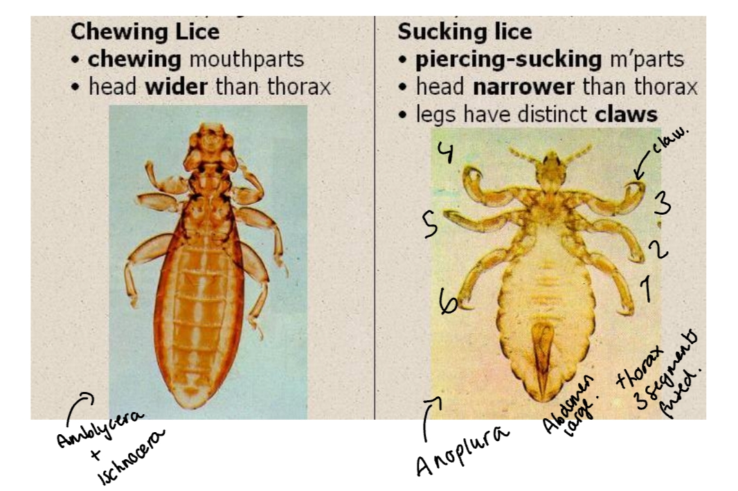
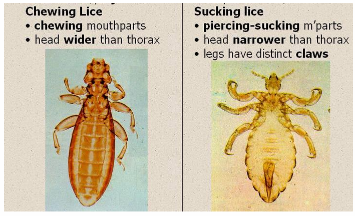
10. Lice of mammals (Anoplura).
Lice = Insects, in Order Phtiraptera.
suborders:
Anoplura (Sucking lice) - feeds on blood, mammals
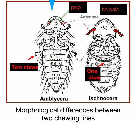
Amblycera & Ischnocera (biting/chewing lice) - Feed on keratin/skin debris
Characteristic: Permanent, highly host specific. Humans and pets do not share lice species.
Morphology: Small, wingless, strong legs with claws for clinging to hair. Poor eyes.
Suborder: Anoplura
Peducilidae (carries disease, like salmonellosis + typhoid fever) - Pediculus
Haematopinidae (Main one of vet importance!) - haematopinus
Linognathidiae - Linognathus
Hoplopleuridae (mainly in rodents) - Haemodipsus
Epizoo/transmission: Transfer by contact with max. populations in winter.
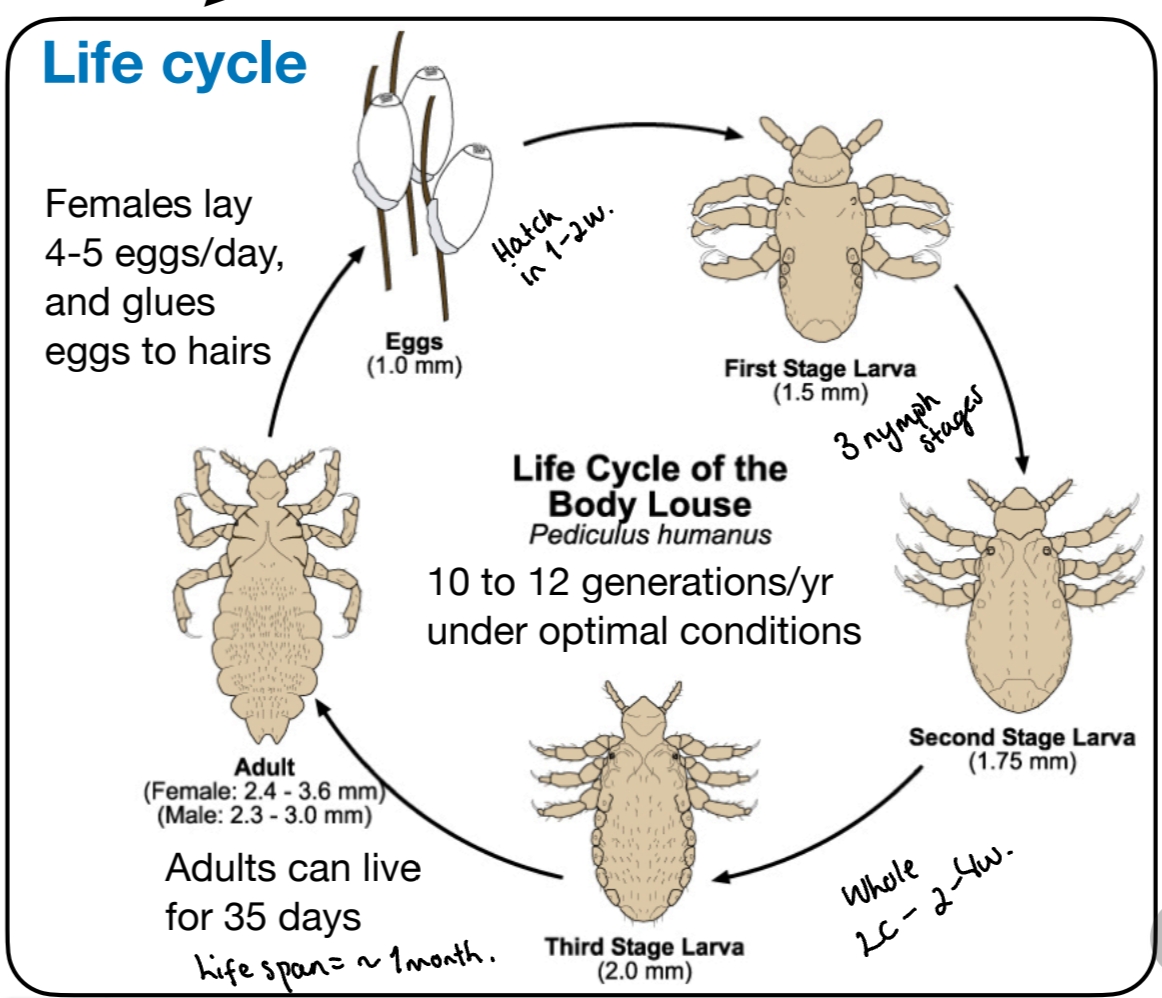
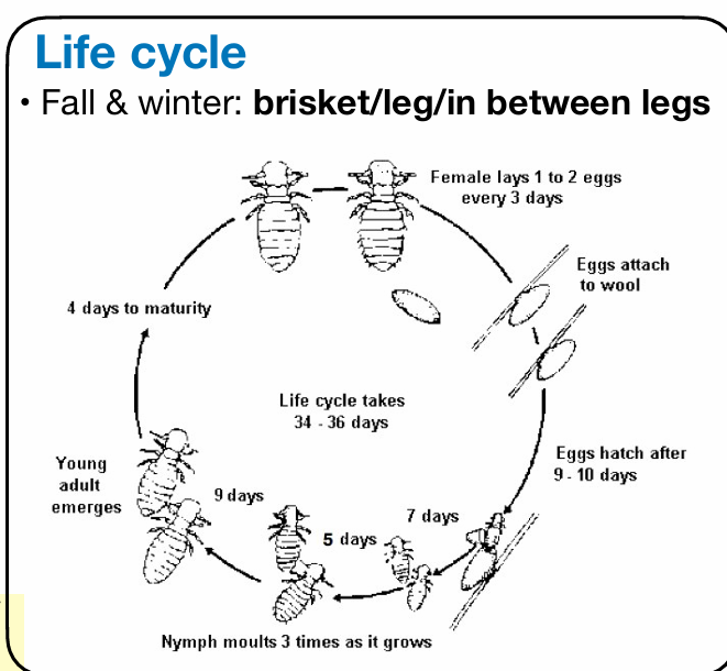
Life cycle: All developmental stages suck blood. Sucking & Chewing lice have similar LC - 3 moults, entire cycle on host, and incomplete metamorphosis!
Can be vectors for Richettsia & Borrelia.
Patho/CS: All cause irritation, hypersensitivity, Pruritus (itching), scratching/rubbing, alopecia, self-trauma, dermatitis, poor coat quality, reduced productivity/body condition. In heavy infestations → anemia.
Species:
Cattle: Haematopinous eurysternus, Linognathus vituli
Sheep: Linognathus ovillus, Linognathus pedalis
Goat: Linognathus stenopsis
Pig: Haematopinus suis
Dogs: Linognathus setosus (some are asymptomatic carriers, others have seborrhea, papules/redness).
Diagnosis: History & signs. samples of skin/hair, adults + eggs under microscope. acetate tapes/skin scrapings. Biopsy
Treatment: Drugs used to treat mange are usually effective against lice too.
pyrethroids - repeated after 10d. Pour-on formulations (deltamethrin + ivermectin in ru.
Clip coat, remove crusts
dogs/cats - insecticidal shampoo, leave on dips 3× in 2w. Permethrin. Spot-on like fibronil, selamectin.Systemic with ivermectin.
Genus - Pediculus in humans:
P. capitis - humans, head louse (all have had them) - spread salmonellosis.
P. humanus - humans, in clothing, body louse, spread infection. - spread typhoid fever.
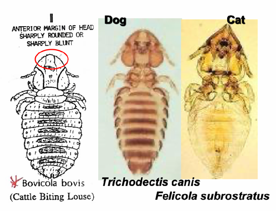
11. (Biting lice of mammals) Ischnocera of mammals
Lice = insects - order Phtiraptera. Includes suborders:
Anoplura (sucking lice, only mammals)
Amblycera and Ischnocera (biting or chewing lice, mammals & birds)
General: They are small, no wings but strong legs with one or two terminal claws. They live attached to the hair of their host. Feed either on skin debris, sebaceous secretions or blood.
ISCHNOCERA (CHEWING)
not host specific, transfer is by contact. Max. populations in winter.
Typical: very active, mobile - patchy alopecia. Thorax is segmented, legs are not adapted for clinging. Feeds on keratin.
Bovicola bovis - little red louse, yellowish tan with dark transverse bands running horizontally across each segment along with some setae (hair-like structures).
Life cycle: Same as sucking lice - Incomplete metamorphosis.
Species based on animals:
CS of biting lice is more focused on skin-impact, more obvious dermatologic signs, while sucking lice have more of a systemic impact (risk of anemia, production loss). But both have pruritus, coat damage and the animal being in poor condition.
Pruritus, scratching, rashes, secondary infection → bad appearance, rough coat, predilection sites of tail, shoulder, back (D.bovicola)
Ruminants & pigs:
Dalmalinia (bovicola) bovis – cattle
Dalmalinia (bovicola) ovis – sheep
Dalmalinia (bovicola) caprae – goat
Dalmalinia equi - horse (only species of lice normally in Norway)
Dogs and cats
Trichodectes canis – dog
Heterodoxus spiniger – dog
Felicola subrostratus – cat
Pruritus, some asymptomatic carriers.
Diagnosis: History & CS. Identification from samples of skin and hair, adults and eggs are seen under microscope. Acetate tapes or superficial skin scrapings can be used. Eggs can be stuck to hairs.
Treatment: chewing lice → topical insecticides best. Sucking → systemic.
Drugs for treating mange are usually effective against lice too.
Pyrethroids (permethrin) - repeated after 10d. (effective for biting)
Pour on formulations and avermectins (ivermectin) in ru
dog/cat - insecticidal shampoo or leave on dips 3 times in 2w.
Fipronil, imidacloprid, metaflumizole/pyriprole, selamectin, are effective.
12. Biting lice of birds (Amblycera + Ischnocera).
Lice are insects, of class Insecta Belonging to order: Phtiraptera. Incl. suborders:
Anoplura (sucking lice)
Amblycera (birds, chewing) and Ischnocera (biting or chewing lice)
General: They are small 0.5-8mm, no wings but strong legs with one or two terminal claws. They live attached to the hair of their host. Feed either on skin debris, sebaceous secretions or blood.
Amblycera + Ischnocera - birds:
Transfer by contact, feeds on keratin.
Morphology of chewing Lice: Dorso-ventrally flattened and wingless. Head not pointed, more somewhat broadened. Mouthpart adapted for chewing (not stylet-like). Very mobile. Thorax is segmented. Legs are not adapted for clinging.
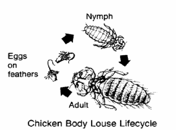
Life cycle: Same as sucking lice, Incomplete Metamorphosis. Entire LC in poultry. 3-5weeks. 3 nymphal stages (moults).
The lice of birds:
Philopteridae, Lipeuridae, Goniodidae.
Most pathogenic: Lipeurus & Menacanthus
Menacanthus stramineus (body lice) - amblycera
Menopon gallinae (quill lice) - amblycera
Lipeurus caponis (wing lice) - ischnocera
Goniocotes gallinae (very small) - ischnocera
Goniodes gigas (bighen lice) - ischnocera
Columbicola columbae - Ischnocera
Pathogenesis & clinical signs:
Picking on feathers, damage, dropping and pulling of feathers.
Reduction of tooth (feather), weight loss, anemia, young birds can die.
Heavy → decr. reproductive potential in males, egg prod. in females, less weight gain in growing chickens, Skin lesions → sec. bacterial infections.
Diagnosis: History & clinical signs. Identification from samples of skin and hair, adult and eggs seen under microscope. Acetate tapes or superficial skin scrapings. Sucking lice are slow to move, easy to catch. Eggs can be stuck to hairs/feathers.
Treatment & Control: same as in mammals, preparations based on cypermethrin, permethrin. Repeat after 14 days.
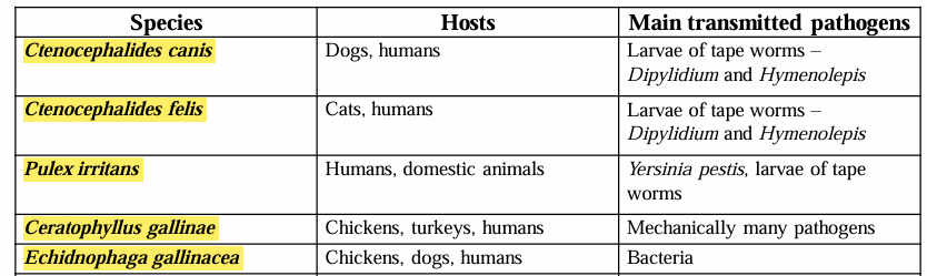
13. Fleas (Siphonaptera) of mammals and birds.
Class: Insecta
Order: Siphonaptera
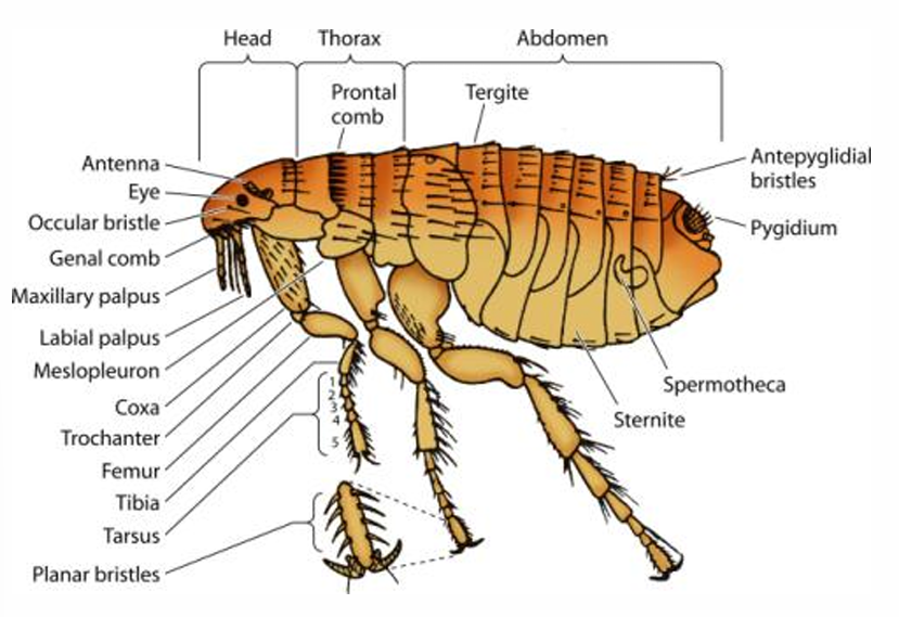
Morphology: Small, wingless, flattened laterally - allowing them to move easily through fur/feathers, covered in bristles, piercing-sucking mouthparts, strong and large legs for jumping, bigger abdomen, thick, dark brown chitinous covering, eyes reduced/absent.
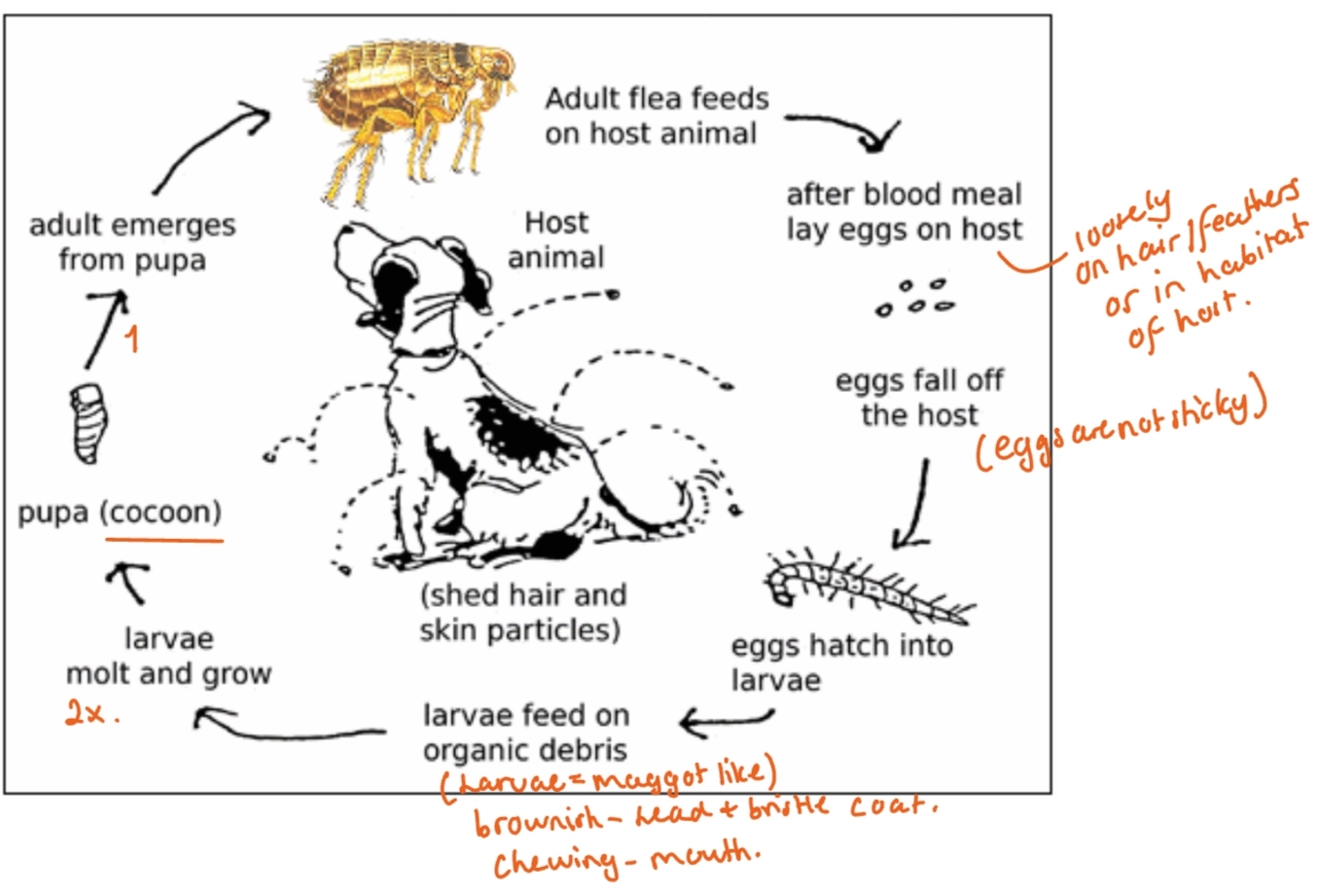
Life cycle: Complete Metamorphosis.
Fleas do have preferred hosts, but not strictly host-specific. Fleas can live for months without a blood meal, during that time, it is desperately trying to find a host. Once it finds a host, it will never purposely leave it.
Pathogenesis and clinical signs:
Fleas cause mild lesions (itching, erythema, small papules) at bite sites in non-allergic animals, showing usually minimal or no dermatological signs.
In flea-allergic animals, the same bite triggers a stronger response, leading to flea allergy dermatitis (FAD) after repeated infestations sensitize the host to allergens in flea saliva.
in dogs: FAD is typ. seen in animals 3-5 years of age, presenting as acutely pruritic, crusted papules with erythema and areas of acute moist dermatitis (hot spots). Non-allergic dogs may still develop anemia, tapeworm infection, hot spots and alopecia from scratching.
In cats: non-allergic individuals may show no signs despite fleas (same as in dogs), whereas flea-allergic cats (with no specific age), can show papulocrustous lesions on dorsom (Miliary dermatitis), self-induced symmetrical alopecia, eosinpholici granuloma complex and facial pruritus.
Diagnosis: History and clinical signs. Identification of fleas or flea “dirt”. Intradermal allergy or in vitro allergy testing with flea allergens. Response to strict flea control programme. Biopsy.
human lesions: pruritic papules on lower legs usually
Diff. dx: allergy esp. food, pediculosis, malassezia dermatitis, demodicosis, superficial pyoderma, psychogenic alopecia (cat)…
Treatment/Prevention:
Control program: Eliminate fleas from all animals, Break the flea LC in the environment (treat such as bedding carpets), control the FA, manage secondary infections
Topical spot-on or spray therapy - fipronil, nitenpyram (systemic), imidacloprid, metaflumizole, pyriprole, selamectin.
Anti-inflammatories: Glucocorticoids (given to help control pruritus), prednisolone, methylprednisolone acetate (cat only), ATBs (given for sec. pyoderma). Antihistamines (limited usage as FAD reaction involves many inflammatory mediators, not just histamine).
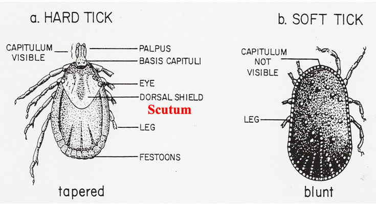
14.Hard ticks - morphology, life cycle, significance
Class: Arachnida, subclass: Acaria (ticks and mites)
Order: Ixodida (Metastigmata) - ticks
Family: Ixodidae (hard ticks)
Species: Amblyoma, Anocentor (boophilus), Dermacentor, Haemophysalis, Hyalomma, Ixodes, Rhipicephalus
Note: has 2 distinct body regions - cephalothorax + abdomen), no wings and antennae, nymph + adult stage - 4 pairs of legs => this differ Acari (tick, mites) from insecta!
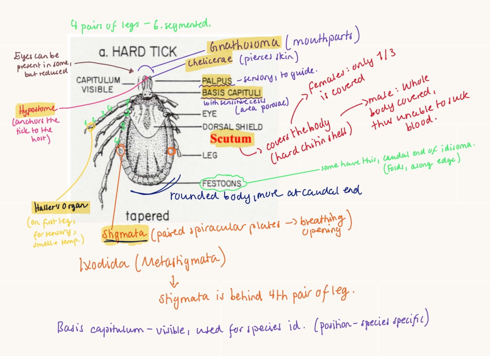
Morphology:
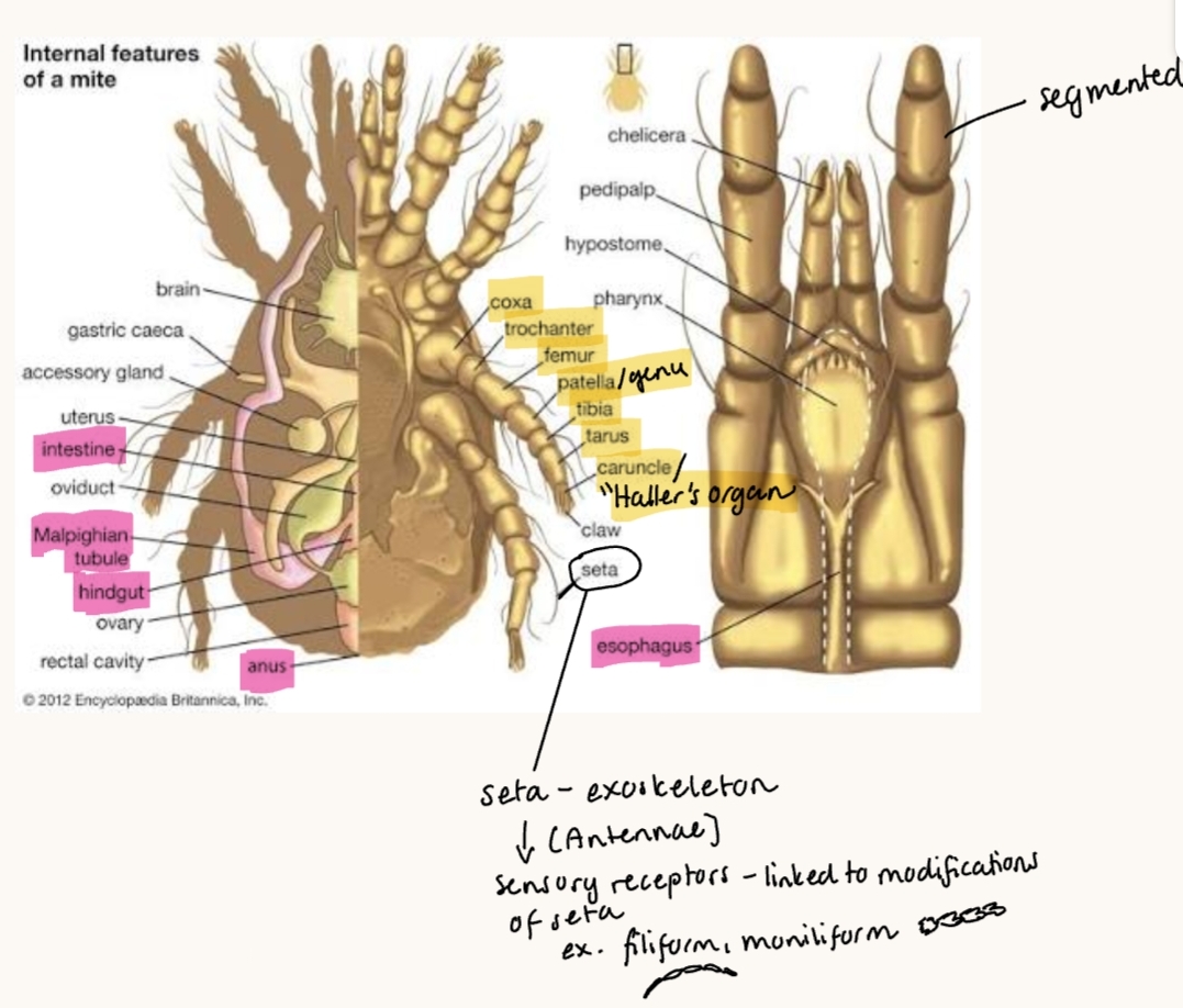
2 body parts: Capitulum (Gnathosoma) + Idiosoma
Scutum - hard chitin shell.
Female: covers 1/3 of idiosoma
Male: covers whole body, thus unable to suck blood
Moutparts (Gnathosoma): Hypostome, chelicerae, palps, basis capitulum with area porosae (for fluid, smell). Adapted for piercing and sucking.
Legs: 4 pairs, 6-segmented, larvae 3 pairs of legs.
Haller`s organ on first leg → for smell, temperature
Dorsoventrally flattened when unfed, becomes bean-bag like when fed. Round/oval, more rounded at end.
Stigmata - posterior of the 4th pair of legs - for respiration. (through cuticle)
Internal organs: in caudal part of idiosoma. Genital opening in ventral midline, anus posterior. Salivary glands can secrete toxins. Some have colored enamel-like areas on body (ornate ticks).
Eyes can be present in some, reduced.
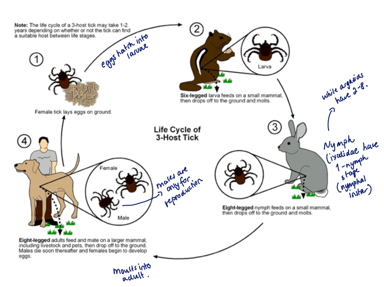
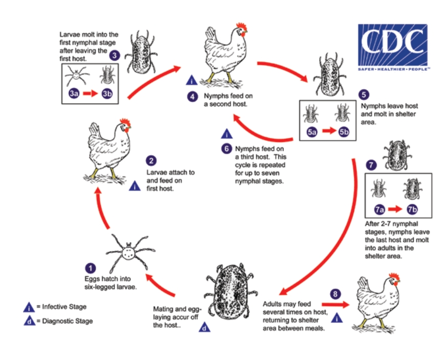
According to the number of hosts, the ticks are divided into:
Three-host (all stages in different hosts) - ixodes, haemaphysalis, dermacentor
Two-host - Rhipicephalus, hyalomma
One-host - Amblyomma, Boophylus (Anocentor)
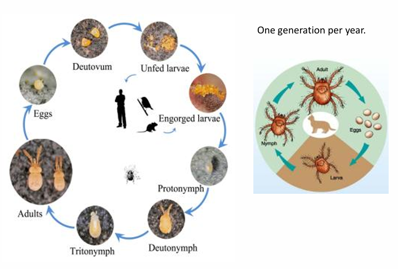
Life cycle: Incomplete metamorphosis.
egg → larvae → nymph → adult. Not permanent.
The medical importance of ticks: Cause skin diseases & allergies + are vectors of pathogens.
Skin issues - Tick bites cause itching, inflammation, swelling (dermatosis)
Blood loss (anemia) - too many ticks feeding on one host
Ear infestation (otoacariasis) - ticks can get into the ear canal → infection
Higher risk of other infections - tick bites makes it easier for others to infect
Tick paralysis, spread of diseases (can transmit bacteria, virus etc.)
Factors that make ticks effective vectors: feed on blood for a long time, feeds slowly (gives time for pathogen transfer), bites many different animals, lives for a long time, able to keep pathogens through life stages or pass to offspring (trans-ovarial/stadial), tough and hard to kill, high egg count, minimal natural enemies.
Pathogenesis & clinical signs:
Local lesions at bite site, erythema, itching. Hyperemia, necrosis, ulceration, bleeding - inflammation, thickening of skin, formation of nodules/granulomas, complication by sec. infections (myiasis).
hyalomma + rhipicephalus release neurotoxins → blocks neuromuscular function → paralysis.
give non-specific toxicosis (general illness by tick toxins), or specific toxicosis (condition caused by tick toxins, ex. tick paralysis)
Diagnosis: fed females are seen under microscope
Treatment: Acaricides (chemical that kills ticks), used in forms of sprays, spot-on, collars and dips. ex. permethrin, flumethrin.
Genus - with species:
Ixodes:
Ixodes ricinus (common sheep tick) - Biggest, most common in EU. - Transmit Borrelia, causing lyme disease.
I.Hexagonus (hedgehog tick) - transmits borrelia
Rhipicephalus (Africa, EU)
R.sanguineus (brown dog tick) - vector for babesiosis
Haemophysialis (Africa, Asia, EU) - most important in SK.
H.Punctata (red sheep tick)
Dermacentor (ornate ticks, in Asia, EU, africa, America) - vector for babesiosis, esp. B.canis
D.reticulatus (ornate sheep tick)
D.variabilis, D.andersoni, D.marginatus
Hyalomma (strippers - striped legs, in africa, Asia, EU)
H.anatolicum - for theileria annulata, equi + lestoquardi.
H.marginatum - for T.equi + babesia occultans
Amblyomma (large ornamented ticks) - not EU.
A.americanum, variegatum (3 host species).
15. Soft ticks (Argasid ticks) – the most important species.
Order: Ixodida
Fam: Argasidae (soft ticks)
Genus: Argas, Ornithodoros, otobius
Morphology:
Differs from hard ticks by: no scutum, we cannot see gnathosoma from dorsal view, more rounded bodies.
Stigmal plate is bw. 3rd and 4th pair of leg.
Geo: Arid souther regions (dry hot areas, like deserts, warmer parts of the world).
Life cycle: Multihost cycle for argasid.
Unlike ixodidae, they have 2-8 nymphal stages, each of which needs a blood meal. (lasting 20-30min. - not several days like hard ticks)
Adult males are for mating and can suck blood during their life (not only for mating as the hard ticks)
Nymphal stages feed several times (hard ticks only once)
SPECIES:
1) Argas
a) Argas Reflecus (pigeon tick): Bite at featherless skin areas (inner thighs, under wings, neck) → small red-blue swollen spots (bruised bumps).
Pigeons behave restless at night (ticks feed at night), weak, poor flying ability.
If there are repeated bites/many ticks → blood loss → severe anemia → can lead to death in nestling (young) birds
Dg: check nests, resting places, look for presence of tick
Control: Acaricides
b) Argas Persicus (Chicken tick): Anemia, weight loss, depression, toxemia (release neurotoxins) and paralysis. reduced egg production. Red spots on skin where tick have fed.
2) Otobius
O. lagophilus (rabbits)
O. megnini: Larvae + nymphs are found inside the ear, adults are difficult to find as they breed in hidden cracks of barns, fences + trees. Only larvae + nymphs infests hosts.
Otobius revolve around ear - irritation, pain and secondary bacterial infection due to the larval and nymph feeding in the ecternal canal. Signs: Head shaking/itching, thick + waxy exudate. But also GIT signs, nervous signs. In horse, O.megnini can cause Colic-like signs (tremors, spasms).
Dg: tick is seen on or inside ear
Treatment: remove tick, dipping, spraying the animal, treating the environment, Acaricide.
3) Ornithodoros
O. Lahorensis
O.moubata
O.moubata porcinus
O. savignyi
Cause local skin signs, blood loss, irritation, toxicosis - paralysis → death of animals (O.savignyi).
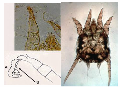
16. Sarcoptosis and notoedrosis of mammals.
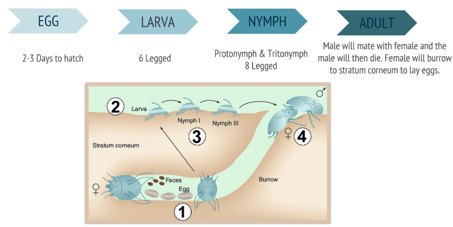
Burrowing mites → cause mange (scabies). Under class arachnida, subclass: Acari (mites, ticks). These are stationary, permanent parasites, adapted to living in the epidermis - forms tunnels under skin.
Order: Sarcoptiformes (Astigmata)
Family: Sarcoptidae
Genus: Sarcoptes
species: S. scabiei (all-host) - under sarcoptosis, Notoedres cati (cat), N.muris (rat), N.musculi (mouse) - rest is under notoedrosis.
Sarcoptes scabiei var. - (strains of same species) - host-adapted form/host specific, so can be in dogs (s.scabiei var. canis) etc. pigs, cattle, small ru. Zoonotic!
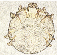
Morphology: small, round, short legs with varying number of ambulacrum (sucker at the end of leg for grip), 1st and 2nd leg pair from 3rd and 4th are widely separated, Triangular scales and transversial striations. Short gnathosoma - chewing function.

Life cycle:
Adult male + female mate (for life due to semen pouch) → female tunnel into skin (epidermis) → lays 3-5 eggs per day → six-legged larvae → nymph stages (proto- & trito-nymph) → adult. Prefers hairless skin areas.
Pathogenesis and clinical signs: In general it starts with Severe pruritus, erythema, formation of papules, later - hyperkeratosis, crusting/scabs of skin. Vertical skin folds (cattle). Lesions - tips of ears, elbows less haired areas, then generalizes. Highly contagious - by body contact.
Can be chronic - weight loss, abnormal behavior, can lead to death. Reduced growth rate & feed efficiency.
papular rash in humans (S.scabiei).
Diagnosis: History + clinical signs, Skin scrapings (until capillary bleeding) + microscopy, Biopsy (if unclear), Serology (ELISA - for allergies, raised allergen-specific LgE & LgG), Rule out other diseases.
Differential Dx: Flea allergy dermatitis, Malassezia dermatitis, atopic dermatitis
Treatment: Antiparasitic therapy - Selamectin, Moxidectin (also macrocyclic lactones like ivermectin in practice). Amitrax (not eq). Dipping tanks (sheep).
Treat all contacts + environment (insecticides - permethrin)
Symptomatic - glucocorticoids (prednisolone), never before dg. (can mask). Treat sec. infections.
good prognosis (unless chronic)
No vaccines available.
NOTOEDROSIS
Notoedric mange (feline scabies, of rabbits and rodents)
Morphology: Similar to S. scabei, but it is smaller.
Life cycle: Similar to S. scabei.
Pathogenesis and clinical signs: Start with itching, first lesion on the edge of ears, rapid spread to forehead, neck. Cause thick skin and wrinkles. Cause self-mutulation - excoriations and alopecia. (looks more dramatic, crusty esp. face)
Diagnosis: Same as S. scabei. Diff. dx: allergies, otodectes cynotis, autoimmune skin diseases..
Treatment: Amitraz, selamectin, fipronil, lime sulphur. Glucocorticoids may be useful in severe pruritic animals. Pyoderma can occur as a rare secondary complication, treated with antibiotics
17. Cnemidocoptosis and cytoditosis
Order: Sarcoptiformes (Astigmata)
Family: Cnemidocoptidae (acarinosis/disease of mites of legs, beaks and skin) & Cytoditidae (acarinosis of the resp. organs + subcutaneous tissue) - These families of burrowing and penetrating mites cause bird diseases.
1) Cnemidocoptosis - Cnemidocoptidae (chewing mites) - species:
Cnemidocoptes mutans – keratinization of legs, scaly leg
C.gallinae – loss of feathers
C.pilae – scaly face, tassel foot
C.prolificus
Morphology: Similar in general to S. scabiei, but rounder, no spines or scales, short legs. chewing mouth. Claw-like structures and tactile hairs instead of suckers (on legs).
Life cycle: Females (viviparous) - 1 larval and 2 nymphal stages, before adult stage. Female lays larvae in tunnels in the epidermis, 6 legged-larvae crawl on skin surface, burrow into superficial layers → molt to nymphs (proto- & trito-) → adult → return to surface to breed.
Pathogenesis & clinical signs:
mites burrow in the stratum corneum → legs and feet (scaly leg), beak, eyelids
Skin lesions - irritation, exudate that dries, forming hard, crusty (calcerous-like) mass. Can lead to beak deformation (C.pilae), difficult to eat, drink, sometimes also affecting breathing. Mites can migrate and cover the eyes. Severe pruritus. Long lasting → weight loss.
Diagnosis: microscope by skin scraping
Treatment: removing the scales with glycerine. Acaricides (Ivermectin, amitrax, pyrethroids), supportive vitamin A. Treatment repeated for 8-10 days.
2) Cytoditosis - Cytoditidae with species:
Cytodites nudus – Cause acarinosis of the respiratory organs and subcutaneous tissue of birds.
Morphology: Oval. Gnathosoma small and rounded. Legs are longer.
Clinical signs: Often without clinical signs, but can show breathing difficulties, granulomatous bronchitis. Transmissable through coughing or contaminated water.
Affects trachea, bronchi, lungs, air sacs, but also liver, kidney serosa
Diagnosis: only post mortem
Treatment: not known
18. Demodicosis mange of animals
Demodectic mange (Demodicosis).
Order: Trombidiformes (Prostigmata)
Family: Demodecidae – burrowing mites
Demodex canis – Inhabits hair follicle, sebaceous duct and gland
D. injai – Inhabits hair follicle, sebaceous duct and gland
D. cornei – inhabit stratum corneum
D. cati – rare in cat, D. gatoi
D. phylloides – pig, D. foliculorum – man, D. bovis, D. equi, D. ovis, D. caprae
Morphology: Elongated body, wormlike. Capitulum is horse-shoe shape with short and wide mouthpart. 4 pairs of short legs, situated behind gnathosoma. Thick in the fronts of body. Transverse ridges, setae. They feed on cells, sebum and epidermal debris. Located in hair follicles and sebaceous glands with “upside down” position. Very species specific.
Life cycle: Eggs → larvae (6-legged, 1-2 larval stages) → moult into 8-legged nymphal stages → adult.
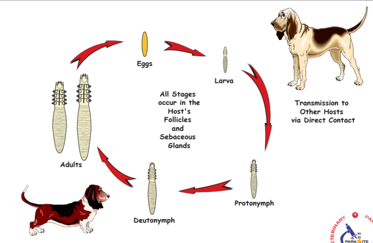
Pathogenesis and clinical signs: Not contagious (or Zoonotic). It is part of normal skin flora in 80% of most healthy canines. Demodex spp. are found in other animals (people too). Pathogenic when immune system is reduced. Mites will start to proliferate in hair follicles → inflammation → secondary bacterial infections, destroying the follicles. Generally, erythema, thick skin..
There are 3 forms in dogs:
Localized form: starts around eyes, nose and ears. Grey-red alopecic spots - Less than 5 patches. Demodectic otitis, periocular scaling. NON-PRURITIC! Can recover spontaneously within 1-2months.
Generalized form: More than 5 patches, lesions extend to head, neck, leg and trunk - juvenile onset. Adult onset - usually linked with a severe internal disease (reticuloendothelial neoplasia, endocrine disorders etc.)
Pododemidocosis: Single foot (local) or 2 or more feet (general)
Worldwide distribution.
Cattle: Outbreaks are seen mainly in tropical areas. In cattle, we see small nodules localised on neck and dewlap, with thick yellowish substance with a large amount of mites. No pruritus. Economical issue - downgrading leather.
Sheep/goat: D.obis/aries. Rare. Small nodules on lips/lower jaw.
Horse/Pigs: Rare, clinically alike cattle.
Diagnosis: history&CS, skin scrapings - reach into hair follicles and sebaceous gl. Squeeze skin to extrude the mites from follicles before scraping. Hair plucking can be done in case of podo, biopsy.
Treatment: Antiparasitic (Amitraz, ivermectin, milbemicin)
local: most cases reosolve after 6-8 weeks
Topical: lime sulphur, selenium sulphide, follicular flushing agents (benzoyl peroxide).
Livestock - treatment not always needed, can use organophosphates.
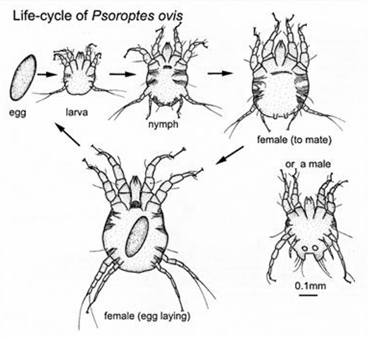
19. Psoroptosis of animals.
Order: Sarcoptiformes (astigmata)
Family: Psoroptidae (Non-burrowing mites!)
Psoroptes ovis – Sheep scab, cattle (lumbus, arms belly - located)
P. cuniculi – rabbit
P. equi
Morphology: Oval body, long segmented legs (3 segments pedikula), funnel shaped suckers at the end (ambulacrum), mouth part is pointed → pierces skin to suck lymph. Abdominal tubercles of male are rounded.
Life cycle: All stages occur around edges of oozing lesions. On host about 10 days. Survive off the host for 2-3w.
Female → eggs on skin → larvae → protonymph → tritonymph → adult.
Pathogenesis and clinical signs:
general: areas of thick hair, but untreated will spread over entire body. itch → alopecia & Skin trauma. Severe → anorexia, emaciation, anemia.
Sheep scab: most severe ectoparasitic infection of sheep, contagious - direct contact, esp. during cutting season by equipment. Starts at neck, shoulder and flanks → papules, serous exudate, dry yellowish crusts, inflammation, releasing wool - alopecia, thickening of skin. Cause itching, rubbing, restlessness, impaired growth/weight loss.
Eq: skin of ears → droopy, lop-eared posture, margins show scaling/thickening, cause tail-rubbing, severe self-inflicted trauma. Rabbits: mainly inner ear (pinna), intense itching, thick, brown, waxy and scaly crusts, shaking head. Cattle: back, shoulder, tail, intense itching, thick scabs, alopecia.
Typical of P.ovis - abrasive yellow scabs, confessed skin (flaking woolfell).
Diagnosis: CS of sheep scab, definitive dg needs the visualization of lesions + confirmation of mites being present. Superficial skin scrape (without capillary bleeding).
Treatment: Dipping (use of cypermethrin dips) or by use of macrocyclic lactone injectables – ivermectin, moxidectin and doramectin. Control of environment is also important. - Sheep.
for others: Pyrethroids batchs, best is giving ivermectin/dora/moxi injection/pour-on. Eq: equalan, Rabbit: ivermectin. Control environment.
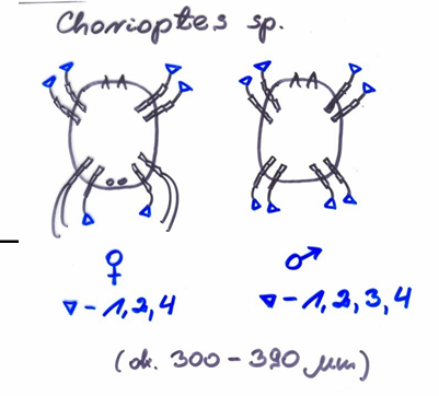
20. Chorioptosis of animals.
Order: Sarcoptiformes (astigmata), subclass: Acari (mites)
Family: Psoroptidae (scab mites)
Genus: Chorioptes
Non-burrowing mites! = Fotskabb/foot mange/scrotal mange
Chorioptes bovis
Ch. Ovis
Ch. Equi
Ch. Caprae
Morphology: Resembles Psoroptes, but body is smaller, but larger than Sarcoptes. Long legs. Do not pierce skin, but feed on skin debris (mouth parts are rounder). Cup-shaped suckers (ambulacrum), Square abdominal tuberkules in male. Unjointed pedicels/non-segmented pedicula.
Life cycle: Same as Psoroptes. Lasts 3 weeks. Release exudate, feed on debris. Eggs are laid on skin at margins of lesions → hatch into larvae → feed for several days → molt to nymphs, feed for a few days → final molt to adult.
Pathogenesis and clinical signs:
Infests the wool-less areas, esp. lower parts of hind legs and scrotum, Can decrease fertility by causing inflammation of scrotal skin.
Ch.bovis → does not pierce skin, but feeds on debris → yellow-brown lesions with haemorrhagic fissures resulting from allergic reactions to the mites or mite by-products.
Intense itching → foot stamping and biting.
Eq: sticky scales, strong pruritus, restlessness, stinging, limb cracking.
Not Zoonotic! Less severe compared to psoroptic or sarcoptic mange. Typ. cutaneous lesions with crusts, scales and thickening skin on the legs.
Diagnosis: Skin scrapings under microscope.
Treatment:
in eq: control by oral ivermectin - effective at reducing mite populations, but cannot eliminate them. Topical treatment include 3-whole body baths, 5 days apart, using selenium sulphide shampoo, and two treatments, 2-3w apart with fipronil spray.
clip long hair, removal of scabs, scrub affected areas with insecticidal shampoo/powder. Oral ivermectin paste/moxidectin. But it may live in environment for 69d. Topical washes = main approach.
21. Otodectosis and cheletiellosis of carnivores.
CHELETIELLOSIS:
Order: Thrombidiformis (prostigmata)
Family: Cheylettiellidae
Genus: Cheyletiella (walking dandruff, non-burrowing mites)
All species are zoonotic!
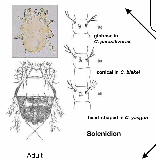
C. Yasguri – dogs
C. Blakei – cats
C. Parasitovorax – rabbits
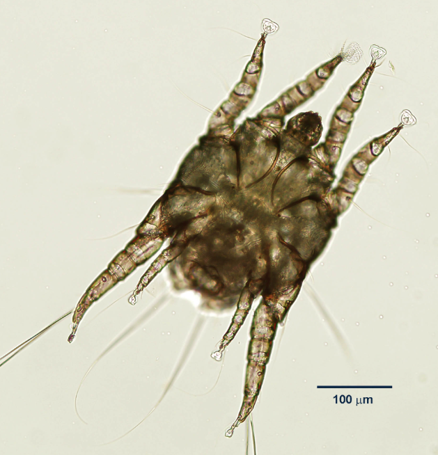
Morphology: Oval with narrow neck. Palps enlarged, end in hook-like claws (mouthparts). Mouth for sucking/piercing. 8 Legs that end in combs instead of claws. Solenidion on first pair of legs (sensory organs for host detection). Moves fast and are very active. Worldwide.
Life cycle: Strong claws allow them to cling to fur and migrate quickly to epidermis. Egg - Larva - 2 nymph stages - Adult.
Pathogenesis and clinical signs: Cause scaling, pruritic dermatitis, most severe on the dorsum. Occurs in overpopulation and poor sanitation.
The mites live in keratin on the skin`s surface and feed on tissue fluids/keratin debris). The entire LC is thought to occur on the host.
Diagnosis: Use transparent adhesive tape (for fast-moving parasites) or glue on a slide. Visual examination of hair coat, presence of walking dandruff.
Treatment:
Lime-sulphur and pyrethrin rinse for cats, puppies and rabbits
Pyrethroids for dogs.
Topical treatment with amitraz and ivermectin. Environmental treatment, cleaning and insecticide sprays, important for eliminating infestation.
OTODECTOSIS:
Order: Sarcoptiformes (astigmata), Family: Psoroptidae Genus: Otodectes (together with chorioptes, psoroptes)
Non-burrowing mites - ear mite. Potential Zoonosis.
Otodectes cynotis
Morphology: Legs - short pedicles with terminal sucker discs, short pretarsi, piercing mouthparts, feeds on lymph and epidermal debris. Bell-shaped pulvilli.
Location: External ear canal. But can also be seen in other parts of body. Common infestation site is tail and head.
Life cycle: Egg → larvae → 2 nymph stages → adult. Last 3w. Eggs attached to skin of ear.
Pathogenesis and clinical signs: A common disease in dogs and cats, especially kittens. Highly contagious between animals - mainly by contact, not species specific.
Pruritus & Otitis externa (erythematous) → shaking of head, scratching of ears. Exudate in ear - brown, grainy, like coffee grounds, sometimes blood is seen. Lining of ear canal is often reddish. Blackish crusts and cerumen build-up - predisposes to sec. otitis.
Diagnosis: Direct otoscopic examination, demonstration on smear, no stain needed. Skin scraping or scotch tape impressions
Treatment: Regular examination of ear canal, cleaning - oil, borax, salicylic, application of acaricidal preparation and special ear drops. Amitraz, selamectin, ivermectin, moxidectin.
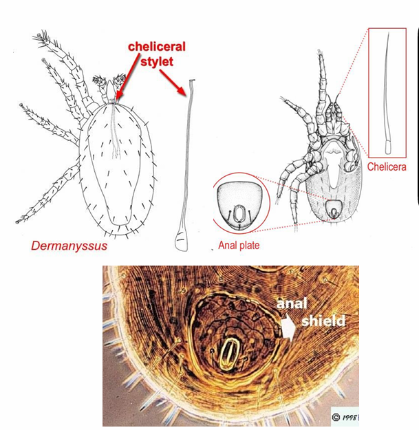
22. Dermanyssidosis
Order: Gamasida (mesostigmata)
Family: Dermanyssidae (Dermanyssus, Liponyssoides)
Dermanyssus gallinae – birds. (poultry red mite)
but also D.avium, D.hirundinis, passerinus, chelidonis, sanguineus
Morphology: Transient form between ticks and mites. They are hairy. Long limbs for active moving. Stigma between 3rd and 4th leg. Larval stages do not have stigma, they breath through cuticle. They are temporally hematophagous ticks, and night feeders.
Life cycle: same as others. Egg – larva – 2 nymphal stages– adult. Males and females suck blood. Lasts 7-10 days. Population incr. in winter. Mites transmit bw. farms, and it may survive for long periods without host.
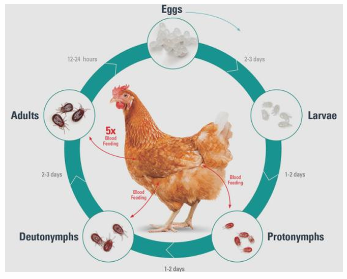
Pathogenesis and clinical signs: Stress/pecking agitation, mortality if high infestation, spread of poultry pathogens of bacterial and viral origin (cholera, dengue fever)
Anemia, weight loss, allergic reaction in humans, agitation, irritation, decr. egg production, decr. egg quality through shell-thinning and blood spotting.
Diagnosis: Finding mites on surface
Treatment: Synergized pyrethroids (carbamates and organophosphates are forbidden in EU). Cleaning with boiling water and acaricides. Individual treatment with acaricide.
23. Trombiculosis.
Order: Trombidiformes (prostigmata)
Genus: Trombicula (neotrombibula)
Family: Trombiculidae (chiggers - parasites of terrestrial vertebrates)
Neotrombicula autumnalis
Morphology: Only larval stages are parasitic! Nympha and adults are non-parasitic. Very small <0,2 mm, yellow, long haired legs, legs have 3 large claws, head has hard hypostome.
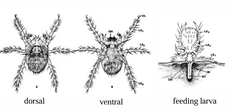
Life cycle: egg – larva (parasitic) – nympha - adult. Feeds by extra-intestinal digestion.
Pathogenesis and clinical signs: Cause dermatitis, vectors of various pathogens (rickettsia spp.)
N. autumnalis has extra-intestinal digestion, larval hypostome penetrates skin and release cytolytic enzymes that digest the tissues. The larvae sucks up digestied tissues (lasts 3-4 days).
Infect cats, dogs, humans. Predilection sites are areas with thin skin (paws, inguinal region, around eyes, around ear canal).
Mild to severe skin reactions, irritation, pruritus, crusts/papules/erosions visible. Sec. alopecia.
Humans: papules with crust in middle, form brown color pigments around (sensitivity reaction to the release of cytolytic enzymes)
Larvae are sometimes grouped together in clusters of small, bright orange/red dots on the skin.
Diagnosis: Anamnesis, clinical findings, microscopic evidence - red larvae.
Treatment and control:
fipronil, selamectin - cats
permethrin - pyriproxyfen combination - dogs
Prophylaxis - should avoid known trombicula foci during season. (seasonal problem, larva suck blood in middle of august to sept. - start repellent/prophylaxis treatment accordingly)
“Synthetic pyrethroids, repellents”