Tissue Preparation for Light Microscopy
1/9
There's no tags or description
Looks like no tags are added yet.
Name | Mastery | Learn | Test | Matching | Spaced |
|---|
No study sessions yet.
10 Terms
What are the 5 stages of tissue preparation?
Fixation
Processing
Embedding
Sectioning
Staining
Fixation
What
Aim
Factors affecting fixation
Types of fixation
What: A treatment with chemical agents to preserve it by slowing down natural breakdown, thus ensuring that the structure maintains close to original
Aim:
Prevent autolysis and bacterial attack
To fix tissue so volume and shape won’t change
To leave tissue as close as their living state as possible
Factors affecting fixation:
pH
Temperature
Penetration of fixative:size of tissue sample
Volume of fixative:tissue
Fixation time (24-48 hours)
Types:
Bouin’s solution
Acetic acid
Formaldehyde 10%
Ethanol
Methanol
Picric acid (bone)
Processing
What
Needs to be firm so
But also needs to be soft enough because
Stages
What: When tissue is placed in a supportive material (like paraffin wax) to hold it steady
Needs to be firm: So tissue can be sliced very thinly
But also needs to be soft: So it doesn’t damages the knife or tissue
Stages:
Dehydration
Clearing
Impregnating
Embedding
Processing: Dehydration
What
Uses
Aim
Delicate specimens are dehydrated in graded ethanol series:
What: Part of processing that removes fixatives and water from tissue and replaces them with dehydrating fluid
Uses: Hydrophilic compounds such as ethanol, methanol, acetone
Aim: Minimize tissue distortion from diffusion currents
Graded ethanol series: 10%-20%-50%-95%-100% ethanol
Processing: Clearing
What
Choices of clearing agent depends on
Examples
What: When the dehydrating fluid is replaced with a fluid that is mixed well (miscible) with both dehydrating fluid and embedding medium
Choices of clearing agent depends on:
Type of tissue
Processor system used
Safety factors
Cost and convenience
Speedy removal of dehydrating agent
Ease of removal by molten paraffin wax
Minimal tissue damage
Examples:
Xylene
Toluene
Chloroform
Benzene
Embedding
What
Most common wax
Precautions while embedding
What: Process by which tissues are surrounded by a medium (agar, gelatin or wax) which when solidified provides sufficient support during sectioning
Most common wax: Paraffin wax
Precautions while embedding:
Wax is clear of clearing agent
No dust particles present
Immediately after tissue embedding, wax must be rapidly cooled
To reduce wax crystal size
Embedding: Paraffin Wax
What
Characteristics compared to dried protein
Melting point
What it does to sample
What: Polycrystalline mixture of solid hydrocarbons
Characteristics: 2/3 density and more elastic than dried protein
Melting point: Ranges from 39C -68C
What it does to sample:
Improves ribboning
Increases hardness
Decrease melting point
Improve adhesion between specimen and wax
Sectioning
What
Equipment
Best ribbon section size
After sectioning:
What: To section solidified paraffin-embedded tissue
Equipment: Microtome and blade
Best ribbon section size: 4 to 5 µm
After sectioning:
Spread tissue in flotation water bath at 42C
Select best section before mounting
Put into dryer hotplate at 37C overnight
Staining
Routine staining consist of
Special staining consist of
Immunohistochemistry staining consist of
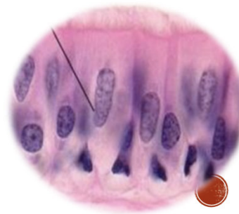
Routine staining: Hematoxylin and Eosin (H&E)
Special staining: PAS, Giemsa or fluorescent
Immunohistochemistry staining: Uses antibodies such as immunoperoxidase and immunofluorescent
Staining: H&E Routine Staining
Principal
Hematoxylin
Type of dye
Stains
Color
AKA
Eosin
Type of dye
Stains
Color
AKA
Principal: Chemical attraction between tissues and dye
Hematoxylin:
Type of dye: Basic
Stains: Acidic structures like nucleus, chromatin, ribosomes
Color: Purplish blue
AKA: Basophilic
Eosin:
Type of dye: Acidic
Stains: Basic structures like cytoplasm
Color: Red or pink
AKA: Eosinophilic
