Gross Anatomy of the Brain
1/116
Earn XP
Description and Tags
Anatomy
Name | Mastery | Learn | Test | Matching | Spaced | Call with Kai |
|---|
No analytics yet
Send a link to your students to track their progress
117 Terms
Central nervous system
brain and spinal cord
Peripheral nervous system
ganglia and nerves
Subdivisions of CNS
Cerebral Hemispheres x2
Basal Ganglia
Diencephalon (thalamus and hypothalamus)
Brainstem
Cerebellum
Spinal cord
Fissures or sulci
depressions
in the cortex
Lobes of cerebral hemisphere
Frontal
Parietal
Temporal
Occipital
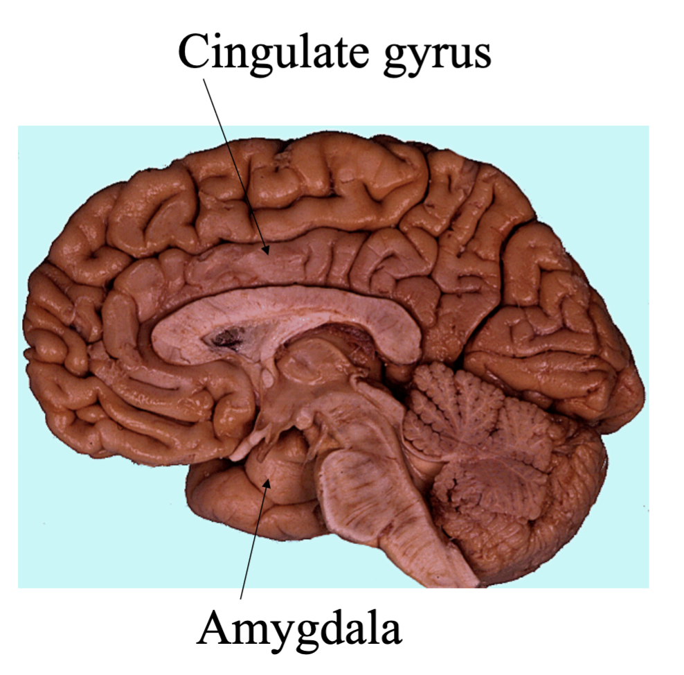
Limbic
Insula
Limbic lobe
On the medial surface the parahippocampal gyrus is separated from the temporal lobe by the collateral sulcus.
The cingulate gyrus from the frontal and parietal lobes by the cingulate sulcus
Insula Lobe
Lobe covered by frontal, parietal and temporal lobe (covering parts termed the operculum)
Strongly involved in sensory processing
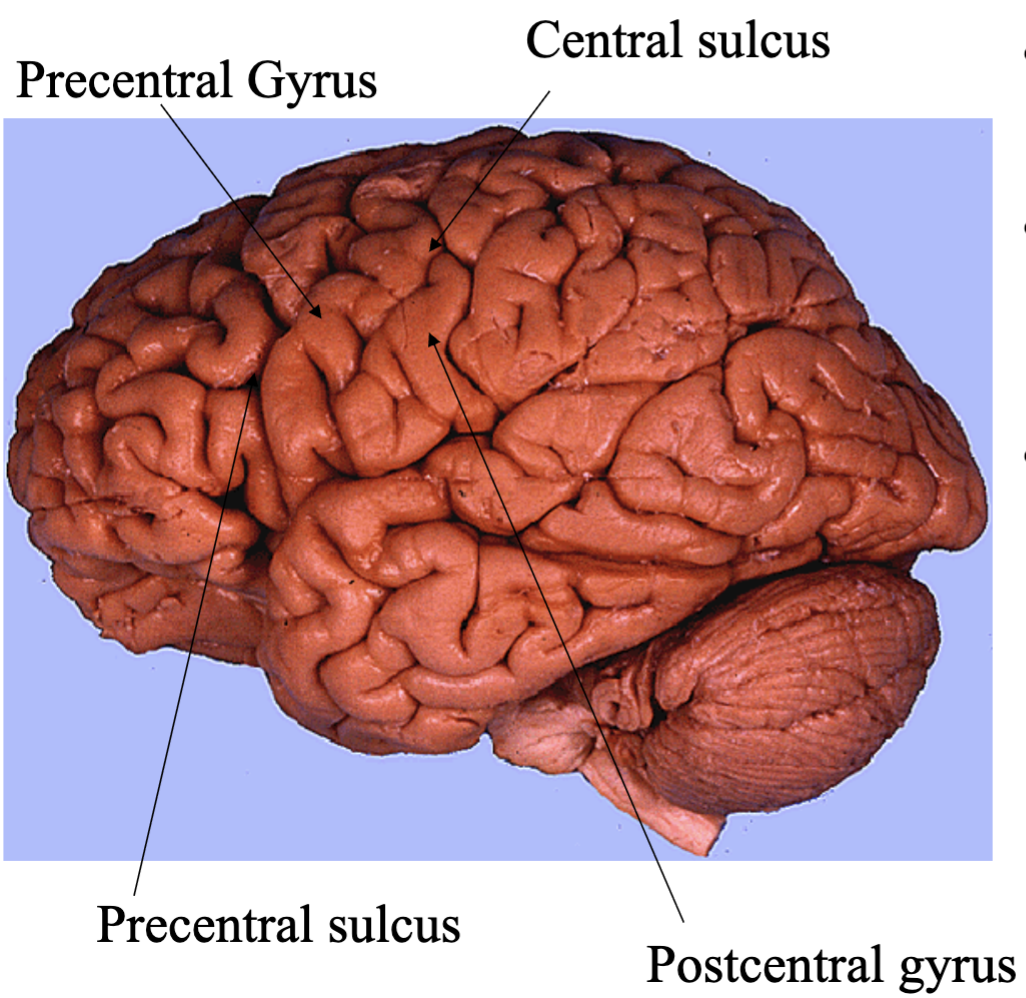
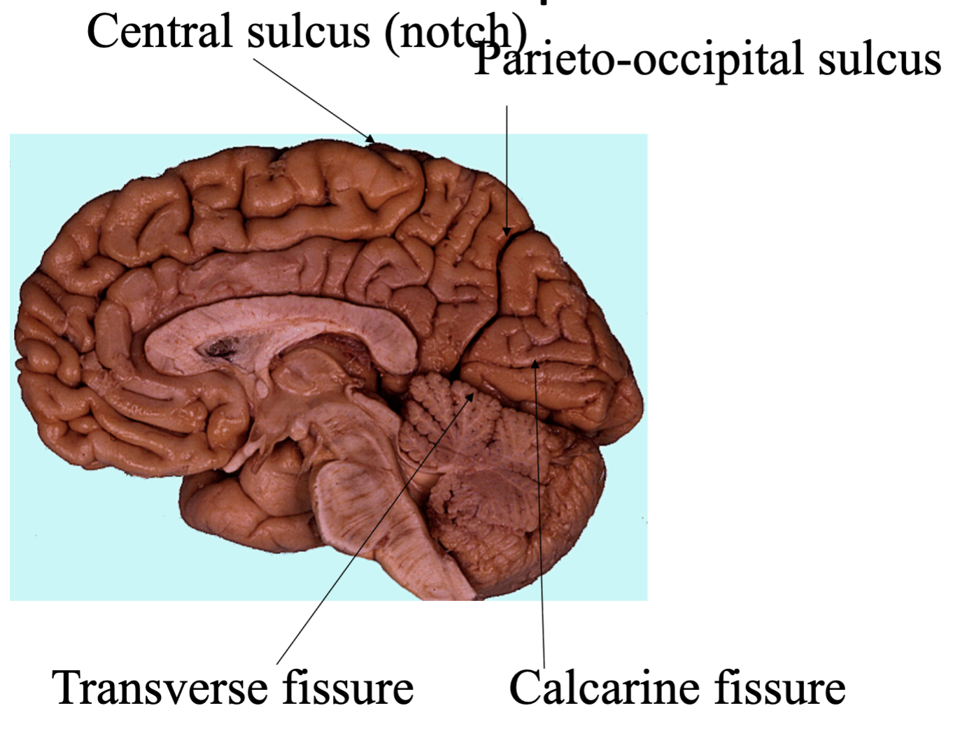
Central sulcus
Central sulcus separates frontal from parietal lobes
Precentral and Post central gyri are adjacent to the central sulcus
Precentral sulcus is anterior to the Precentral gyrus and likewise the sulcus posterior to the postcentral gyrus will be the post central sulcus
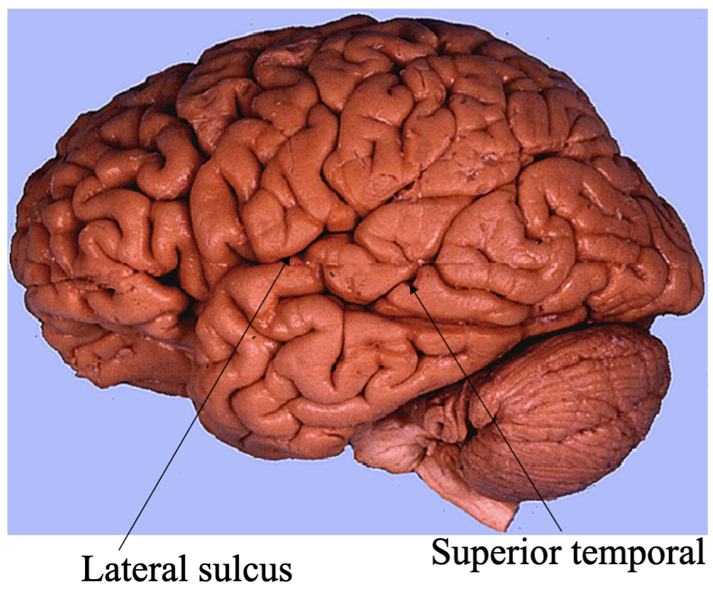
Lateral Sulcus (Sylvian Fissure)
The Lateral Sulcus (or fissure) separates superior temporal gyrus from the frontal and parietal lobes
Superior temporal sulcus is found centrally on the temporal lobe
Parieto-occipital sulcus
Parieto-occipital sulcus Separates parietal and occipital lobe
Calcarine fissure important for visual fields
The Cuneus is above and the Lingual gyrus below
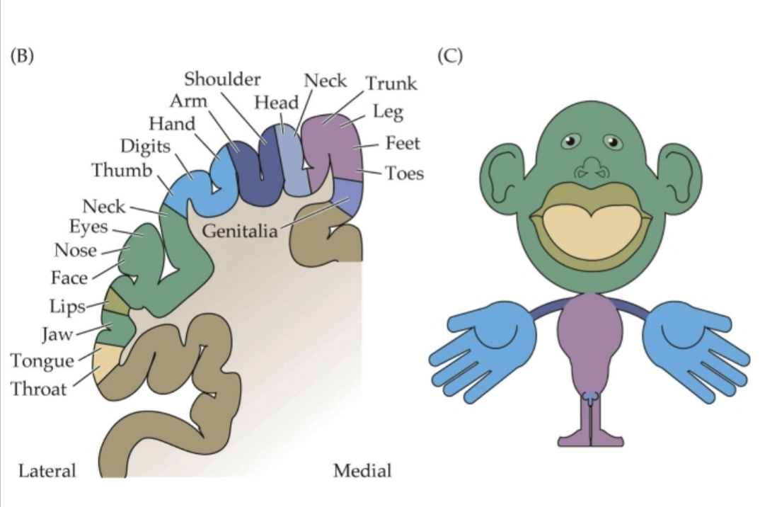
Primary sensory (somatosensory)
Parietal lobe
Post central gyrus
Organized somatotopically (homunculus)
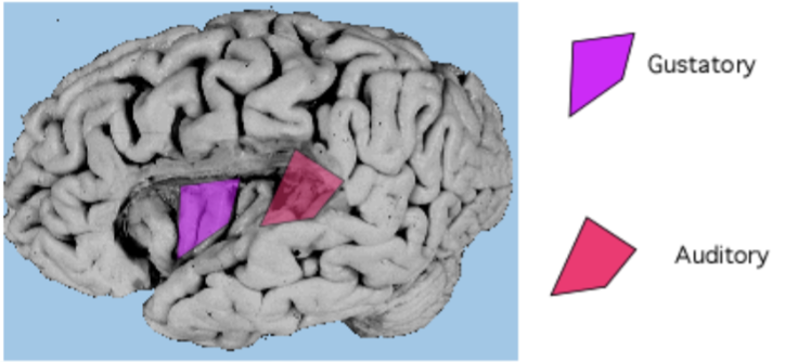
Where is the primary sensory (gustatory) located?
Insula (and operculum)

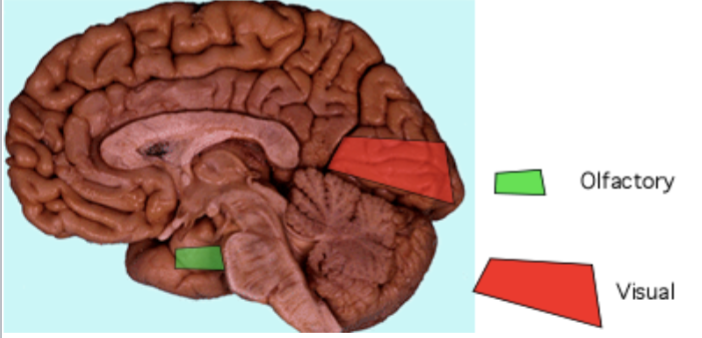
Where is the primary sensory (olfactory) located?
Olfactory Bulb
Where is the primary sensory (auditory) located?
Superior temporal gyrus and transverse temporal (Heschl’s gyrus)
Where is the primary sensory (visual) located?
cortex around the calcarine fissure (lingual gyrus and cuneus
Where is the primary motor lcoated?
Precentral gyrus
Layer V of cortex are the upper motor neurons

Where is the secondary motor located?
Posterior portions of middle and superior frontal gyri
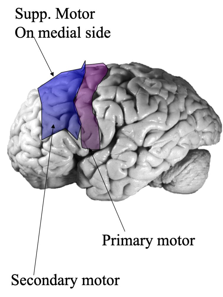
Where is the supplemental motor located?
Medial portion of superior frontal gyrus

What is the Association cortex?
Have no primary motor component
Have only higher order sensory processing
Integration of signals
Multimodal neurons (respond to more than one sensory cues)
Motor planning
Learning and memory
Social awareness
Abstract thought
Broca’s (motor) speech area
Opercular and triangular parts of the inferior frontal gyrus
Important for the speech production
Wernicke’s (motor) speech area
Posterior part of superior temporal gyrus (Brodmanns area 22 temporal lobe)
Important for speech and language comprehension
Supramarginal gyrus (area 40 and angular gyrus 39) are also important for language
Importance of prefrontal area
Social awareness, working memory and decision making
Has been implicated in restraining “animal instincts”
Involved in motivation and has high level of dopamine input from Brainstem ventral tegmental area (VTA)
Orbitalfrontal cortex is one area involved in higher sensory processing integrating all senses to from a sensory perception
What can lesions in prefrontal area result in?
Absence of “willpower” (abulia)
Confabulation
Depression
Mania
Utilization Behavior
Importance of parietal area
Visual association area (parietal eye fields)
Spatial association (non-dominate side)
Lesions in this association area are also associated with neglect
Higher visual processing
The “where” of an object
What is hemineglect?
Can result from damage the association cortex
Patient will ignore or “neglect” to notice one side of sensory space
Usually contralateral
Importance of temporal area
Auditory association
Area is bilaterally connected to hippocampus
Higher visual and auditory processing
The what of an object including recognizing features and object naming (or object recognition through sounds)
Face recognition has been linked to temporal lobe association cortex.
Prosopagnosia: inability to recognize and identify faces
Importance of occipital area
Involved in visual processing
Occipital association cortex is continuous with the parietal and temporal association areas
Dorsal processing stream integrates with motion sensitive areas (the “where” of an object
Ventral stream integrates object recognition areas (the ”what”) of an object
What is subcortical white matter?
Commissural fibers
Projection fibers
Internal capsule (mostly up down)
Fasciculi (mostly longitudinal)
What are commissures
connect the left and right hemispheres to each other
Corpus collosum
What are Anterior Commissures?
Interconnects olfactory bulbs and amygdala
What are Posterior Commissures?
Important for pupillary light reflex
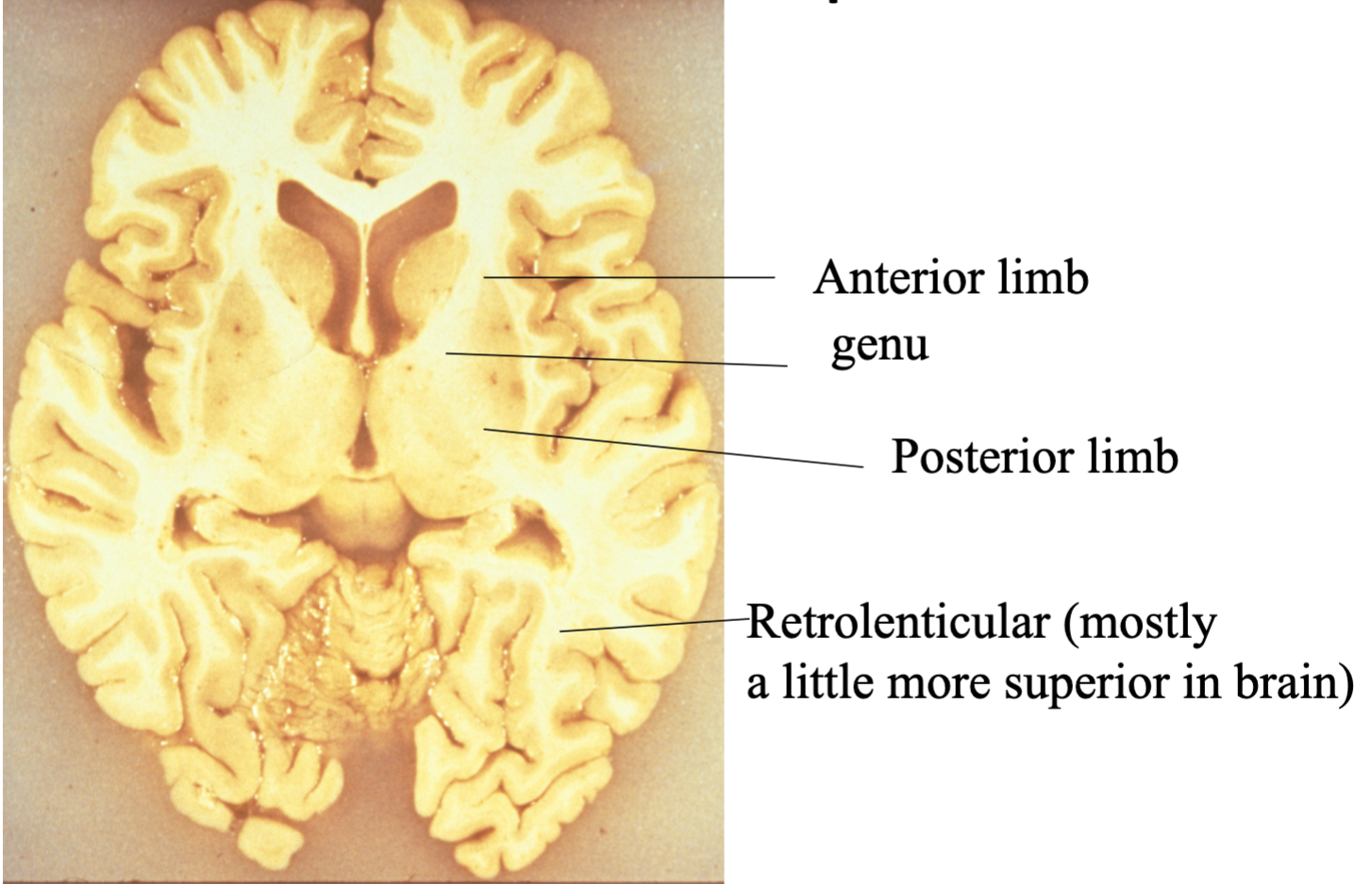
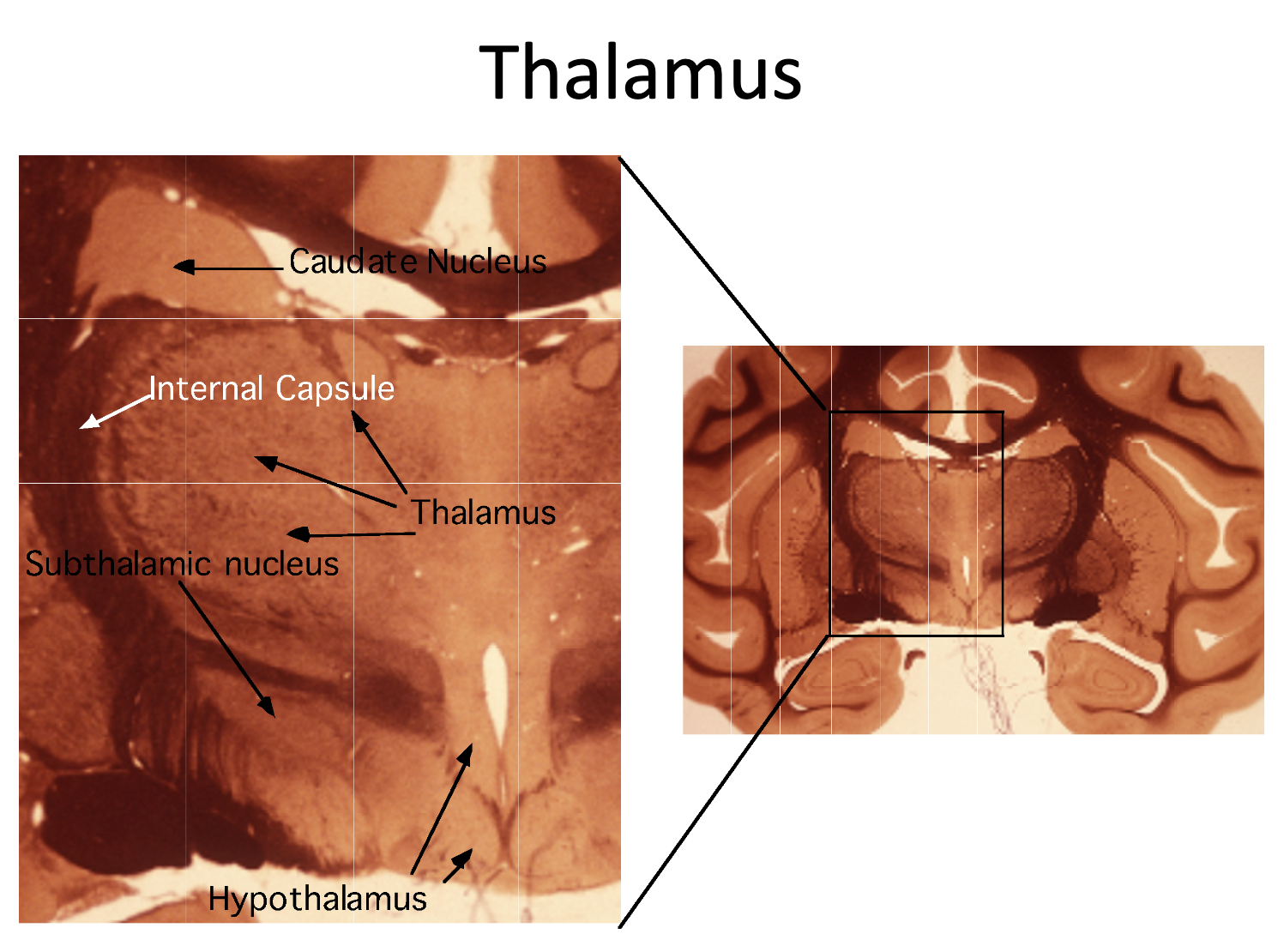
What is an internal capsule?
Are projection fibers that physically encapsulate the thalamus.
These are projections to and from cortical and subcortical structures
What is Diencephalon
Connects the forebrain with the rest of the CNS.
It is located caudal to the forebrain and rostral to the Midbrain
Consists of thalamus, hypothalamus, epithalamus and pretectum.
What is the thalamus
Bulk of Diencephalon
Relay between sensory to cortex and motor and cortex
Primary Function: conscious sensation and conscious movement
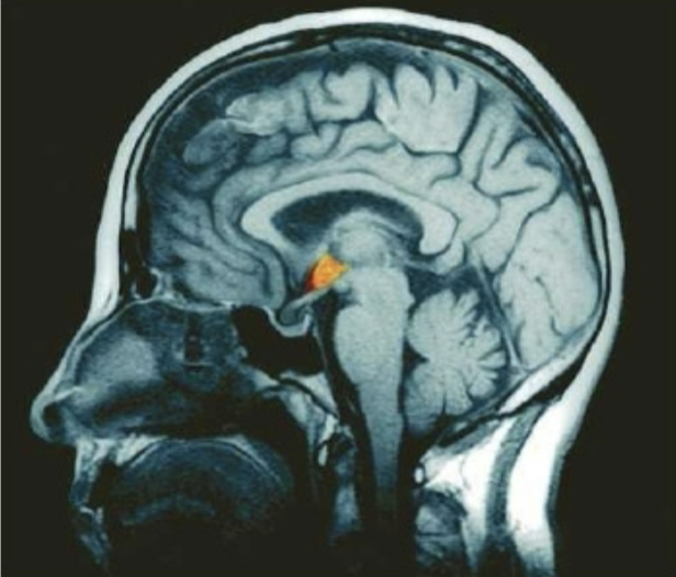
What is the hypothalamus?
Primary function: autonomic and instinctual functions
Hormonal
Autonomic
Behavioral
Receives input from many areas including circulation
What is the primary basal ganglia?
Primary function: is to initiate wanted movement and inhibit unwanted movement
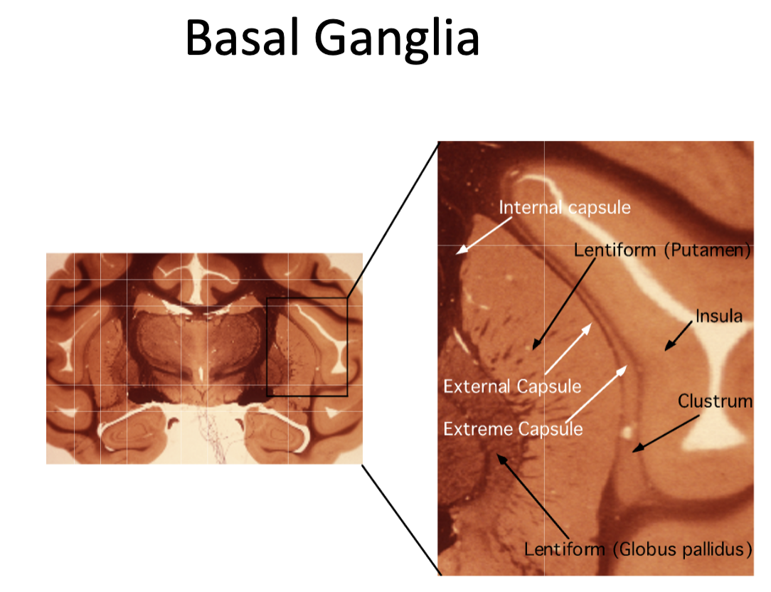
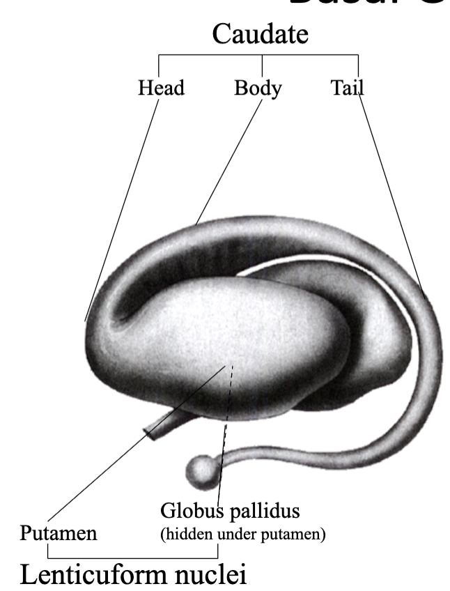
What are the Three Main Nuclei of Basal Ganglia
Caudate
Putamen
Globus pallidus
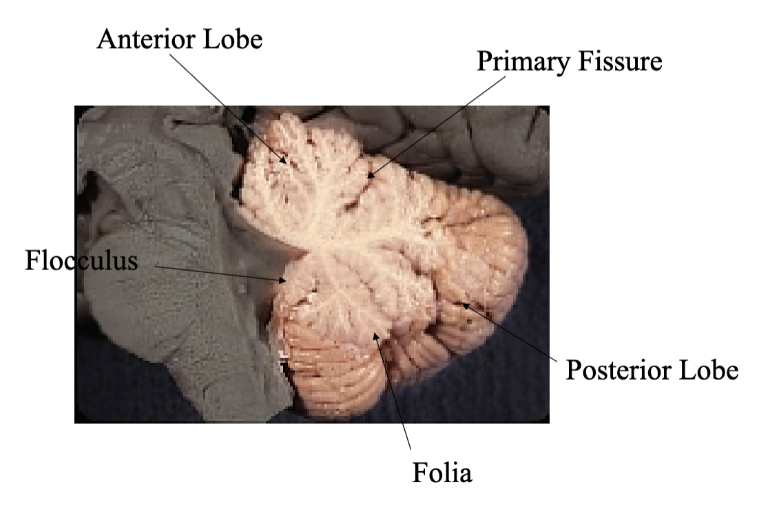
Anatomical parts of cerebellum
Floccular nodular lobe
Anterior Lobe
Posterior Lobe
Functional parts of cerebellum
Floccular nodular lobe: balance
Vermis: Correcting movements of trunk
Paravermis: correcting movements of extremities
Hemispheres: timing movements
Functions of the brainstem
Conduit for ascending and descending tracts
Nuclei for second order sensory neurons (Cranial and spinal nerves)
Nuclei for lower motor neurons of the cranial nerves
Nuclei for critical neuroransmitter and neurohormones
Nuclei for life supporting autonomic functions
Parts of the brainstem
Midbrain
Pons
Medulla
Longitudinal Divisions of the Brainstem (Tectum)
dorsal (roof) of the brainstem
mostly midbrain tectum (superior and inferior colliculi)
includes meduallary velum
(dorsal columns and nuclei in the medulla are tectum but are usually always referred to by their names)
Longitudinal Divisions of the Brainstem (Tegmentum)
intermediate longitudinal zone
contains most ascending fibers, cranial nerve nuclei and reticular formation
Longitudinal Divisions of the Brainstem (Base)
ventral brainstem
contains descending fibers and related nuclei
dominated by the cerebral peducles (midbrain), Base of pons and pyramids (medulla)
What are the Rostral Caudal Divisions of the brainstem
spinal cord-medulla transition
caudal (closed) medulla
middle and rostral (open) medulla
caudal pons
caudal midbrain
rostral midbrain
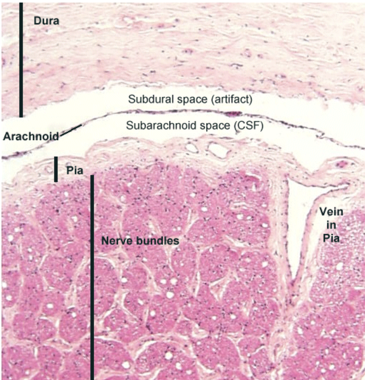
What are the 3 meninges?
Dura
Arachnoid
Pia
What are meninges?
Thick dural layer
Thin arachnoid layer
CSF filled space
Thin pia layer
What is the the dura layer?
Two dural layers
Periostial
Meningeal
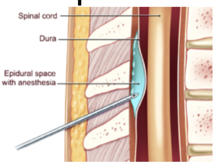
What are Spinal Meninges
One Layer of dura
Meningeal
Periostial becomes vertebrate perostium
(Note: Unlike in the cranium the vertebral column has a true epidural space)
Epidural puncture
Epidural injection is possible because of the true space between vertebrae and dura mater
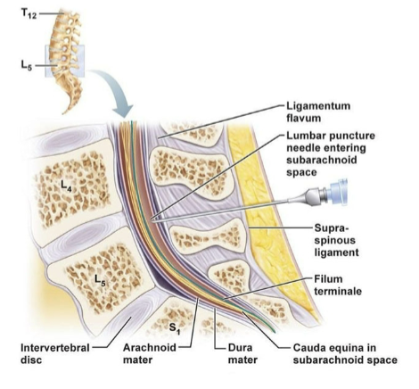
Lumbar puncture
Lumber punctures go deeper. Through the dura and arachnoid and into the subaracnoid space
What are Infoldings?
At important fissures of the brain the menigeal layer of dura folds in on itself.
(Falx Cerebri and tentorium cerebelli can be seen here)
Where the meningeal layer pulls away from the periostial layer, and folds in sinuses are created
These sinuses drain venous blood and CSF into the jugular vein
Importance of sinuses and infoldings
Dural venous sinuses interconnect and drain through the jugular foramen into the internal jugular vein
Importance of the lateral ventricle
where most CSF is made
Where is CSF formed?
the ventricles by the choroid plexus
about 350-500ml a day
25 ml in ventricles rest in subarachnoid region
turns over 2-3 times a day
What is CSF?
clear liquid
colorless
cell free (5 cells per ul)
ionic makeup different from blood
lower in protein and glucose
lower in K+ and CA++
higher in MG++ and CL-
Importance of choroid plexus
in every ventricle
not in cerebral aqueduct or central canal
largest amount in atrium of lateral ventricle (glomus)
Whats actively makes CSF?
a blood filtrate, actively made by choroid epithelium in the choroid plexus
What is the function of CSF?
adds buoyancy to brain
provides stable ionic environment
gylmphatic system: acts as lymphatic system form CNS
Importance of buoyancy
top a brain in isotonic saline (equivalence of being surrounded in CSF)
brain affected by gravity
Importance of ventricular circulation
CSF enters the subarachnoid space from the fourth ventricle
CSF circulates around in the subarachnoid space until it can be cleared
What is a spinal tap done?
Lumbar cistern
largest pocked of CSF
where the spinal cord ends (conus medullaris) but the arachnoid and dura continue and house spinal route (cauda equina)
What are the 2 reasons a spinal tap is done in the lumbar cistern?
Where most CSF is pooled
Low risk of hitting and thus damaging spinal cord
Where is a large portion of CSF drained?
Through the arachnoid granulations in the superior sagittal sinus
Where are arachnoid granulations?
in spinal nerve roots
Where else can CSF leak out of?
cranial and spinal nerves
CSF exiting where large blood vessels enter an exit meninges might be regulated by glia cells
Where does some CSF drain into?
the nasal cavity
What is rhinorrhea?
runny nose caused by excessive CSF leakage
often due to trauma
can be due to space occupying lesions or congenital malformations
What is hydrocephalus?
“water on the brain”
symptom of excess CSF in the ventricular system
can be congenital or acquired
What is noncommunicating hydrocephalus?
Caused by a blockage of CSF flow out of the ventricular system
Caused by a blockage or or stenosis of with in the ventricular system
most commonly at the cerebral aqueduct
Excess CSF in the ventricular system
Some ventricles are enlarged, some are normal
Key is that the CSF can not get out of the ventricular system
What is communicating CSF
CSF NOT cleared fast enough through the subarachnoid space
Excess CSF in CNS
All ventricles are enlarged
Caused by a decrease or impaired filtration of CSF through arachnoid granulations
Choroid tumors causing an increase in CSF production
Key is that the CSF has freedom to flow out ventricular system and into subarachnoid space
Since in some cases CSF pressure, as measured by spinal tap, is “normal” this can be called Normal Pressure Hydrocephalus
Ex Vacuo: excess CSF in the ventricular system as a result of decreased brain mass
What would cause an enlarged third ventricle?
Stenosis or blockage at the cerebral aqueduct —> enlarged lateral and third ventricle
Normal size fourth ventricle
What is hydrocephalus ex-vacuo?
ventricular enlargement due to decrease in brain tissue
particularly basal ganglia
Seen in degenerative diseases:
Parkinson’s
Alzheimer’s
Huntington’s
Normal pressure
How does spina bifida cause hydrocephalus?
Arnold-Chiari malformation
Herniation of the cerebellum blocking the flow of CSF out of the fourth ventricle
compress the foramen
Rated type I, II and III on level of severity
Chiari II usually accompanies a myelomeningocele
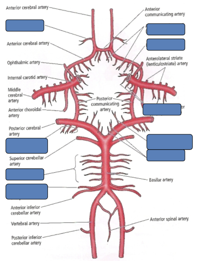
What arteries supply the CNS?
Internal carotid
Vertebral arteries
Importance of internal carotid artery
branch of common carotid
supplies most of cerebellum
Importance of vertebral arteries
Branch of subclavian
Supply brainstem, cerebellum, posterior cortex
In arterial plexi, where are arteries more dense?
more dense in gray matter, than white matter
What are the branches of the internal carotid?
Anterior cerebral artery (ACA)
Middle cerebral artery (MCA)
Importance of posterior communicating artery
branched off internal carotid first
vertebral supply
Arteries to know in the circle of Willis
Anterior cerebral (ACA) x2
Anterior communicating x1
Middle cerebral (MCA) x2
Lenticulostriate x many both sides
Anterior Choroidal x2 (first of lenticulostriate
Posterior communicating x2
Posterior cerebral (PCA) x2
Superior cerebellar x2
Basilar x1
Anterior inferior cerebellar (ACIA) x2
Posterior Inferior cerebellar (PICA) x2
Anterior spinal artery x1
Importance of anterior cerebral artery
Medial surfaces of the frontal and parietal lobes and the cingulate gyrus
Importance of lenticulstriate arteries
branches off the MCA that supply Deep structures
Basal ganglia
Internal capsule
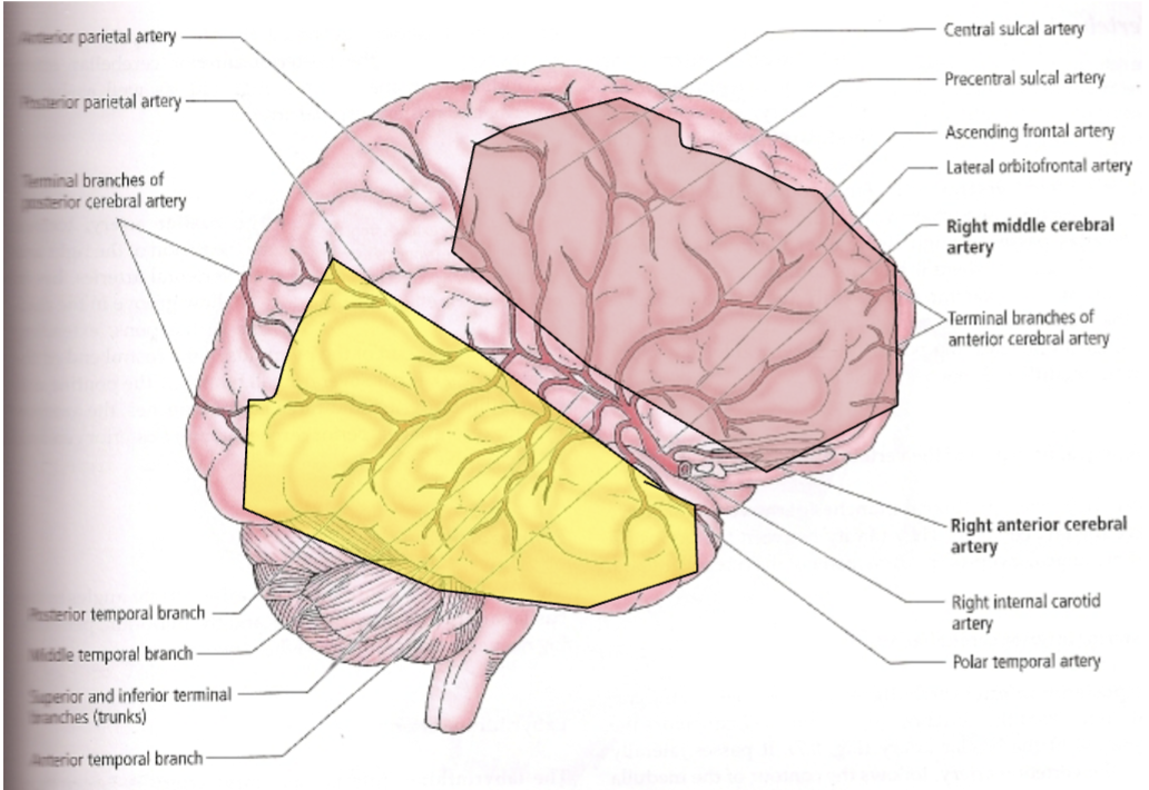
Importance of middle cerebral arteries
Cover most of the lateral surface of the brain
Superior branches (pink) cover frontal and parietal lobes
Inferior (yellow) cover temporal and occipital lobes
Importance of posterior cerebral artery
covers the inferior and medial temporal and occipital lobes
Perforating arteries supply thalamus and parts of internal capsule
lesion: visual defect
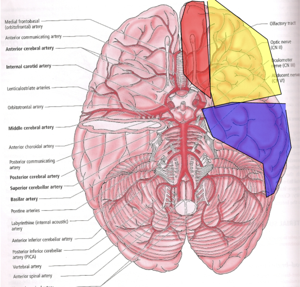
What supplies blood to the inferior surface of the brain?
Covered by parts of most main arteries
ACA (red)
MCA (yellow)
PCA (blue)
What is barrel syndrome?
brachial diplegia and sometimes bilateral paresthesia due to watershed infarcts in the anterior and middle cerebral artery watershed territory
What causes barrel syndrome?
Blockage in internal carotid
Areas of the cortex that have overlaped coverage (ACA and MCA)
If there is a decrease in blood flow to two adjacent arteries these areas are the first to become ischemic
Why are arteries relationship to cranial nerves important?
Neuroanatomically (being able to locate CNS structures)
Clinically aneurysms at key locations may compress cranial nerves causing dysfunction
Ballooning of arteries
Bulging of arteries become space occupied
neurological deficits
Important arteries for spinal cord blood supply
Radicular arteries: arise from the aorta along the vertebral column and help supply the cord and anastomose with the spinal arteries
Segmental medullary arteries: similar but doesn’t anastomose
Artery of adamkiewicz: described as the largest radicular artery
is variable from person to person but general originates from a posterior intercostal artery in the lower thorax
widely assumed now that in most people it is in fact a segmental artery and is mostly responsible for the anterior lumbar and sacral cord
Major anastomotic artery of spinal cord
stroke in anterior spinal cord, paralysis in top ½ of body
Arterial supply of CNS
lateral: upper body
medial: lower body
Importance of superficial cerebral veins
Superficial veins are on the surface of the cerebrum and mainly drain into the superior sagital sinus (SSS)
Importance of Superficial Middle cerebral vein
Can be found traveling along the lateral fissure
Interconnects multiple sinuses
Can drain down, or up to SSS
Importance of Superior and Inferior Anastomotic veins
Interconnect superficial intermediate vein to the superior sagittal sinus or transverse sinus respectively
Can drain to transverse sinus or up SSS
Importance of Dural Venous sinuses
Channels that form between two dural layers
Valveless veins that drain most venous blood and CSF from the brain
Know unpaired (midline) sinuses
Superior sagittal (SSS)
Inferior sagittal (ISS)
Straight Sinus
Confluence
Occipital
(sometimes paired)
Know paired sinuses (have right and lefr)
Cavernous
Transverse
Superior Petrosal
Inferior Petrosal
Sigmoid
What is the main drain for the brain?
interior jugular vein
Importance of deep cerebral veins
Drain deep structures and primarily drain into the great vein of galen then the straight sinus