3.2.1.3 methods of studying cells
1/22
There's no tags or description
Looks like no tags are added yet.
Name | Mastery | Learn | Test | Matching | Spaced |
|---|
No study sessions yet.
23 Terms
What is centrifugation?
A technique used to isolate different organelles in a cell → separates structures of different density
What is cell fractionation?
The process used to separate cellular components while preserving individual functions of each component
What is step 1 of centrifugation?
Homogenisation → cells are first broken open by grinding a tissue in an ice, cold isotonic buffer solution using a blender (homogeniser) → releases the organelles
Explain why an ice cold, isotonic, buffer solution is used
Ice cold → low kinetic energy minimises enzyme reactions within the cells that might cause self digestion (autolysis) of the organelles
Isotonic → prevents the osmotic movement of water in and out of organelles that might cause them to burst / shrivel
Buffer → maintains pH so proteins, especially enzymes, aren’t denatured
What is step 2?
Filtration → resulting suspension is filtered to remove cell debris e.g cell wall that haven’t burst in homogenisation → passed through a gauze
Explain the ultracentrifugation procedure that happens next
Homogenate is poured into tube + placed into centrifuge
Centrifuged at low speed → densest organelles e.g nuclei form a pellet / sediment at the bottom of tube
Supernatant liquid above sediment contains rest of organelles + can be spun at a higher speed for a longer period of time → second most densest organelle forms pellet
Procedure is repeated, increasing the speed + duration of centrifugation to obtain a series of pellets in order of decreasing density
What is the order of the organelles in decreasing density → order they’re isolated
nuclei, chloroplasts, mitochondria, endoplasmic reticulum, ribosomes, soluble proteins remain in supernatant
Naughty Clever Monkeys Like Eating Raspberries
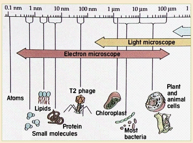
Define resolution
Ability to distinguish between 2 separate points
How does a light microscope work
Light rays by lenses pass through specimen usually mounted on a glass slide
How must the specimen be prepared for light / optical microscopes
Thin → light can pass through it + a single layer of cells is visible
Stained → structures are visible
Which type of specimens can be looked at with a light microscope
Dead + living samples
What magnification + resolution do light microscopes have?
Magnification - 1500
Resolution - 200nm → relatively poor so small structures aren’t visible
How does an electron microscope work?
Uses beam of electrons focused by electromagnets → electrons have a shorter wavelength than light → greater resolution of 0.05nm
Why is a vacuum required for electron microscopes
Electrons are absorbed by air molecules → only dead specimens can be looked at
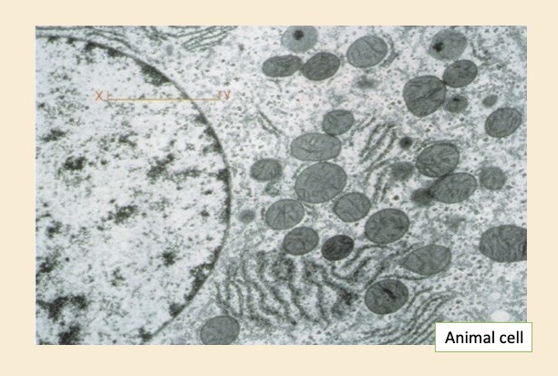
Why are electron microscopes important?
They can investigate the fine structure (ultrastructure) of a cell
What happens if you have a low resolution but a high magnification
Images become blurred
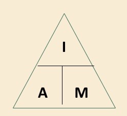
How to calculate magnification?
image / actual
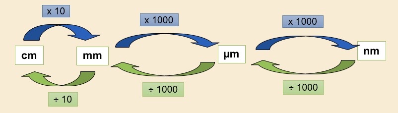
How to convert between units?
What are the 2 main types of electron microscopes?
Transmission electron microscope
Scanning electron microscope
How does a TEM work?
Beam of electrons is transmitted through specimen
Specimen must be thin + stained using electron dense substances e.g heavy metal salts
These substances deflect the electrons in beam + pattern that the remains electrons produce as they pass through the specimen is converted to an image
How does a SEM work?
Specimen is coated with a thin film of heavy metal e.g gold
Electron beam is scanned to and fro across specimen
Electrons are reflected from surface are collected + produce an image on a viewing screen
Compare SEM + TEM