Body Systems
1/31
Earn XP
Description and Tags
Health Term 2 2025
Name | Mastery | Learn | Test | Matching | Spaced | Call with Kai |
|---|
No analytics yet
Send a link to your students to track their progress
32 Terms
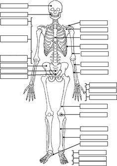
Label the diagram (Yes/No)
Yes Calcaneus
What are the sections of the vertebrae in order and how many pieces are there in each section?
Cervical (7)
Thoracic (12)
Lumbar (5)
Sacrum (5 fused together)
Coccyx (4 fused together)
List the functions of the skeletal system.
Allows movement, shape and protection, mineral storage, production of blood cells.
Describe the skeletal function of ‘allows movement’.
The skeleton allows movement of the body. Bones provide surfaces for muscles to attach to and allow muscles to pull on them to produce movement.
Describe the skeletal function of ‘shape and protection’.
The skeleton provides the body with its shape, and also protects internal organs, reducing the impact of injuries. For example, the ribs protect the heart and lungs, the cranium protects the brain and the spine protects the spinal cord.
Describe the skeletal function of ‘mineral storage’.
Bones store minerals such as calcium, iron, potassium and phosphorus. The minerals are released into the blood when needed.
Describe the skeletal function of ‘production of blood cells.
White and red blood cells are produced in the bone marrow.
What do red and white blood cells do?
Red blood cells carry oxygen to our cells and is red because of the haemoglobin, which is a protein containing iron.
White blood cells fight off infections.
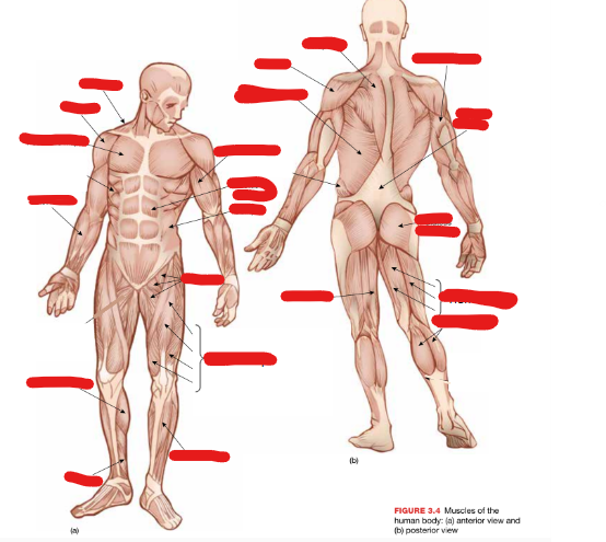
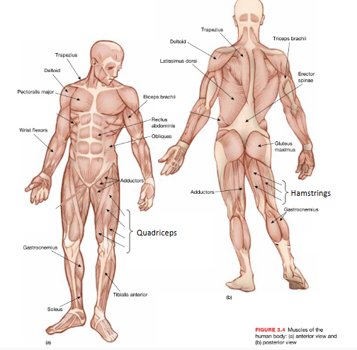
Label the muscles. (Y/N) And how many muscles are there?
Y 600
What are the functions of the muscular system?
Create movement, posture, and heat production.
Describe the muscular function of ‘create movement’.
Muscles are responsible for our movements. Muscles pull on bones to create movement.
Describe the muscular function of ‘posture’.
Skeletal muscles provide the force needed to stabalise the body. Their flexibility and strength are key to maintaining proper posture.
Describe the muscular function of ‘heat production’.
When muscles contract, they generate heat, which is vital to maintaining our body temperature.
What are tendons and ligaments?
Tendons connect bones to muscles and ligaments connect bones to bones. Tendons are fibrous connective tissues that attach muscles to bones, enabling movement, while ligaments are connective tissues that connect bones at joints, providing stability.
What is the difference between extension and flexion?
Flexion is a movement that decreases the angle between the joints, whereas extension is a movement that increases the angle between the joints.
Explain reciprocal inhibition. Include agonist, antagonist, lengthening, shortening.
Reciprocal inhibition refers to how muscles work in pairs to create movement. While one muscle in the pair contracts, the other relaxes. The muscle that contracts is the agonist and it shortens. The muscle that relaxes is the antagonist, which lengthens.
Describe an example of reciprocal inhibition.
An example of reciprocal inhibition is the biceps contracting while the triceps relax during arm flexion. In this movement, the biceps are the agonist, which shortens, while the triceps are the antagonist and lengthens as it relaxes. Together, they pull on the radius and ulna through the tendons, which decreases the angle at the elbows.
Explain how the process of reciprocal inhibition allows flexion at the knee to occur. [4 marks]
Reciprocal inhibition is the process where a pair of muscles work together to create movement. When flexing the knee, the quadriceps act as the antagonist which relaxes and lengthens, while the hamstrings act as the agonist, which contracts and shortens. When the agonist muscle contracts, it pulls on the tibia and fibula at the tendons to decrease the angle at the knee joint.
What are the functions of the circulatory system?
Circulates blood
Delivers nutrients to body cells (oxygen)
Removes waste (carbon dioxide) from body cells
Maintains stable body temperature
What are the types of blood vessels?
Arteries, veins and capillaries.
Arteries
Arteries generally carry oxygen rich blood and always carry blood away from the heart. They have elastic walls so that they can expand and accommodate more volume. They also have the thickest walls out of the three.
Aorta
The aorta is the largest artery in the body. It carries oxygen-rich blood from the heart (pumped from the left ventricle) to the rest of the body.
Capillaries
Capillaries are the smallest type of blood vessel, with walls that are only one cell thick. They carry both oxygenated and deoxygenated blood, exchanging nutrients and waste in the body. There are millions of capillaries throughout the body, and they branch out from arteries, eventually forming into veins.
Veins
Always travel into the heart, and generally carries deoxygenated blood from the body. Has thinner and less elastic walls compared to the arteries and has valves to stop blood from flowing the wrong direction.
How does blood travel around the body?
In arteries, it is the heart that moves the blood around the body. As the heat beats, it pumps blood out of our heart at high pressures. Meanwhile, the veins rely on the skeletal muscles contracting as well as the valves that stop blood from flowing the wrong direction, to push it to the heart.
Describe the pathway of blood.
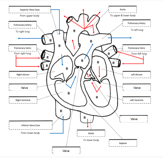
Deoxygenated blood is first brought into the right atrium, through the superior and inferior vena cava. The superior vena cava is responsible for blood from the top half of the body, while the inferior vena cava is responsible for the bottom half. After the deoxygenated blood enters the right atrium, it is pumped through a valve and into the right ventricle. Then, it travelled through another valve called the pulmonary valve into the pulmonary artery, which splits left and right to send the deoxygenated blood into its respective lung. There, gas exchange occurs where oxygen enters the blood as CO2 leaves, and the blood returns rich with oxygen, through the pulmonary vein, and into the left atrium. There, it is pumped into the left ventricle through a valve before it travels through a valve to the aorta and the oxygenated blood is distributed across the body. (Don’t forget, the valves close after the blood is pumped through them to stop the blood from flowing back the way it came). Oxygen is exchanged for CO2 in the blood as the body’s cells take the nutrients through a process called gas exchange. The then deoxygenated blood begins its journey back to the heart via the veins.
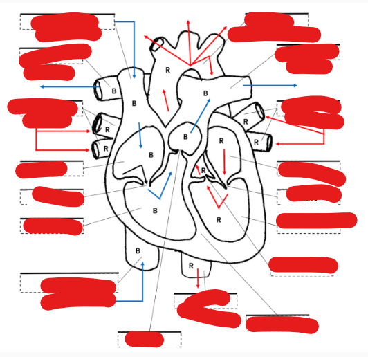
Label the diagram of a heart. (Yes/No)
Yes
What are the functions of the respiratory system
Draw in air from the atmosphere
Transfers oxygen from air into the blood
Removes carbon dioxide from the blood
Expels heat in air exhaled
Allows the vocal cords to create speech as air is exhaled.
What does breathing rely on?
Breathing relies on the tendency of air particles to naturally move from areas of higher pressure to areas of lower pressure.
Describe inspiration.
Inspiration is also known as inhalation, and is the process of drawing air into the lungs. During inspiration, the diaphragm contracts, moving downwards as the intercoastal muscles (rib muscles) also contract, increasing the size of the chest cavity. Together, this causes the pressure in the lungs to decrease, resulting in air being drawn in as the gas moves from high to low pressure areas.
Describe expiration.
Expiration is also known as exhalation, and is the process of expelling air from the lungs. During expiration, the diaphragm relaxes, moving back up as the intercoastal muscles (rib muscles) also relax, decreasing the size of the chest cavity. Together, this causes the pressure in the lungs to increase, resulting in air being forced out as the gas moves from high to low pressure areas.
Describe gaseous exchange, what it relies on, and what it does in the two examples that it occurs in the body.
Gaseous exchange is the process where gases move across a surface without the use of energy. Gas exchange relies on the tendency of gas molecules to move from areas of high concentration to areas of lower concentration. Between the alveoli and the capillaries at the lungs, deoxygenated blood becomes oxygenated, and carbon dioxide moves from the capillaries to the alveoli as oxygen moves from the alveoli into the blood stream, carried by haemoglobin. This is because in the capillaries, the blood is very low in oxygen, while in the alveoli, there is a higher level of oxygen, causing it to diffuse into the blood stream. The same is true for the carbon dioxide, which moves out of the blood and is expelled into the air in the next exhalation. Between capillaries and skeletal muscles and muscle cells, the same process occurs, except the exchange occurs between the capillaries and the muscle cells instead of the alveoli.