PNS: Muscle
1/76
Earn XP
Description and Tags
Write the correct answer to the question
Name | Mastery | Learn | Test | Matching | Spaced |
|---|
No study sessions yet.
77 Terms
What is the skeletal muscle in contact with?
The skeletal muscle is in contact with bones, allowing for voluntary movement and stability of the skeleton.
What controls skeletal muscle contraction?
The nervous system, specifically motor neurons, controls skeletal muscle contraction by transmitting signals that trigger muscle fibres to contract. It’s under conscious, voluntary control.
What is the NMJ?
It is a place where the axon terminals of motor neurons synapse with individual muscle fibres to form this specialized junction.
What kind of proteins overlap filaments together?
These proteins are called troponin and tropomyosin, which regulate the interaction between actin and myosin during muscle contraction.
What is the sarcoplasmic reticulum?
The sarcoplasmic reticulum is a specialized endoplasmic reticulum in muscle cells that stores and releases calcium ions, which are essential for muscle contraction.
Larger muscles have…
dozens of fibres.
Smaller muscle have…
fewer fibers than larger muscles.
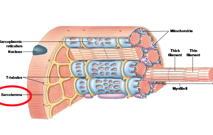
What are muscle fibres made up of?
Myofibrils, which are bundles of myofilaments.

In a muscle fibre, what is the sarcolemma also known for?
The cell membrane that surrounds it.

What is the role of the invaginations of the sarcolemma in the T-tubule system?
It allows the spread of action potential deep into the muscle. It transmits the action potentials, enabling muscle fibres to respond.
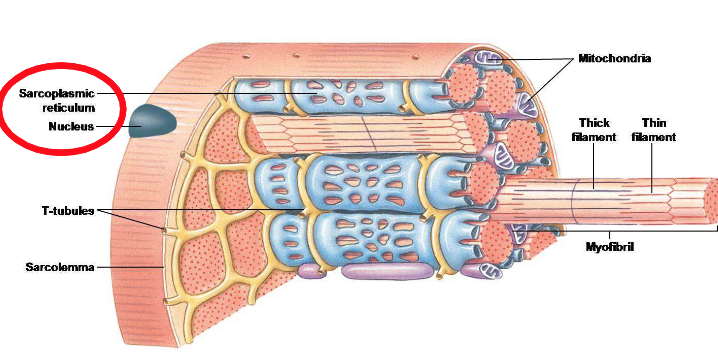
Why does the skeletal muscle fibre have the fastest contraction time out of all three muscle types?
The sarcoplasmic reticulum controls how fast Ca²⁺ appears and disappears, which directly determines how fast a skeletal muscle fibre contracts and relaxes.

Why is motor unit one slow and oxidative?
It only has slow-twitch fibres. These fibres are more fatigue-resistant, utilize aerobic metabolism, and are efficient for prolonged activities. This indicates that it’s a small motor unit.
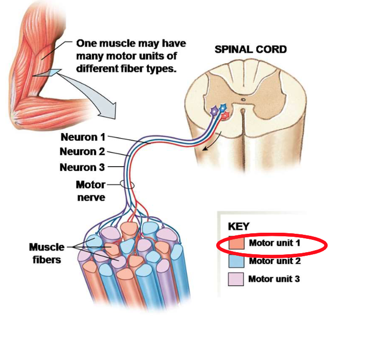
Having small motor units….
allow us to have small and concise motor movements.

Having larger motor units…
allow a large development of force and less control of gradations in force.

If motor units two and three have faster-twitch fibres than motor unit one, does this mean that they are large motor units?
Yes, larger motor units typically contain fast-twitch fibres, which are geared for rapid and powerful contractions but are less fatigue-resistant. They have fewer mitochondria, which results in lactic accumulation.

To win an Olympic marathon, do you need slow- or fast-twitch fibres?
You need slow-twitch fibres, as they are more efficient for endurance and prolonged aerobic activities.
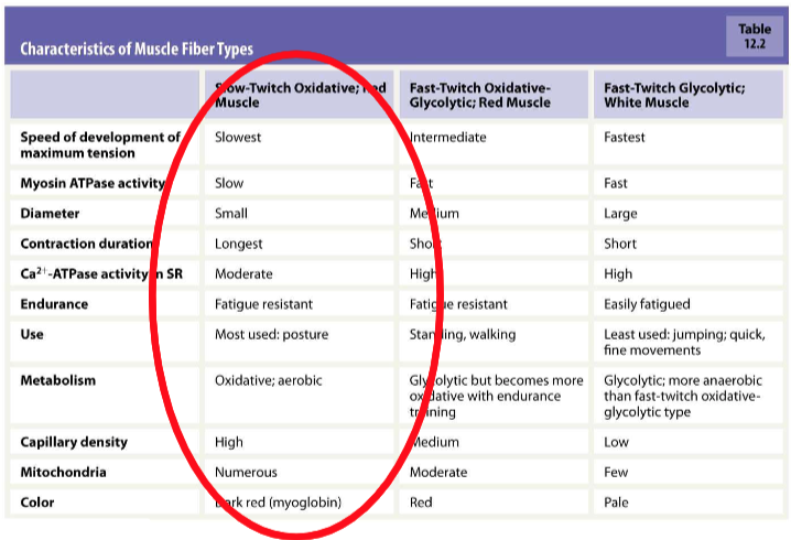
Muscles that are involve with low-level force production, such as walking and maintaining posture, need…
slow-twitch muscle fibres, which are more endurance-oriented and fatigue-resistant.
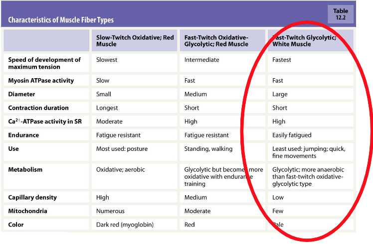
If you want to win a 100-metre sprint, you'd better be born with 80% of…
fast-twitch muscle fibres, as they are essential for explosive power and speed.
Fast twitch-Oxidative fibres are somewhere between…
slow-twitch and fast-twitch glycolytic fibres in terms of fatigue resistance and force generation.
Large-diameter motor neurons innervate…
have high excitatory synaptic input to fast-twitch muscle fibres, facilitating rapid and powerful muscle contractions.
Small-diameter motor neurons are…
more easily excited by the summation of EPSPs than large-diameter motor neurons.
All motor neurons release…
acetylcholine at the neuromuscular junction, facilitating muscle contraction.
All motor neurons synapses are…
excitatory, and it depends on the summation of EPSPs/IPSPs.

Explain step one of the communication of NMJ.
Action potential arrives at the terminal bouton of the motor neuron.

Explain step two of the communication of NMJ.
Calcium ions enter the terminal bouton, triggering the release of acetylcholine.

Explain step three of the communication of NMJ.
Acetylcholine binds to nicotinic receptors on the motor end plate.
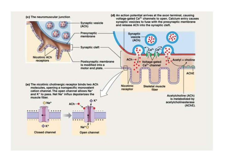
Explain step five of the communication of NMJ.
ACh triggers the opening of small cations Na2+ and K+. The net movement of sodium is inward.

Explain step six of the communication of NMJ.
Muscle action potential is generated, leading to muscle contraction.
Are peripheral tissues exposed to circulating toxins?
Yes, they can be affected by toxins in the bloodstream.

Explain step-by-step how curare-form drugs affect the poisoning of NMJ.
Curare-type drugs block ACh receptors at the NMJ. They prevent opening of Ca2+ channels at the endplate. This inhibition stops muscle action potentials, allowing certain structures to remain dilated and leading to paralysis.
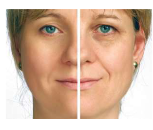
What happens when Botox is applied on your face?
Botox inhibits ACh release at the neuromuscular junction, causing temporary muscle paralysis in the treated area, which reduces the appearance of wrinkles, as it smoothes out muscles.

The long, thin filament of a muscle fibre is made up of…
actin.
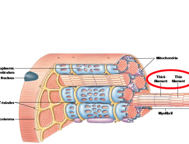
The thick filament is made up of…
myosin, which is in charge of making numerous cross-bridges between actin during muscle contraction.
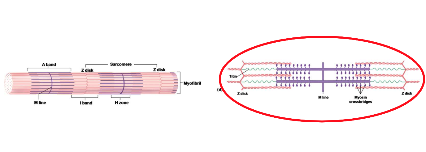
Sarcomeres help actin and myosins...
to contract and relax during muscle movement.
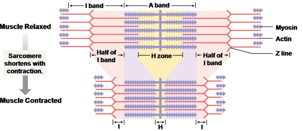
So if sarcomeres shorten in length…
Actin and myosin don’t change in length, but instead they slide past one another.
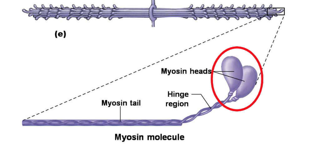
Myosin heads are known as the…
actin binding site.
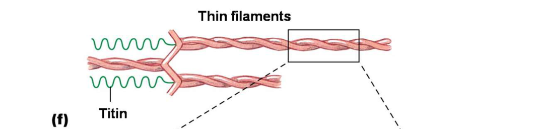
The thin myofilament is made up of…
2 strands of actin filaments.
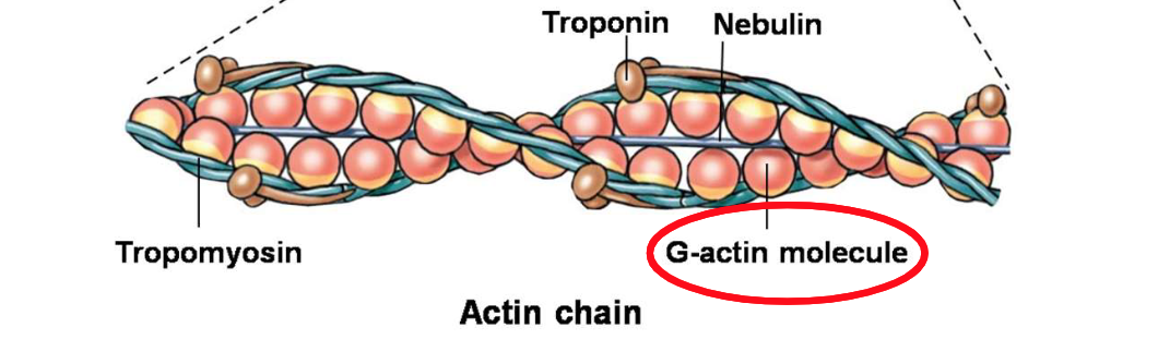
The Guanine-Actin molecule is the…
binding site for myosin heads in muscle contraction.
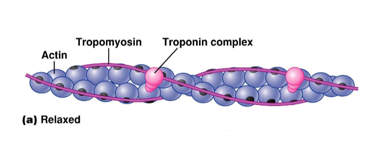
What is the role of tropomyosin and troponin?
Troponin holds tropomyosin over the myosin and actin binding site.
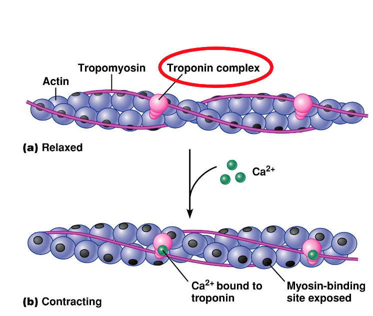
What happens when calcium binds to troponin?
Calcium binds to troponin, which shifts tropomyosin off actin’s myosin-binding sites, allowing contraction. This process exposes the binding sites for myosin heads to attach to actin, facilitating muscle contraction.
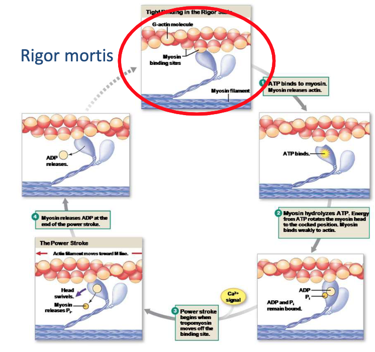
What is rigour mortis, and how does it affect the body?
Rigour mortis is the post-death stiffening of muscles due to chemical changes in muscle tissue. It occurs because ATP production ceases, preventing myosin heads from detaching from actin filaments. Muscle proteins break down and relax again.
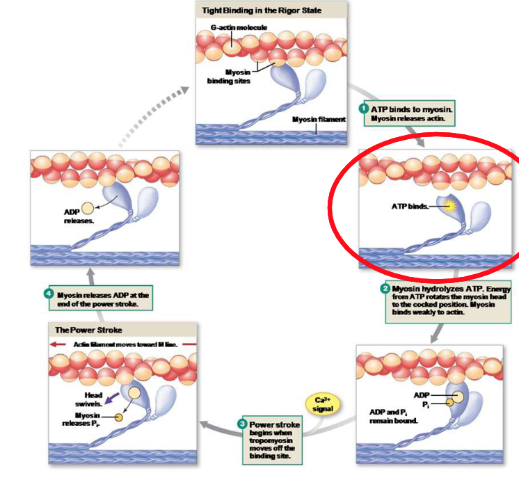
Explain the first step of the crossbridge cycle.
ATP is bounded, so the myosin detatches from actin.
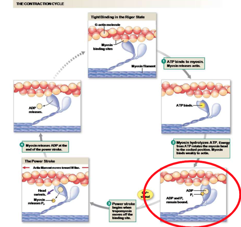
Explain the second step of the crossbridge cycle.
Bounded ATP is now hydrolyzed. Myosin now has high affinity for actin. Ready to go for another power stroke, as the myosin head pivots and pulls actin. This action generates force.
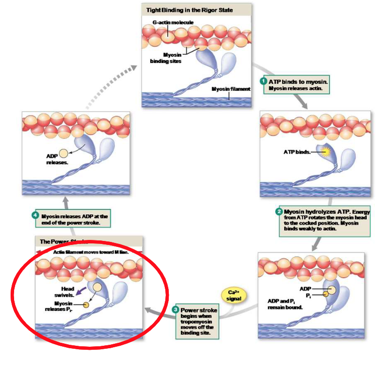
Explain the third step of the crossbridge cycle.
Ca2+ is present. Tropomyosin moves away from the binding sites on actin, allowing the myosin head to attach to actin, and a power stroke can occur.
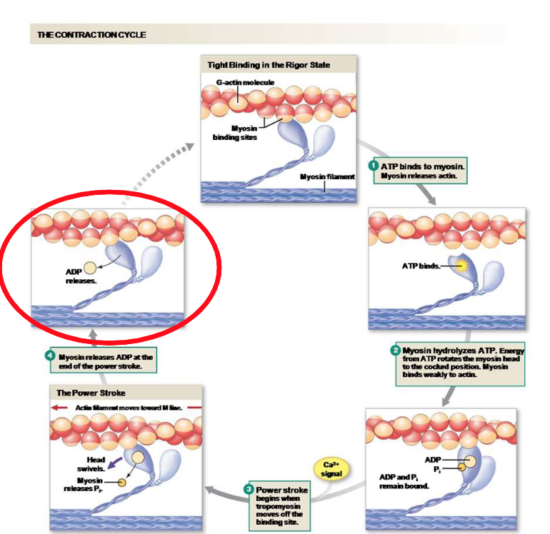
Explain the fourth step of the crossbridge cycle.
Once the crossbridge cycle is completed, the myosin head is in its low state for actin. ATP is hydrolyzed to ADP because of the last stroke. Myosin detaches from actin, and the cycle can restart.

Explain the first step of excitation-contraction coupling.
AP arrives at the somatic motor neuron.
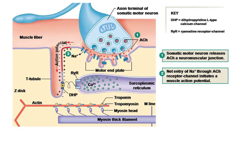
Explain the second step of excitation-contraction coupling.
Voltage-gated Ca2+ channels open in the axon terminal, releasing Ca2+ to trigger the release of acetylcholine (ACh) into the synaptic cleft.

Explain the third step of excitation-contraction coupling.
Binding of ACh triggers depolarization of the motor end plate and an AP in the muscle cell.
Explain the fourth step of excitation-contraction coupling.
Action potential in the t-tubule alters the conformation of the DHP receptor.

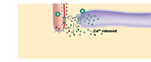
Explain the fifth step of excitation-contraction coupling.
DHP receptor activation leads to the opening of ryanodine receptors, releasing Ca2+ from the sarcoplasmic reticulum.
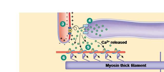
Explain the sixth step of excitation-contraction coupling.
As Ca2+ enters the cytoplasm, Ca2+ binds to troponin, shifting tropomyosin and exposing myosin-binding sites on actin, initiating contraction and facilitating cross-bridge formation.
Explain the last step of excitation-contraction coupling.
When Ca2+ is removed from the cytoplasm, leading to muscle relaxation as tropomyosin re-covers the myosin-binding sites on actin.
To generate a muscle force, how many twitches do you need working together?
Multiple twitches in rapid succession are required to increase the force of contraction.
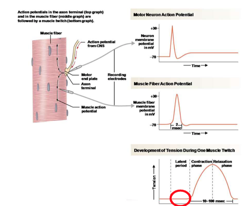
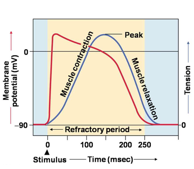
Explain the process of the twitch contraction based on this image.
The AP is travelling down the T-tubules, and Ca2+ is being released by the sarcoplasmic reticulum to initiate the cross-bridge cycle.

Why could there be a delay in AP for twitch contraction?
It could be due to the time taken for the action potential to travel down the T-tubules and trigger calcium release from the sarcoplasmic reticulum.
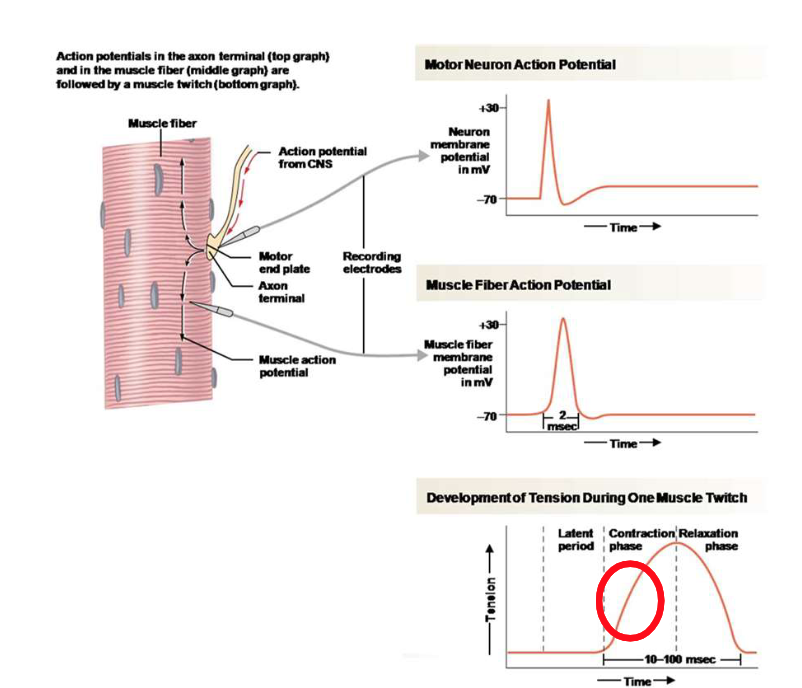
During the contraction phase…
Tension is produced, and the force is steadily rising.
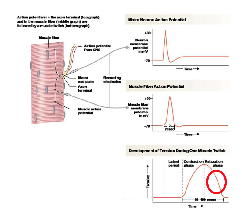
During the relaxation phase…
Calcium ions are being pumped back to the sarcoplasmic reticulum. As intracellular Ca2+ falls, fewer crossbridges are allowed to interact with actin, so the tension falls to 0.
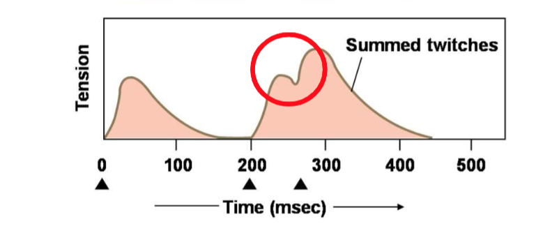
With multiple AP in a muscle fibre….
It results in frequent stimulation of the muscle as tension goes up. The successive twitches are on top of each other, and the contractile force rises.
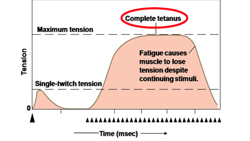
When summation leads to complete tetanus…
the muscle fibers maintain a steady and maximal contraction without relaxation between stimuli.
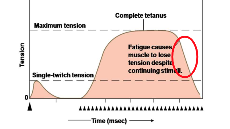
If the muscle fatigues...
Tension falls rapidly. This occurs when the muscle is unable to maintain the required force due to a depletion of energy sources or accumulation of metabolic byproducts.
Where is the smooth muscle found?
Smooth muscle is found in the walls of hollow organs such as the intestines, blood vessels, and the uterus. It regulates involuntary movements like digestion and blood flow.
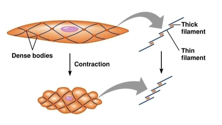
Is the smooth muscle arranged in sarcomeres?
No, smooth muscle does not have sarcomeres; instead, its fibres are arranged in a lattice-like structure that allows for contraction.
The smooth muscle contractions are much…
slower, uses less energy and is sustained than skeletal muscle contractions. This allows for prolonged, involuntary control of various organ functions.
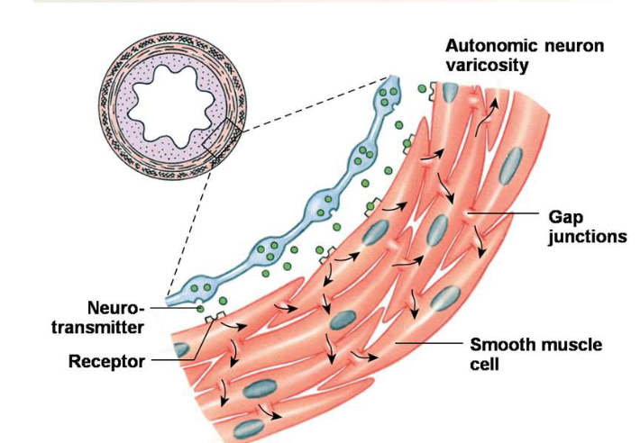
Where are single-unit muscles found in the body?
They are found in the intestinal tract and blood vessels where they facilitate local contractions.

Single-unit muscles need…
a certain amount of muscular tension to be maintained to facilitate continuous contraction and function.
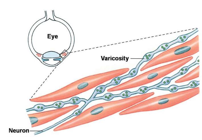
The Multi-Unit Smooth Muscles are…
composed of individual muscle fibres that operate independently, allowing for precise control of contraction in locations such as the iris of the eye and the walls of larger airways. They prohibt our eye lens from changing spontaneously.

Explain the excitation-contraction coupling in a smooth muscle cell.
A Ca2+ influx in the plasma membrane triggers release of the Ca2+ sarcoplasmic reticulum. Ca2+ binds to calmodulin, activating myosin light chain kinase (MLCK) to phosphorylate myosin, enabling contraction.
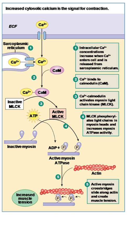
How do you relax the smooth muscle cell after excitation-contraction coupling?
Relaxation occurs through the removal of Ca2+ ions, leading to the dephosphorylation of myosin by myosin light chain phosphatase, which inhibits contraction.
Explain the step-by-step process of the contraction of the heart.
Na+ voltage-gated channels open a lot quicker than the Ca2+ channels. Ca2+ enter from the ECF. AP is generated by the increased permeability of Na+, followed by a slower increase from Ca2+ channels, leading to the contraction of cardiac muscle fibers.
The cardiac contractile cell AP lat for almost as long as the concentration and relaxation, but if you want to fire another action potential in a close succession…
Tetanus will be generated. By the time you’ll be able to fire another action potential, the cardiac muscle will be in a relaxed state, so summation and tetanus cannot occur.
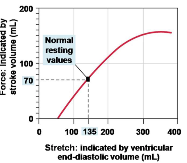
The greater volume of blood in the cardiac chambers (relaxation of the heart)…
the greater the developing force of contraction.

The more you stretch the muscle…
the greater the force, the more muscle will generate due to more overlap of myosin and actin.

Based on this image, what happens if you remove Ca2+ from the myofilament?
The muscle will relax. Without calcium, myosin cannot bind to actin, leading to muscle relaxation. The calcium pump in the SR will remove the calcium ions from the cytosol, which causes detachment of cross-bridges and lowers the level of active force.

Digitalis strengthens when…
calcium levels increase in the heart muscle cells, enhancing contraction force.

The increase of intracellular levels of sodium ions…
leads to increased calcium levels in cardiac muscle, which enhances contraction.
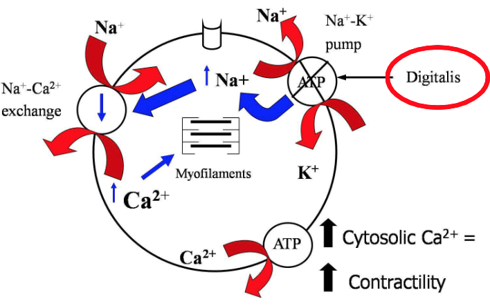
The increase in force of contractile function inhibits the…
Na+ and the K+ ATPase pump in cardiac cells, leading to increased intracellular sodium and calcium concentrations.

A decrease in sodium ion influx leads to…
a reduced intracellular calcium concentration, decreasing contractile force.