Biology - Chapter 15: Nervous Coordination and Muscles
1/98
Earn XP
Description and Tags
Name | Mastery | Learn | Test | Matching | Spaced | Call with Kai |
|---|
No analytics yet
Send a link to your students to track their progress
99 Terms
How is a resting potential maintained across the axon membrane in a neurone? (3)
1. Sodium-potassium pump actively transports 3 Na+ ions out and 2 K+ ions in
2. Membrane more permeable to K+ and less permeable to Na+ as K+ channels are open, while Na+ channels are closed
Why is the speed of transmission of impulses faster along a myelinated axon than a non-myelinated axon? (3)
1. Myelination provides electrical insulation so depolarisation occurs at the nodes of Ranvier only
2. This means the impulse jumps from node to node - saltatory conduction
3. In contrast, in non-myelinated axons, depolarisation occurs along the whole axon
Describe the events involved in transmission across a cholinergic synapse (6)
1. Action potential causes the presynaptic neurone's membrane to be depolarised
2. Calcium channels open and Ca2+ ions enter
3. This causes the synaptic vesicles to fuse with the presynaptic neurone's membrane and release ACH
4. ACH diffuses across the synaptic cleft
5. It binds to complementary receptors on the postsynaptic membrane
6. Na+ ions enter the post synaptic neurone by facilitated diffusion, resulting in the depolarisation of the postsynaptic neurone's membrane
ACH
Acetylcholine
How do synapses ensure synaptic transmission is unidirectional? (2)
1. Neurotransmitter only made in presynaptic neurone
2. Receptors only on postsynaptic neurone
Why is it important that neurotransmitters are transported back out of synapses?
If not removed, they keep binding to receptors so would keep causing action potentials / depolarisation
What happens if myelin sheaths are damaged? (3)
1. Nerve impulse cannot jump from node to node
2. Depolarisation occurs along the entire length of the axon's membrane
3. Nerve impulses are transmitted more slowly
Describe the all or nothing principle
When the threshold is reached, there is the maximum response (any stimulus below threshold value does not generate an action potential)
2 main forms of coordination in animals
The nervous system and the hormonal system
Neurotransmitters
Chemicals that are involved in communication between adjacent neurones or between nerve cells and muscles
How does the nervous system work? (2)
- Nerve cells pass electrical impulses along their length
- Stimulate target cells by secreting neurotransmitters
How does the hormonal system work? (2)
- Produce hormones that are transported in blood plasma to target cells
- Target cells have specific receptors on CSM and are stimulated by change in concentration of hormones
Describe the response by the nervous system (3)
Rapid, short-lived and localised
Describe the response by the hormonal system (3)
Slow, long-lasting and widespread
Compare the nervous system and hormonal system (8)
Nervous system vs Hormonal system
1. Communicate by nerve impulses vs chemicals called hormones
2. Transmitted by neurones vs blood system
3. Transmission is rapid vs slow
4. Travel to specific parts of body vs whole body but only target cells respond
5. Response is localised vs widespread
6. Response is rapid vs slow
7. Response is short-lived vs long-lasting
8. Effect is temporary and reversible vs potentially permanent and irreversible
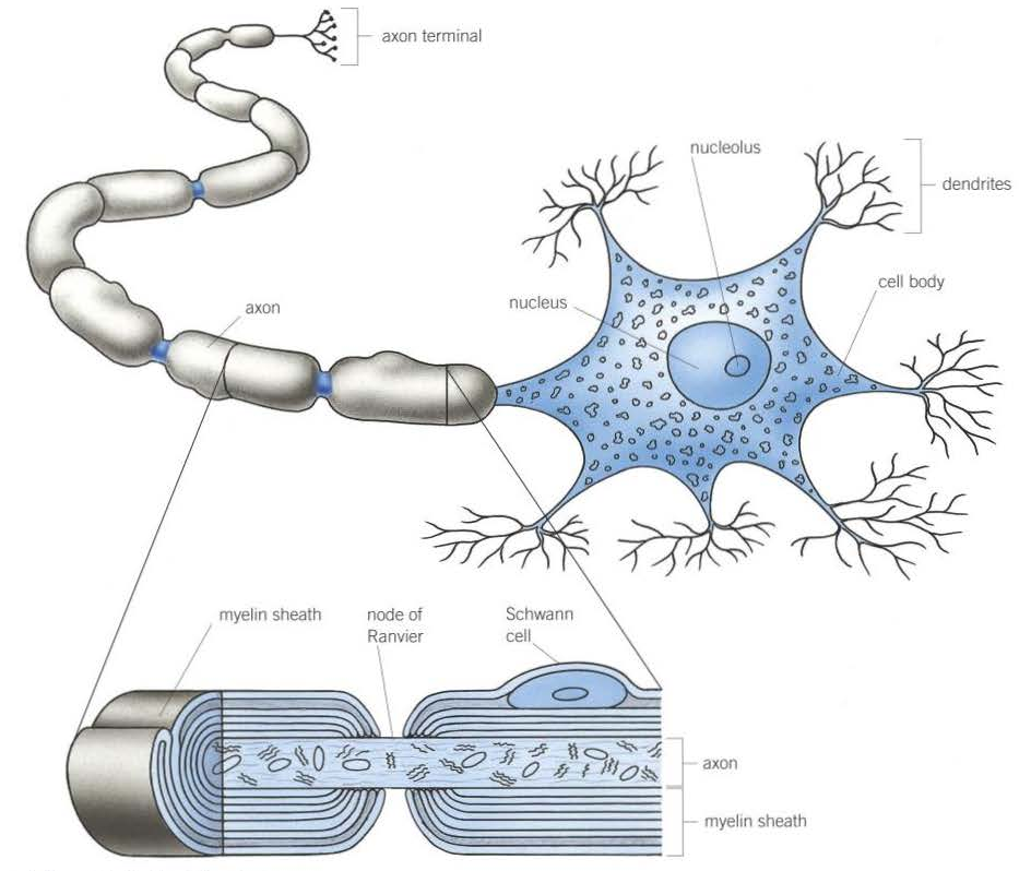
Describe the 6 parts of motor neurone
1. Cell body: contains cell organelles including large amounts of RER - produces proteins and neurotransmitters
2. Dendrons, extend into dendrites: carry nerve impulses towards cell body
3. Axon: single long fibre that carries nerve impulses away from cell body
4. Schwann cells: surround axon to insulate it, carry out phagocytosis and are involved in nerve regeneration
5. Myelin sheath: made up of Schwann cells + lipid called myelin
6. Nodes of Ranvier: between Schwann cells where no myelin sheath
3 types of neurones
Sensory, motor and relay
Resting potential
The state of the axon without an action potential - it is when the inside of the axon is negatively charged relative to the outside
What is the state of the axon called during the resting potential?
It is polarised
What is the state of the axon called during the action potential?
It is depolarised
Action potential
The state of the axon when the energy from a stimulus of a sufficient size causes a temporary reversal of the charges either side of a part of the axon membrane
Voltage-gated channels
Channels in the axon membrane that open or close depending on the voltage across the membrane
Describe the process of an action potential (8)
1. Energy from stimulus causes Na+ channels to open
2. Na+ ions diffuse into axon down electrochemical gradient, depolarising the membrane
3. More Na+ channels continue to open - influx of Na+ ions
4. When max potential reached, Na+ channels close and K+ channels begin to open
5. K+ ions diffuse out of axon down electrochemical gradient, repolarising the membrane
6. More K+ channels continue to open
7. Diffusion of K+ ions results in temporary overshoot of electrical gradient as inside of axon is hyperpolarised (more negative than usual) - refractory period
8. K+ channels close, Na-K pump starts again - resting potential maintained
Depolarisation
Temporary reversal of the charges on the cell-surface membrane of a neurone that takes place when a nerve impulse is transmitted
Repolarisation
The return of the resting potential in the axon of a neurone after an action potential
Saltatory conduction
The passage of action potentials along a myelinated axon in which the action potential jumps from one node of Ranvier to another
How do action potentials pass along a myelinated axon?
Saltatory conduction - action potentials can only occur at nodes of Ranvier as myelin sheath insulates rest of axon, so they jump from node to node
Nerve impulse
The transmission of an action potential along the axon of a neurone
3 factors that affect the speed at which an action potential travels
1. The myelin sheath: acts as electrical insulation so action potentials can only jump from one node of Ranvier to another
2. The diameter of the axon: the greater the diameter, the faster the speed of conductance (less ions leak from axon)
3. Temperature: affects rate of diffusion of ions
Threshold value
The level of stimulus needed to trigger an action potential
2 ways an organism can perceive the size of a stimulus
Number of impulses passing in a given time or by having different neurones with different thresholds
Refractory period
Period after an action potential when sodium voltage-gated channels are closed and the inward movement of Na+ ions stops, so it is impossible for a further action potential to be generated
3 purposes of refractory period
1. Ensures action potentials are propagated in one direction only (i.e. only forwards)
2. Produces discrete impulses
3. Limits the number of action potentials
Synapse
Point where one neurone communicates with another neurone or an effector
Describe the structure of a synapse (4)
1. Synaptic cleft: gap between presynaptic and postsynaptic neurone
2. Synaptic knob: swollen portion at end of axon
3. Mitochondria and endoplasmic reticulum in synaptic knob
4. Synaptic vesicles
2 types of summation
Spatial summation and temporal summation
2 types of synapses
Excitatory and inhibitory
Describe how inhibitory synapses work (4)
1. Presynaptic neurone releases a type of neurotransmitter that binds to chloride ion protein channels on postsynaptic neurone
2. This causes Cl- channels to open and Cl- ions move into the postsynaptic neurone by facilitated diffusion
3. The neurotransmitter also causes K+ channels to open so K+ ions diffuse out of the postsynaptic neurone
4. This causes the membrane of the postsynaptic neurone to repolarise and eventually undergoes hyperpolarisation
2 functions of synapses
1. To allow a single impulse along a neurone to initiate new impulses in a number of different neurones at a synapse, so a single stimulus can create a number of simultaneous responses
2. To allow a number of impulses to be combined at a synapse, so nerve impulses from receptors reacting to different stimuli can contribute to a single response
Cholinergic synapse
Synapse in which the neurotransmitter is ACH
What breaks down ACH and what is ACH broken down into?
Acetylcholinesterase - broken down into acetyl (ethanoic acid) and choline
Where are cholinergic synapses located at in vertebrates?
The CNS and neuromuscular junctions
2 ways drugs could effect the nervous system
1. Stimulate nervous system by creating more action potentials in postsynaptic neurones
2. Inhibit nervous system by creating fewer action potentials in postsynaptic neurones
Spatial summation
A number of different presynaptic neurones release enough neurotransmitter together to exceed the threshold value of the postsynaptic neurone to trigger a new action potential
Temporal summation
With a single presynaptic neurone, there are several impulses in a short time to provide enough neurotransmitter to reach threshold
How does the skeleton enable the contraction of skeletal muscles?
It is incompressible so if a muscle exerts a force, the bone moves via the tendons (instead of the muscle changing shape)
Antagonistic pairs
Pairs of muscles that work in opposition to each other (when one contracts, the other relaxes)
Where is cardiac muscle found?
In the heart
Where is smooth muscle found?
In the walls of blood vessels and the gut
Describe the structure of skeletal muscle
Made of myofibrils, grouped into muscle fibres, then bundles of muscle fibres, then the whole muscle
Why are skeletal muscles made up of many myofibrils grouped together?
To increase their strength
What is special about muscle fibres? (3)
- They share nuclei
- They share a cytoplasm - the sarcoplasm
- They have a large concentration of mitochondria and endoplasmic reticulum within the sarcoplasm
2 types of protein filament that make up myofibrils
Actin and myosin
Describe the structure of actin
Thinner than myosin and made of 2 strands wound around one another
Describe the structure of myosin
Thicker, consists of long rod-shaped tails with bulbous heads that project to the side
5 features of sarcomere/myofibril diagrams

1. I-band: light-coloured band where only thin filaments are present
2. A-band: dark bands where thin and thick filaments overlap
3. H-zone: lighter-coloured region at centre of A band
4. M-line: centre of H-zone
5. Z-line centre of each I band
Sarcomere
The distance between adjacent Z-lines
What happens to sarcomeres when muscles contract?
The sarcomeres shorten
Tropomyosin
A protein that forms a long, thin fibrous strand wound around the actin filament
2 types of muscle fibres, what movement they provide and what they are most-suited for
1. Slow-twitch fibres: slower and less powerful contractions but over a long period of time so suited for endurance work
2. Fast-twitch fibres: faster and powerful contractions but over a short period of time so suited for short bursts of intense activity
How are slow-twitch muscle fibres adapted to bring about movement and why? (3+1)
1. Large store of myoglobin, which stores oxygen
2. Rich supply of blood vessels, which deliver oxygen and glucose
3. Numerous mitochondria, which produce ATP
This maximises the amount of aerobic respiration taking place to prevent the build up of lactic acid
How are fast-twitch muscle fibres adapted to bring about movement? (4)
1. Thicker and more numerous myosin filaments
2. A high concentration of glycogen, which can be broken down into glucose
3. A high concentration of enzymes involved in anaerobic respiration, which provides ATP rapidly
4. A store of phosphocreatine, which rapidly regenerates ATP from ADP in anaerobic conditions
Neuromuscular junction
The point where a motor neurone meets a skeletal muscle fibre
Why are there many neuromuscular junctions spread throughout muscle?
To ensure that muscle contraction is rapid and powerful when simultaneously stimulated by action potentials
Motor unit
A motor neurone and all the muscle fibres it supplies together
How does the arrangement of muscles into motor units affect the force that a muscle exerts?
The larger the force required, the more motor units are stimulated
What happens when a nerve impulse is received at a neuromuscular junction? (6)
1. Synaptic vesicles fuse with the presynaptic membrane, releasing ACH
2. ACH diffuses across synaptic cleft to postsynaptic membrane (i.e. the membrane of the muscle cell) and binds to the complementary receptor sites
3. This changes the permeability of the muscle cell to Na+ ions, which diffuse in rapidly
4. This influx of Na+ ions depolarises the membrane
5. Ca2+ ions released by endoplasmic/sarcoplasmic reticulum, resulting in muscle contraction
6. The ACH is broken down by acetylcholinesterase into choline and ethanoic acid to ensure the muscle is not over-stimulated. The choline and ethanoic acid diffuse back into the neurone, where they are recombined into ACH using energy from ATP produced by the mitochondria
4 similarities between synapses and neuromuscular junctions
1. Both have neurotransmitters transported by diffusion
2. Both have receptors that, on binding with the neurotransmitter, cause an influx of sodium ions
3. Both use a sodium-potassium pump to repolarise the axon
4. Both use enzymes to break down the neurotransmitter
5 differences between synapses and neuromuscular junctions (S vs NMJ)
1. Excitatory or inhibitory vs only excitatory
2. Neurones to neurones/effector organs vs only neurones to muscles
3. Motor, sensory and intermediate neurones involved vs only motor neuones
4. New action potential may be produced along another neurone vs action potential ends here
5. ACH binds to receptors on membrane of post-synaptic neurone vs ACH binds to receptors on membrane of muscle fibre
Sliding filament mechanism
The process of skeletal muscle contraction where actin and myosin filaments slide past each other
What happens to the sarcomere during muscle contraction? (3)
1. I-band gets narrower
2. Z-lines move closer together - sarcomere shortens
3. H-zone becomes narrower
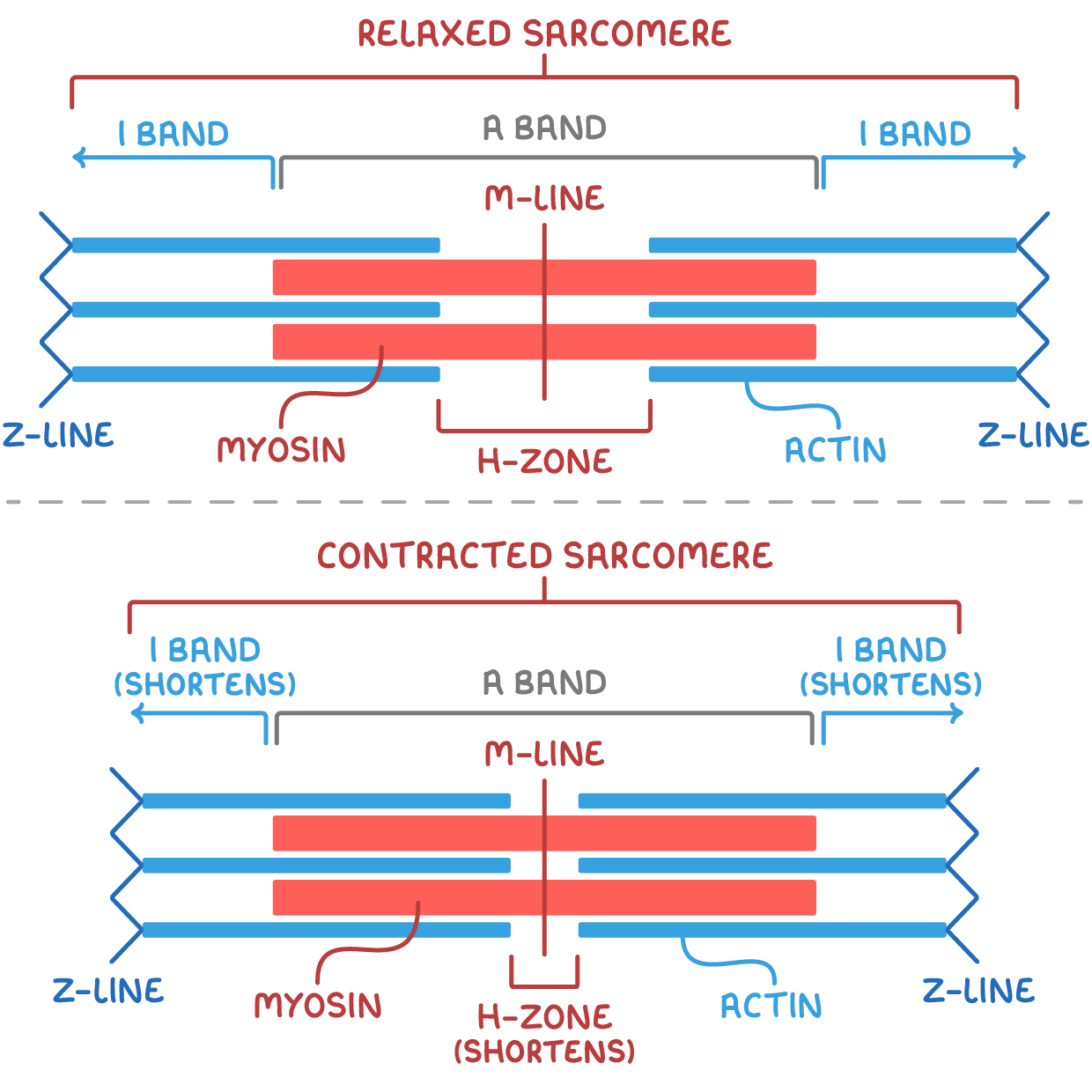

What in the sarcomere does not change during muscle contraction?
The width of the A-band as the myosin filaments have not shortened
3 stages for the sliding filament mechanism of muscle contraction
Muscle stimulation, muscle contraction and muscle relaxation
Where are slow-twitch muscle fibres found?
In calf muscle
Where are fast-twitch muscle fibres found?
Muscles used for fast movement e.g. the biceps
Describe the process of muscle stimulation (4):
1. Action potential reaches many neuromuscular junctions simultaneously, causing calcium ion channels to open
2. Ca2+ ions diffuse into the synaptic knob
3. The Ca2+ ions cause the synaptic vesicles to fuse with the presynaptic membrane, releasing ACH into the synaptic cleft
4. The ACH diffuses across the synaptic cleft, binding to the receptors on the muscle cell-surface membrane so it depolarises
Describe the process of muscle contraction (9):
The action potential causes calcium ion channels on the sarcoplasmic reticulum to open
Ca2+ ions released by the sarcoplasmic reticulum diffuse into the muscle cytoplasm down the diffusion gradient
This causes the tropomyosin blocking the actin binding sites to change shape and pull away, uncovering the binding sites
ATP molecules attach to the myosin heads - the myosin heads then attach to the actin binding sites, forming a cross-bridge - the ATP is hydrolysed to ADP
This releases energy for the myosin heads to bend so they change their angle, pulling the actin filament along as they do so
A new ATP molecule binds to each myosin head so it detaches from the actin filament
The Ca2+ ions activate ATPase, which catalyses the hydrolysis of ATP to ADP
This releases energy for the myosin head to return to its original position
It also causes another ADP molecule to attach to the myosin head and the cycle repeats as long as the concentration of Ca2+ ions remains high
Sarcoplasmic reticulum
The endoplasmic reticulum of the muscle
Why is the movement of actin filaments attached to one set myosin heads in the opposite direction of the actin filaments attached to the other set?
The myosin heads are joined tail to tail in opposite facing sets, so the movement of one set of myosin heads is in the opposite direction of the other set and the actin filaments attached to them also move in opposite directions
What is the effect of the actin filaments moving in opposite directions?
They are pulled towards each other, shortening the distance between two adjacent Z-lines (i.e. the length of the sarcomere) so the muscle shortens, causing movement
Describe the process of muscle relaxation (3):
1. When nervous stimulation stops, Ca2+ ions are actively transported back into the sarcoplasmic reticulum using energy from the hydrolysis of ATP
2. As the Ca2+ ions are being reabsorbed, the tertiary structure of tropomyosin changes and it blocks the actin filament again
3. The myosin heads cannot bind to the actin filaments, so contraction stops - the muscle relaxes
What is energy needed for in muscle contraction? (2)
The movement of the myosin heads + the reabsorption of Ca2+ ions into the sarcoplasmic reticulum by active transport
3 sources of ATP for muscle contraction
1. Aerobic respiration
2. Anaerobic respiration (i.e. glycolysis)
3. Anaerobically using phosphocreatine
How is phosphocreatine used to produce ATP? (2)
1. It's stored in muscle and used as a reserve store of phosphate
2. It is available immediately and so can be used to add phosphate to ADP, forming ATP
How is the phosphocreatine store replenished?
Using phosphate from ATP when the muscle is relaxed
Cardiac muscle
Type of muscle found only in the heart. It is myogenic - it can contract continuously throughout life without stimulation by nerve impulses.
Smooth muscle
Type of muscle found in the alimentary canal and the walls of blood vessels. Its contraction is not under conscious control.
What is the role of tropomyosin in muscle contraction? (2)
Changes shape and moves out of the way when Ca2+ ions bind to it
Allows myosin to bind to actin and form crossbridges
What is the role of myosin in muscle contraction? (3)
Myosin head binds to actin and pulls actin past
It then detaches from actin and moves further along
This uses ATP
What is the role of ATP in muscle contraction? (4)
Binds to myosin, so myosin forms cross bridges with actin
Hydrolysis of ATP results in myosin heads to bend
Causes ‘power stroke’ where myosin pulls on actin
A new ATP molecule attaches to the myosin heads so they detach and move back to their original position
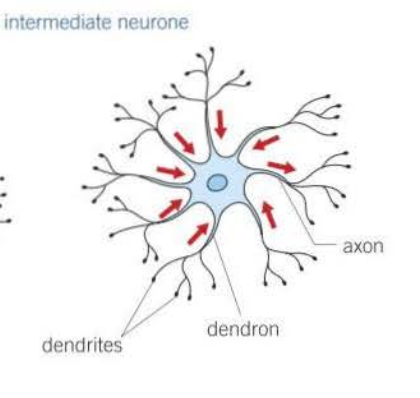
Draw an intermediate neurone
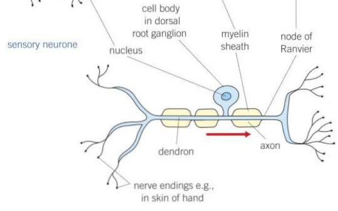
Draw a sensory neurone
What is the role of calcium ions in muscle contraction? (5)
They diffuse into myofibrils from the sarcoplasmic reticulum
Cause tropomyosin to move,
Exposing the binding sites on actin
This allows myosin heads to bind to actin
Also activates ATPase
What would happen if sodium ion channels were kept open? (2)
The neourone would remain depolarised (couldn’t repolarise)
So no action potentials can be transmitted
What happened if the resting potential of a neurone fell to 0 mV?
The electrochemical graduent was not maintained / same concentration of ions either side of membrane
5 factors that would affect the amount of phosphocreatine in muscle tissues
Age
Gender
Muscle mass
Exercise
Ethnicity
Role of ATPase in muscle contraction (3)
It hydrolyses ATP
Because muscle contraction requires ATP
As myosin uses ATP (e.g. to form actinomyosin crossbridge)
What is needed to reform phosphocreatine?
ATP