Gluteal & Posterior Thigh
1/21
There's no tags or description
Looks like no tags are added yet.
Name | Mastery | Learn | Test | Matching | Spaced | Call with Kai |
|---|
No analytics yet
Send a link to your students to track their progress
22 Terms
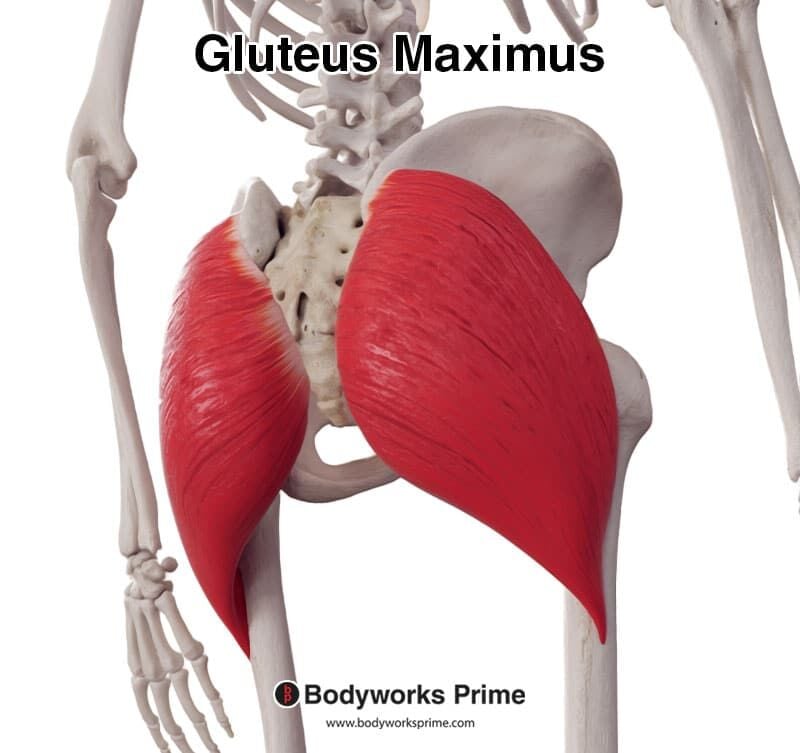
M. gluteus maximus
The gluteus maximus 🍑 is the largest superficial buttock muscle. Extends and laterally rotates the thigh at the hip (N. gluteus inferior). 🔎 On Anatomage: thick red sheet covering posterior pelvis. 💡 "Maximus = Major mover."
M. gluteus medius
Gluteus medius is a fan-shaped muscle partly under G. maximus abducts and medially rotates thigh (N. gluteus superior). 🔎 Slice away the surface to reveal it between iliac crest and greater trochanter. 💡 "Medium = move sideways."
M. gluteus minimus
Deepest gluteal muscle abducts & medially rotates hip. Supplied by N. gluteus superior. 🔎 On Anatomage: smallest red fan directly on ilium. 💡 "Mini = most medial."
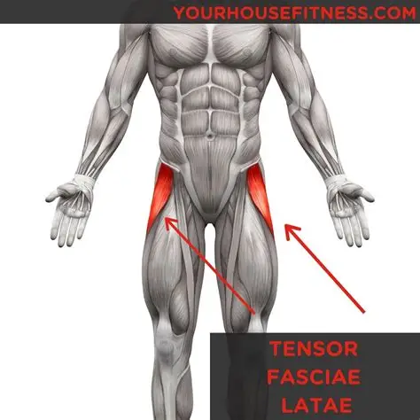
M. tensor fasciae latae
Tightens iliotibial tract and flexes/abducts hip. (N. gluteus superior). 🔎 Lateral thigh, slender band anterior to G. medius. 💡 "Tenses your jeans." 👖
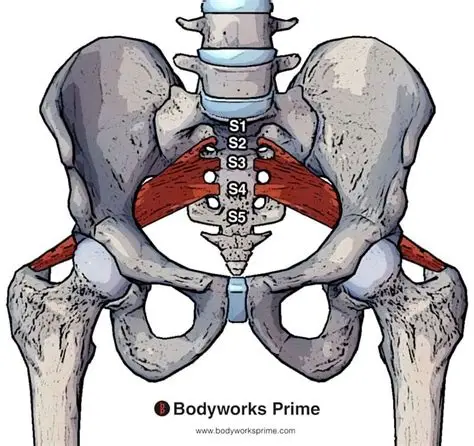
M. piriformis
Pear-shaped muscle passing through greater sciatic foramen laterally rotates & abducts hip. (Branches of S1-S2). 🔎 Locate landmark—above it exits A./N. glutea superior below = inferior bundle. 💡 "Piriform = pear portal."
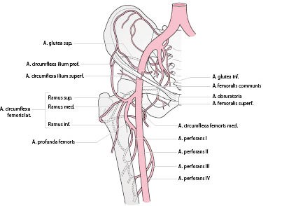
A. glutea superior
Superior gluteal artery supplies G. medius & minimus. 🔎 Seen emerging above piriformis toward iliac crest. 💡 "Superior = super above piriformis."
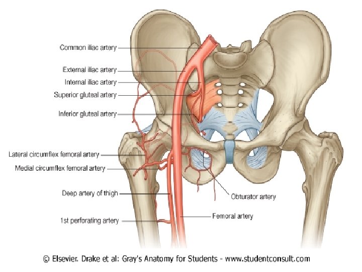
A. glutea inferior
Inferior gluteal artery supplies G. maximus & hamstrings. 🔎 Emerges below piriformis with sciatic nerve. 💡 "Inferior = below piriformis."

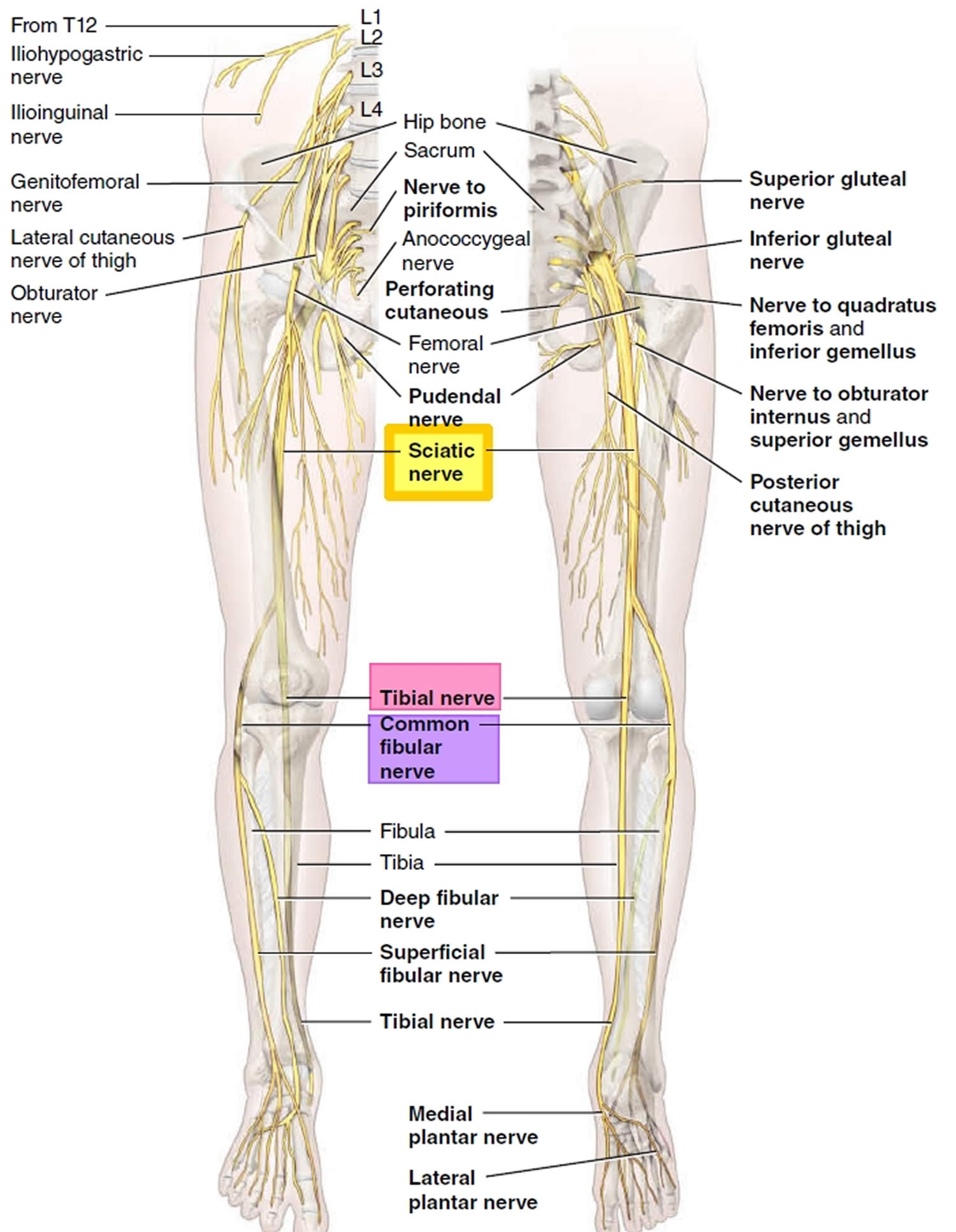
N. ischiadicus (Sciatic nerve)
Largest nerve ⚡ in body exits below piriformis → runs deep to G. maximus → posterior thigh. Splits into tibial & common fibular branches. 🔎 Bright yellow cord mid-thigh. 💡 "Big yellow highway."
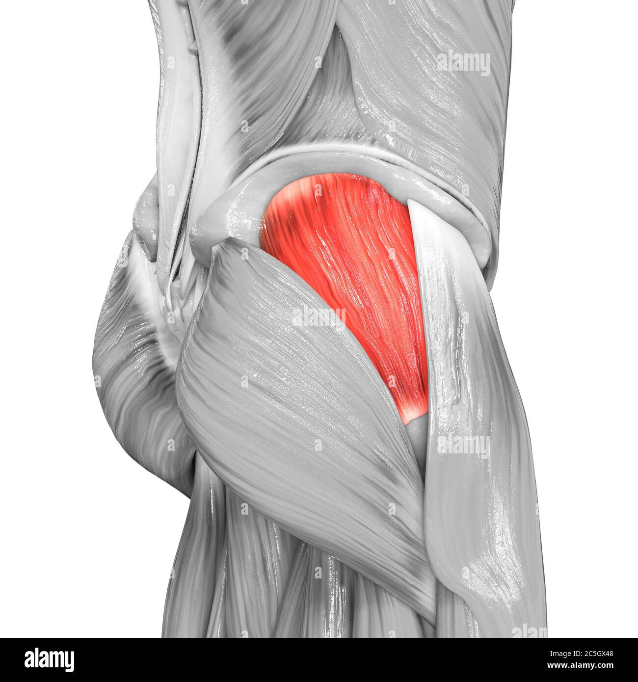
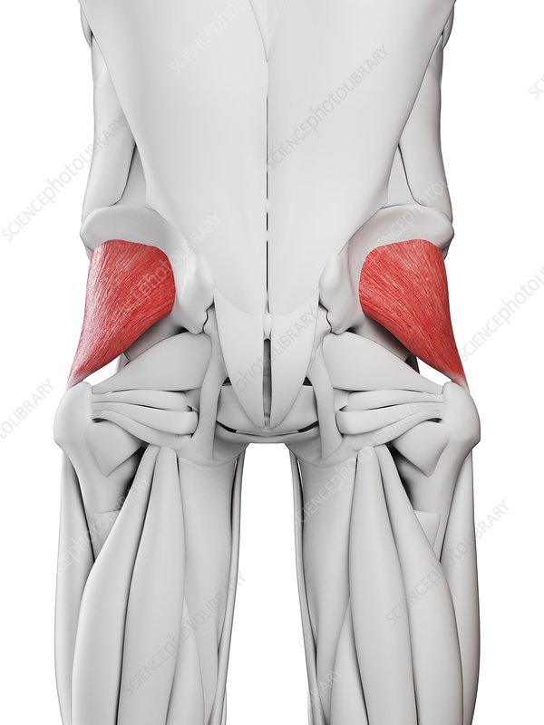
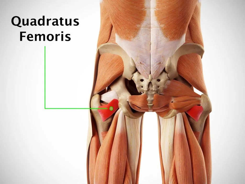
M. quadratus femoris
Flat rectangular muscle between ischial tuberosity & femur laterally rotates hip. (N. to quadratus femoris). 🔎 Below gemelli, short and dark red. 💡 "Quad = square rotator."
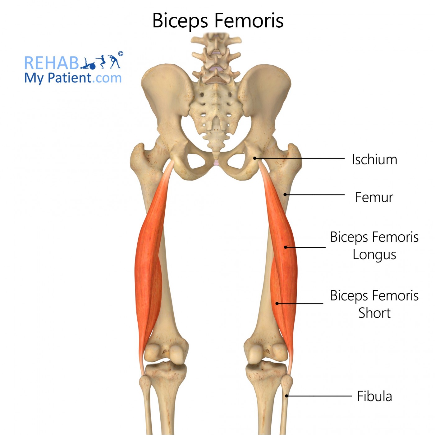
M. biceps femoris (caput longum et breve)
Hamstring muscle with two heads extends hip and flexes knee. (Long = tibial part of sciatic, Short = common fibular). 🔎 Lateral posterior thigh → tendon to fibular head. 💡 "Bi = two heads behind."
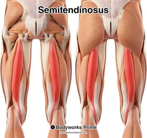
M. semitendinosus
Long cord-like hamstring medial to biceps extends hip & flexes knee. (Tibial nerve). 🔎 Visible as thick strap → pes anserinus. 💡 "Tendon = tail of hamstrings."
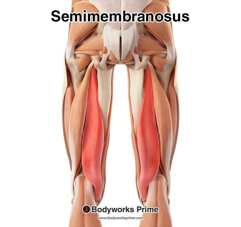
M. semimembranosus
Broad flat hamstring deep to semitendinosus same function. (Tibial nerve). 🔎 Deeper medial sheet on Anatomage. 💡 "Membrane = medial flat."
A. profunda femoris
Deep femoral artery branches posteriorly from A. femoralis → gives perforating branches to hamstrings. 🔎 Red deep vessel between adductors & hamstrings. 💡 "Profunda = deep supply."
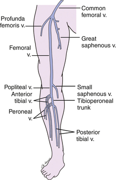
V. profunda femoris
Deep femoral vein 💙 accompanies artery drains posterior thigh → V. femoralis. 🔎 Blue partner vessel. 💡 "Vein very deep."
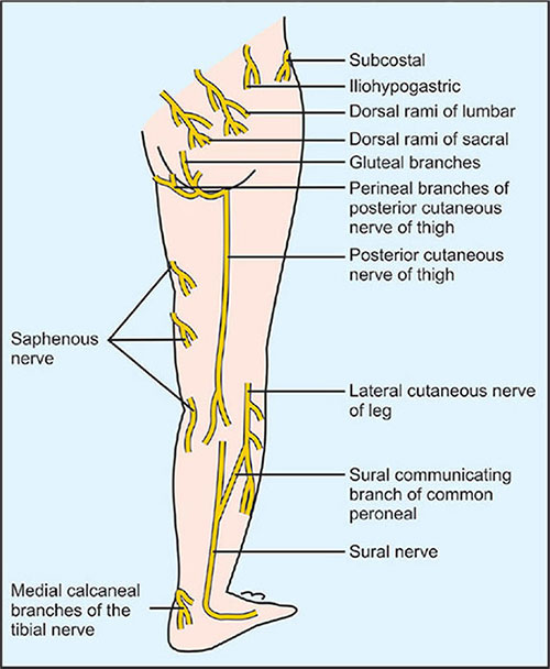
N. cutaneus femoris posterior
Sensory nerve ⚡ to posterior thigh skin emerges below piriformis → beneath fascia lata. 🔎 Thin yellow line superficial to hamstrings. 💡 "Cutaneous = covers skin."
Regio poplitea (Popliteal fossa)
Diamond behind knee bounded by biceps laterally & semimembranosus/semitendinosus medially. Contains A./V. poplitea & tibial/common fibular nerves. 🔎 Find red-blue-yellow bundle posterior to knee. 💡 "NAV reversed deep to superficial."

A. poplitea
Continuation of femoral artery through adductor hiatus divides into A. tibialis anterior & posterior. 🔎 Deepest red vessel in popliteal fossa. 💡 "Popliteal = posterior pivot."

V. poplitea
Popliteal vein 💙 superficial to artery receives small saphenous vein. 🔎 Blue vessel posterior knee. 💡 "Vein on top of artery."

N. tibialis
Large branch of sciatic nerve ⚡ runs through popliteal fossa → posterior leg. Supplies hamstrings & posterior leg muscles. 🔎 Central yellow cord posterior to artery. 💡 "Tibial = to toe plantar."
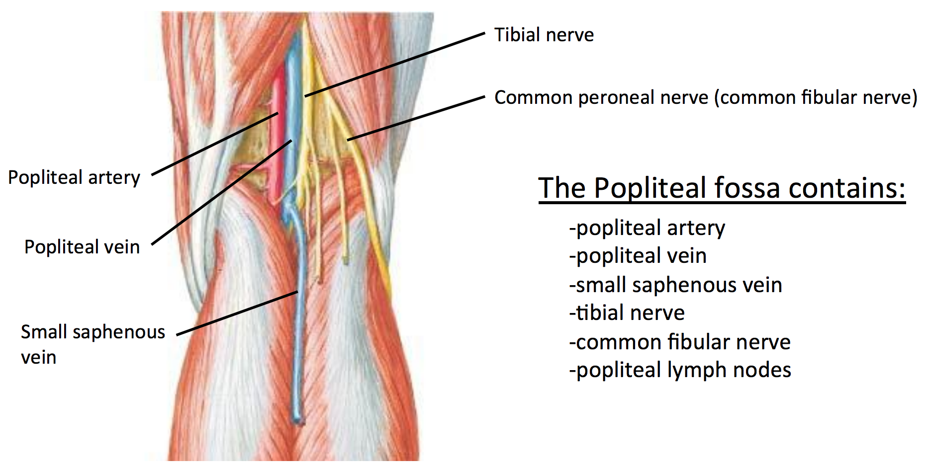
N. fibularis communis
Lateral branch of sciatic ⚡winds around neck of fibula → divides into superficial & deep fibular nerves. 🔎 Yellow cord curving laterally around fibula. 💡 "Common fibular = curve laterally."

V. saphena parva
Small saphenous vein 💙 runs midline posterior leg → drains into V. poplitea. 🔎 Blue surface line up calf. 💡 "Small saphenous = short posterior."

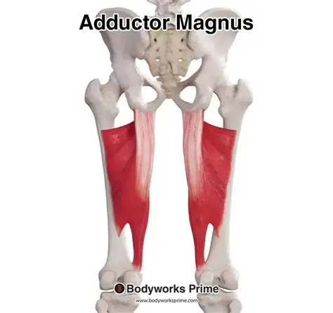
M. adductor magnus (posterior part)
Extends hip and forms adductor hiatus for femoral vessels. (Tibial part of sciatic). 🔎 Deep posterior medial thigh sheet. 💡 "Magnus = massive adductor."