Light Microscopy
1/27
There's no tags or description
Looks like no tags are added yet.
Name | Mastery | Learn | Test | Matching | Spaced | Call with Kai |
|---|
No analytics yet
Send a link to your students to track their progress
28 Terms
Introduction
Review of light microscopy
Light Microscopy review:
The light optical microscope, often referred to as the “light optical microscope,” is a type of microscope that uses visible light and a system of lenses to magnify images of small samples.
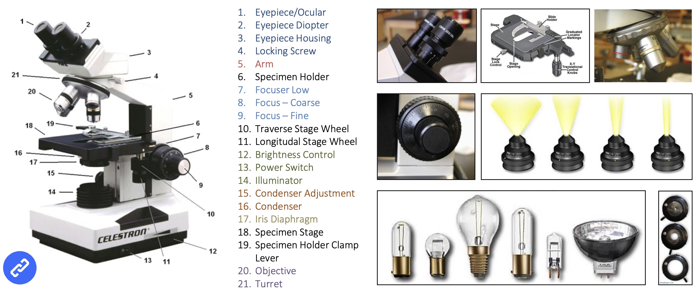
The modern optical microscope
(should be able to name every part!)
white light source is the best (LED), want to make sure that all the colours of the spectrum are of uniform intensity and that will give you your best illumination.
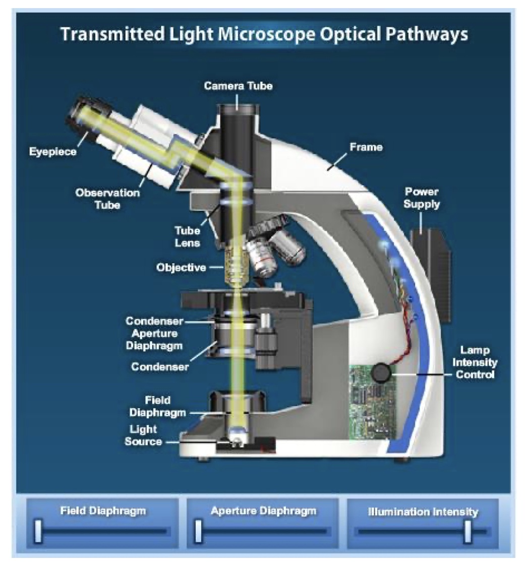
Light Path
The design of an optical microscope must ensure that the light rays are organized and precisely guided through the instrument. The lighting has to be absolutely straight. Mirror placed to bend the light (ergonomics).
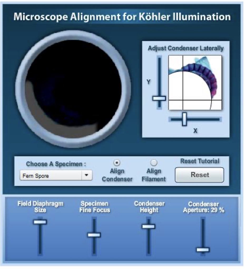
kohler illumination
illumination of the specimen is the most important variable in achieving high-quality images in microscopy and critical photomicrography or digital imaging. The sample has to be illuminated evenly right across.
Conjugate planes
The conjugate planes are critical for establishing proper illumination in the microscope.
four conjugate planes can be brought simultaneously into focus: the field diaphragm, the specimen plane, the intermediate image plane (where the reticule is positioned), and the human eye.
The condenser moves up and down to bring the edge into focus.
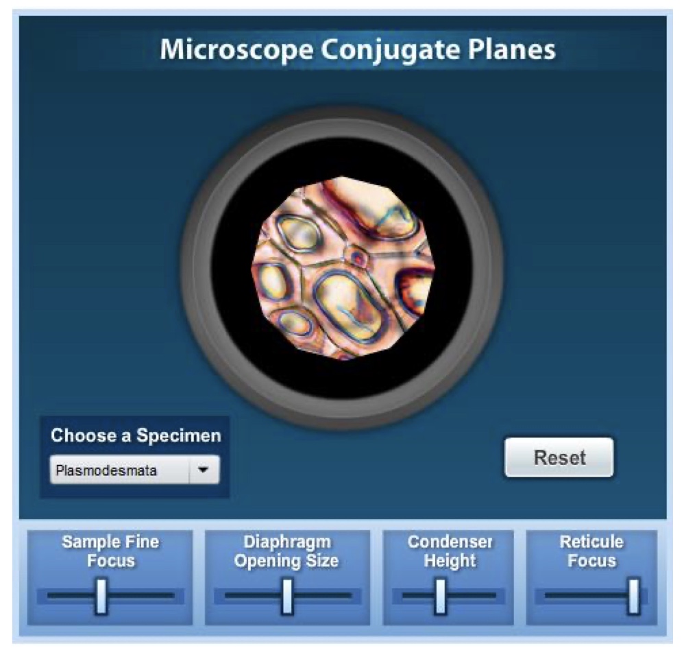
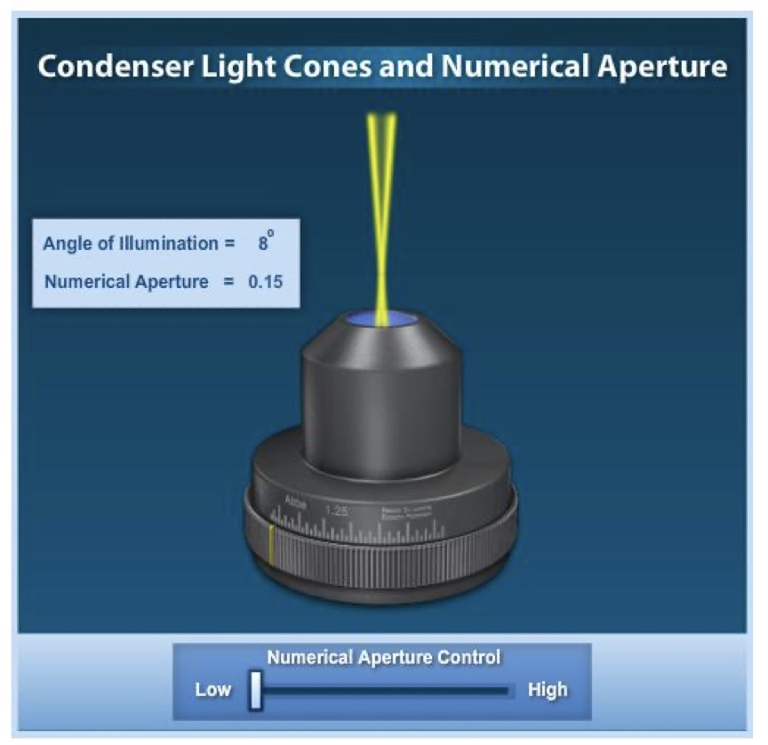
Condenser light
it is critical that the condenser light cone be properly adjusted to optimise the intensity and angle of light entering the objective front lens.
Each time the objective is changed, a corresponding adjustment must be performed on the condenser to provide the proper light cone to match the numerical aperture of the new objective.
open and closing the Irish diaphragm changes the diameter.
if you have a very very thin sample you want to limit the light intensity, if it is thick you need to increase the light intensity to allow it to pass through.
To improve resolution the beam needs to be very narrow, close the iris diaphragm.
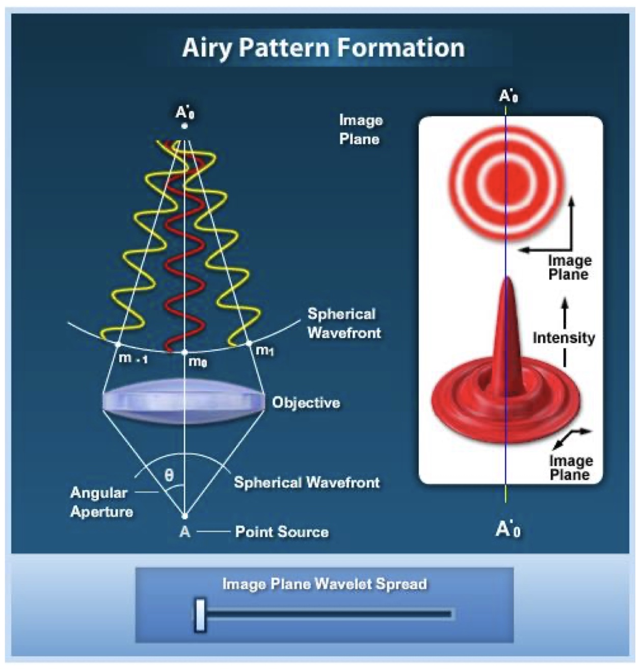
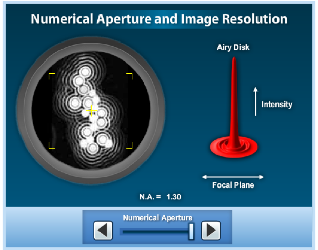
Airy Disk
when an image is formed in the focused image plane of an optical microscope, every point in the specimen is represented by an airy diffraction pattern having a finite spread.
intense central beam, zero order signal is the most intense, 1st order, 2nd order and then 3rd order.
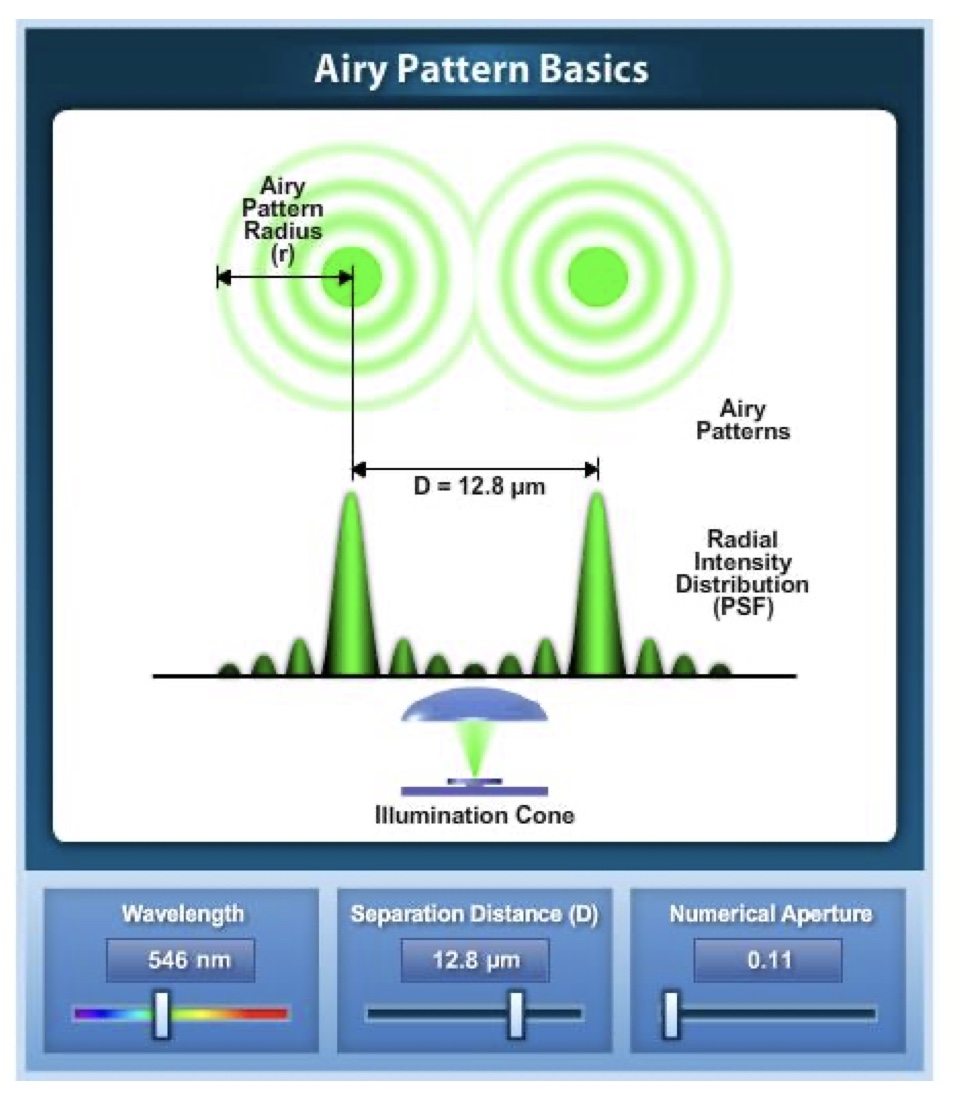
Resolution
The Airy disk pattern size changes with objective numerical aperture and the wavelength of illumination.
The close approach of two airy patterns as they approach the Rayleigh criterion determine the ability to resolve two closely spaced objects in the microscope.
the light order signal depends on the wavelength of light, so the difference between each ring is 1 wavelength of light.
resolution has to be high or you won’t be able to see the sample (e.g DNA is too small, 2 nanometres).
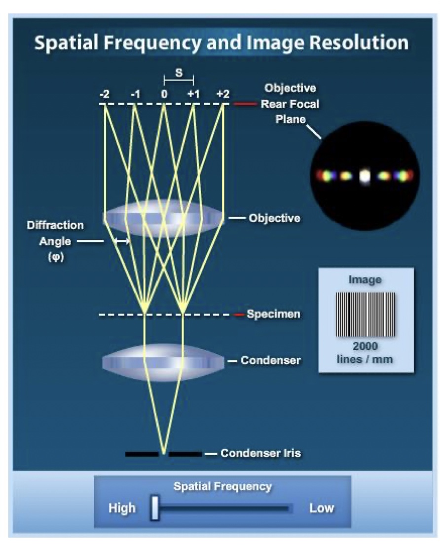
Diffraction Grating
when a line grating is imaged in the microscope, a series of monoscopic images representing the condenser iris opening can be seen at the objective rear focal plane.
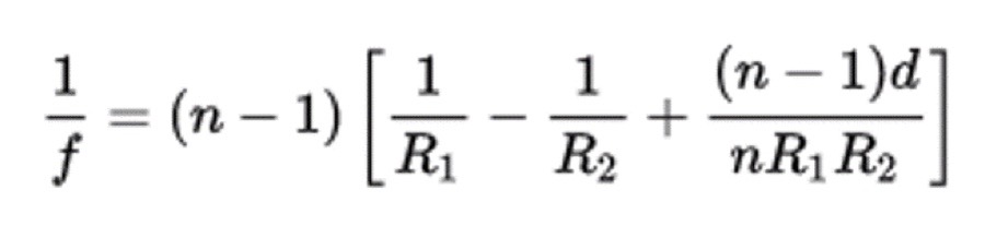
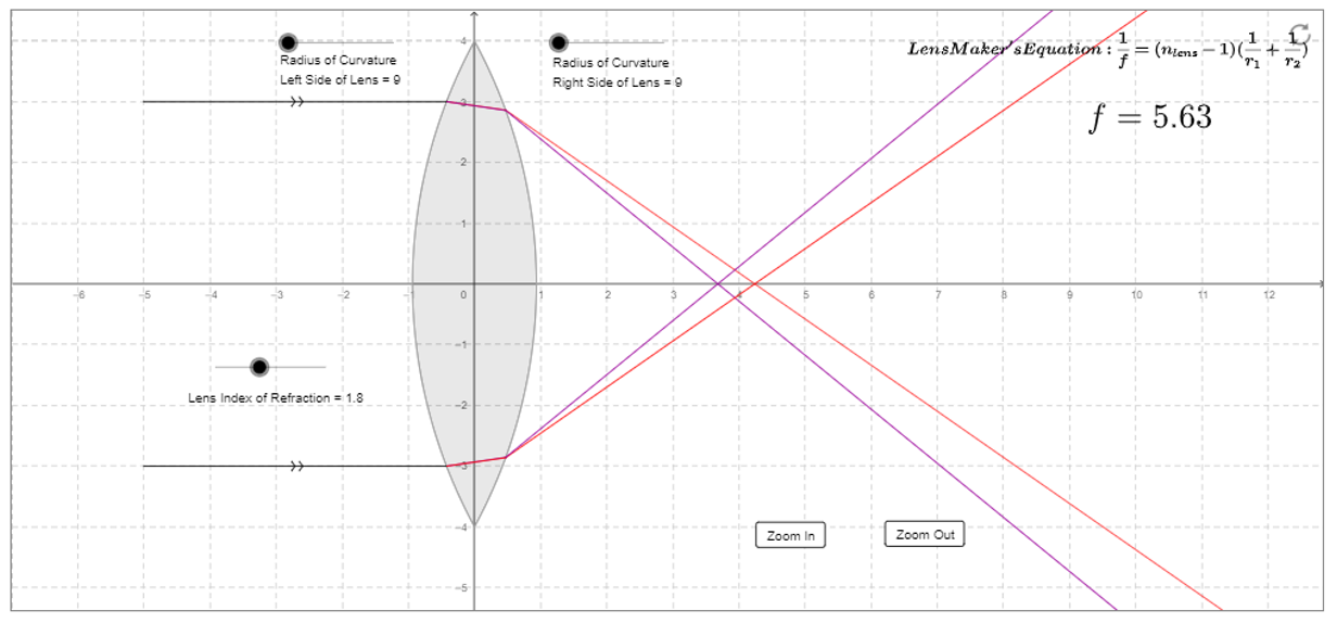
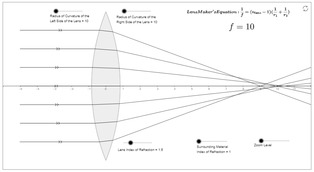
Ray diagrams
lens maker equation (air):
d = thickness of the lens, is a big factor
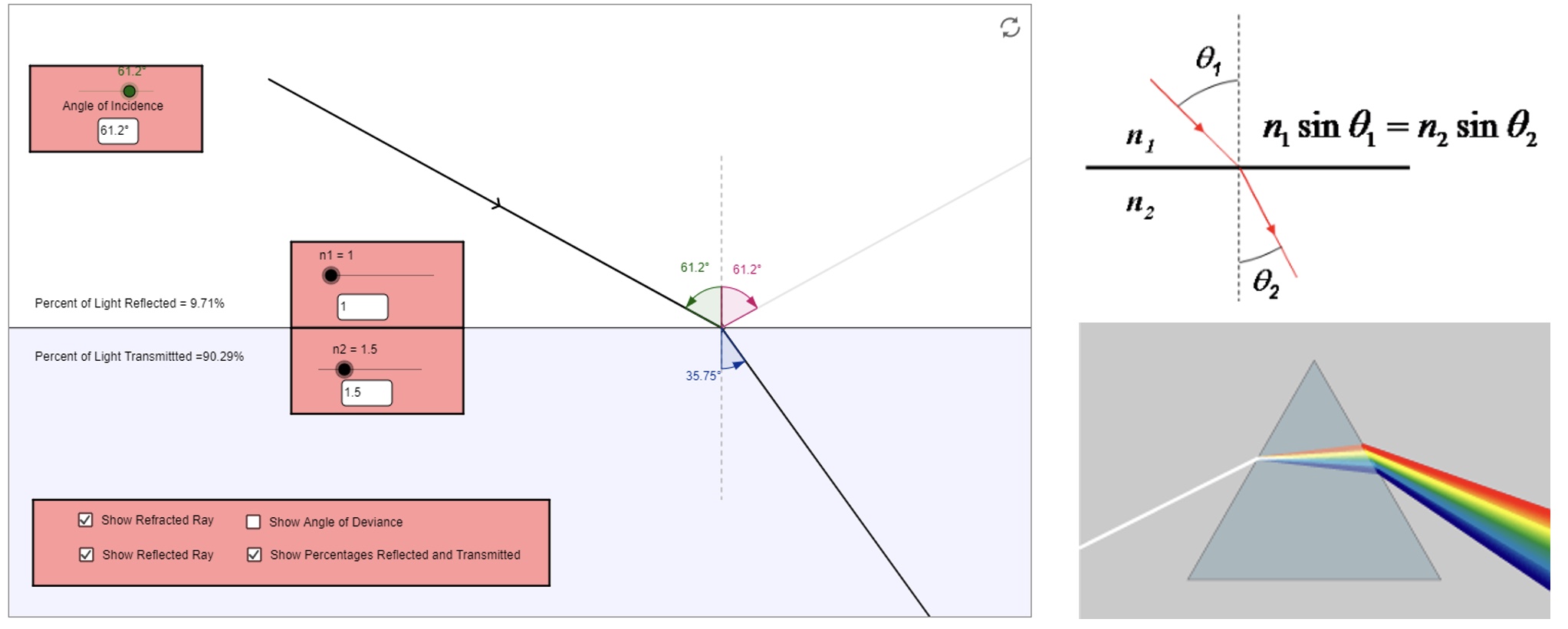
Reflection, Refraction, Snell’s law, dispersion
light is trying to pass through a medium as quickly as possible, the mediums slow it down.
reflected light - tends to be plane polarised.
refracted light - disperses.
snells law tends to work up to 45o. n1 and n2 are refraction.
rainbow = diffraction, red is the least energetic (less bending), blue is most energetic (bends more)
Eye optics
glasses are either concave or convex and they correct the aberrations in your eye!
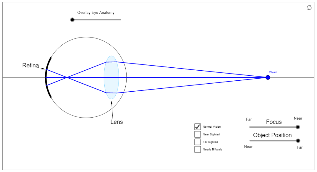
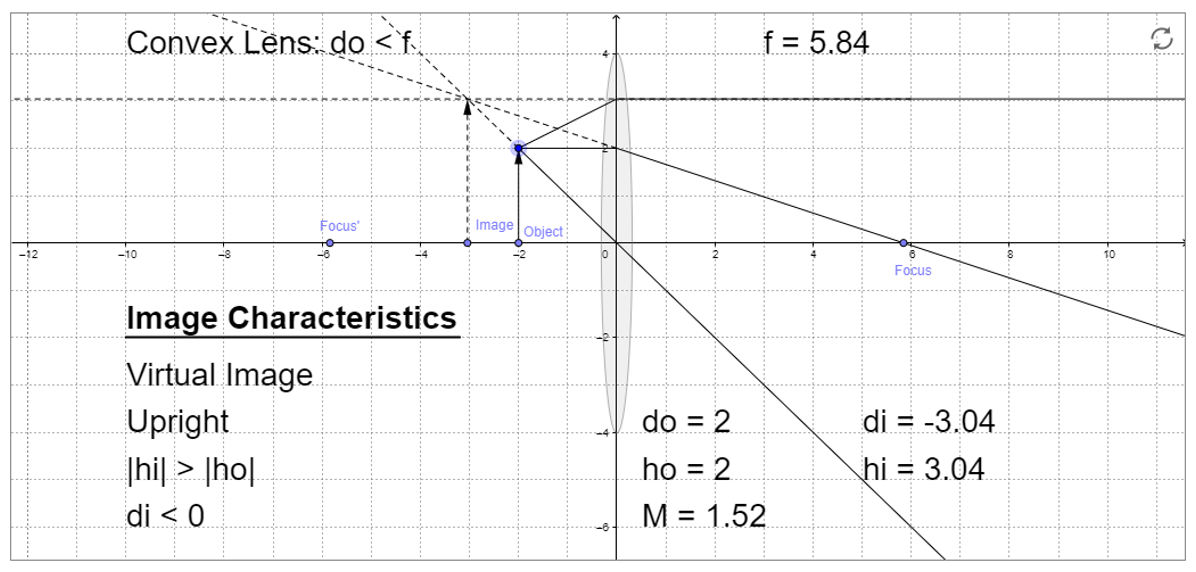
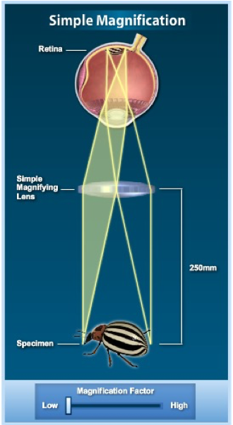
Magnifying Glass
(image appears on the same side of the object = virtual image)
a simple microscope or magnifying glass (lens) produces an image of the specimen upon which the microscope or magnifying glass is focused.
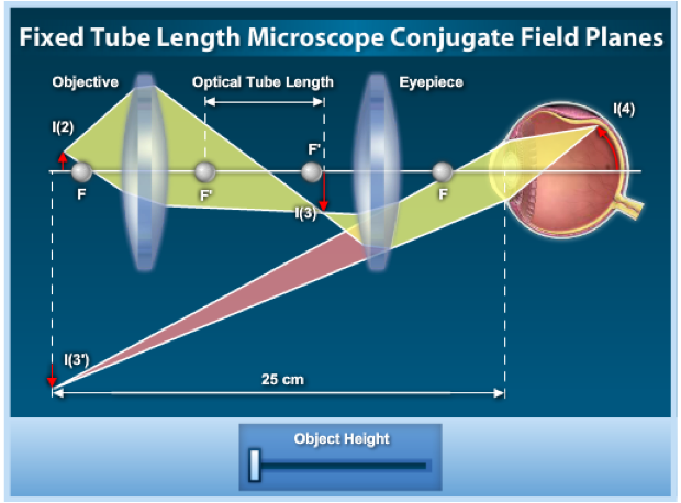
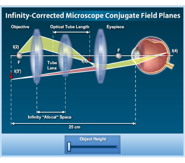
Compound Microscope
the geometrical relationship between image planes in the traditional fixed tube length (usually 160 millimetres) optical microscope is shown.
In most of the imaging steps in the microscope optical train, the image is real and inverted, but a virtual image is also produced on one of the image planes.
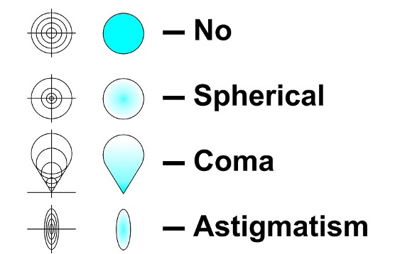
Aberrations
The use of lenses in microscopic systems are subject to aberrations due to the shape of lenses, refraction and tilt. These include:
chromatic
spherical
field curvature
astigmatism
comatic
geometric distortion
focus depth and spherical
Chromatic aberrations
Spherical aberrations
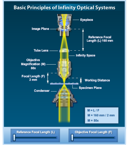
Infinity corrected optics
a majority of modern research microscopes are equipped with infinity-corrected objectives that no longer project the intermediate image directly into the intermediate image plane.
Light emerging from these objectives is instead focused to infinity, and a second lens, known as a tube lens, forms the image at its focal plane.
Infinity corrected optics
(most microscopes)
infinity-corrected microscope optical systems are designed to enable the inception of auxiliary optical devices into the optical pathway between the objective and eyepieces without introducing spherical aberration, requiring focus corrections, or creating other image problems.
Numerical aperture and resolution
the image formed by an objective at the intermediate image plane of a microscope is a diffraction pattern produced by spherical waves exiting the rear aperture and converging on the focal point.
The higher the NA the better the resolution! Image is high, which is why the spots can be seen, if it was lower the airy discs would fuse together.
the best objective you can get are plan apochromat objectives - most expensive.
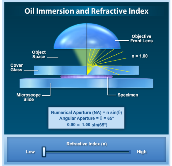
Oil immersion and refractive index
one way of increasing the optical resolving power of the microscope is to use immersion liquids between the front lens of the objective and the cover slip.
The problem with refraction is when light comes out of your sample through the cover slip, it refracts. and when yule got quite a high numerical aperture, high magnification objective, you end up losing some of this signal due to refraction. to avoid this oil (N=1, refractive index)is used to fill the gap between the objective and the cover slip. Air (N=1.5). mounting media (pertex or hydromount N= 1.5) you want it to be as similar to the sample as possible.
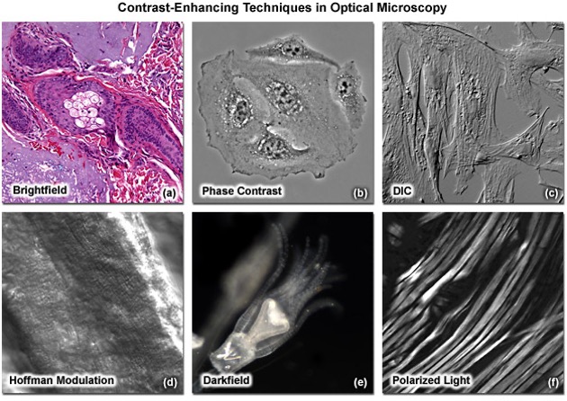
Imaging modes
imaging can be performed in many modes including;
brightfield
darkfield
phase contrast (thin samples)
polarised light
DIC
Hoffman modulation
Rheinberg illumination
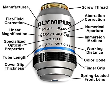
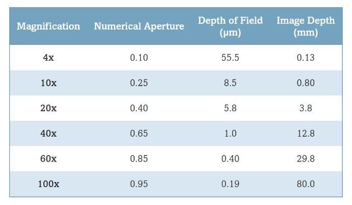
Objective specifications
Microscope objectives are precision optical systems that feature a wide range of magnifications, numerical aperture, immersion media, specialized contrast applications, and other properties.

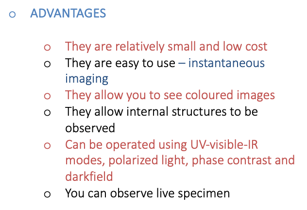
Pros of the compound microscope:
Cons of the compound microscope:

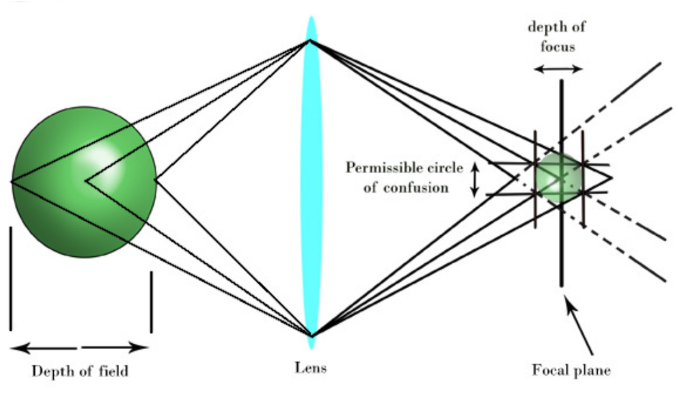
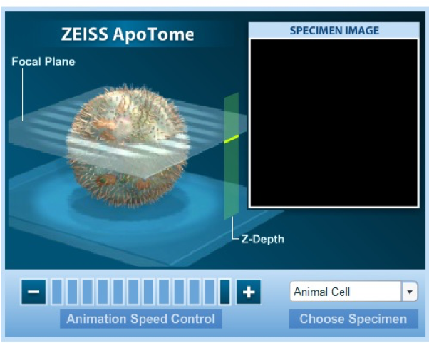
Depth of field/focus
Confocal microscopy
Laser scanning confocal microscopy is capable of producing the highest out-of-focus discrimination of all routine optical sectioning techniques.
Commonly operated in association with a fluorescence microscope.