BIOSCI 107
1/151
Earn XP
Description and Tags
Connective tissues and nerve and muscle tissues
Name | Mastery | Learn | Test | Matching | Spaced | Call with Kai |
|---|
No analytics yet
Send a link to your students to track their progress
152 Terms
What is the purpose of Connective tissue
Binds, supports and strengthens other tissues, major transport system (blood), stored energy reserves (fat or adipose )
How is connective tissue different to epithelia
Not found on body surfaces and has potential to be highly vascular (except cartilage and tendons)
How is connective tissue similar to epithelia?
is supplied by nerves (except cartilage)
What is connective tissue made from?
Cells and extracellular matrix (ECM)
What is Extracellular matrix made from?
Ground substances and protein fibres
What is Ground Substance made from and what is it?
Part of ECM, water, proteins and sugars (Polysaccharides)
How to make Proteoglycans?
Glycosaminoglycans join with proteins
Glycosaminoglycans (GAGS)
Long unbranched polysaccharides
What does hyaluronice acid do (GAGS)
Slippery substance that JOINS, cells together, LUBRICATES joints and MAINTAINS the shape of eyeball
What does Hyaluronidase do
Made by white blood cells, sperm and some bacteria, makes ground substance more liquid
Chondroitin Sulphate
Support and provide features of cartilage, bone, skin, blood vessels.
Keratan Sulphate
Bone, cartilage, cornea
Dermatan sulphate
Skin, tendons, blood vessels, heart valves
Abonormal perioorbital ECM and thyroid disease
Common in younger women, Goitre (Swollen thyroid gland), autoimmune action on fibroblasts and overaction of thyroid, swelling of ECM and increase muscle size and fat.
What increases orbital contents with thyroid disease and abnormal periorbital ECM?
Deposition of GAGS and influx of water
Collagen
Strong and flexible to resist pulling, 25%of body, found in bone, cartilages, tendons and ligaments, parallel bundles
Reticular Fibres
Fine bundles of collagen coated in glycoprotein, Strength and support, forms part of basement membrane, thinner branching to spread through tissue, makes networks in vessels and through tissues ( especially adipose tissue, smooth muscle tissue and nerve fibres)
What are reticular fibres made by?
Fibroblasts
Where does reticular fibres make networks in vessels and through tissue?
Adipose, nerve fibres, smooth muscle tissue.
Elastic Fibres
Fibrous network of thin fibres, can be stretched 150%, skin, blood vessels and lung
Where are elastic fibres found
Skin, Lungs, Blood Vessels
What are elastic fibres made up of?
The protein Elastin surrounded by the glycoprotein fibrillin, giving more strength and stability.
What are the three kinds of connective tissue fibres found in ECM?
Collagen, reticular, elastic
What is Marfan syndrome
A defect in the elastic fibres, resulting form a dominant mutation on chromosome 15, which codes for fibrillin. TGFb increases growth but doe snot bind noramlly to keep it inactive.
What is fibrillin?
Fibrillin is a large glycoprotein which contributes to the structural scaffold for elastin.
What are the two common cell types in connective tissue?
Fibroblasts and adipocytes
Where are fibroblasts located?
Distributed across a wide range of connective tissues (migratory)
What is the function of fibroblasts?
Secretes components of the matrix (fibres and ground substances)
Where are Adipocytes found?
Underneath skin and around organs
What is the function of adipocytes?
Stores fat (triglycerides)
What are the less common cells in connective tissue?
Macrophages, Plasma cells, Mast cells, Leucocytes
Two types of embryonic connective tissue
Mesenchyme and mucous
Two different kinds of connective tissue?
Embryonic and Mature
Mesenchyme CT
Allows other CT to fomr, make up of CT cells in a semi-fluid ground substance containing reticular fibres.
Mucous CT
Has fibroblasts scattered around embedded in jelly like ground substance, supports umbilical cord
What are the three kinds of Loose Connective tissue (MATURE)
Areolar, adipose, Reticular
What is the function of Areolar connective tissue?
Around almost every structure to provide strength, support, flexibility
What are the 3 types of fibres present in areolar connectivw tissue?
Collagen, Reticular, elastic
Where is Adipose CT found
with areolar CT
What does Adipose CT do
Insulation, energy source and temperature control
What do the brown and white adipose in adipose connective tissue do?
Energy storage, heat production
Where is reticular CT found
As a filter for RBC in spleen, and microbes in Lymph nodes
What does reticular CT form?
Reticular cells dominant
What does reticular CT do
acts as a scaffold of organs, binds smooth muscle
What are the 3 kinds of dense connective tissue?
Regular, Irregular, Elastic
What does regular dense connective tissue do?
Forms regularly arranged collagen, slow healing, tension strength along axis of fibres
Where is regular dense CT found
tendons, ligmanets, aponeuroses
Where is Irregular dense CT found?
In sheets called Fasciae
What deos Irregular dense CT do?
Form irregularly arranged collagen around a few cells, tensile strength in many directions.
Where is Elastic dense CT found?
Lung tissue, elastic arteries, bronchial tubes, true vocal cords, some interverbrael ligaments, penis ligament.
What does elastic dense CT do?
forms elastic fibres with fibroblasts between, enables stretching with recoil.
What are the 3 kinds of supporting CT cartilage?
Hyaline, Fibrocartilage, Elastic
Hyaline Cartilage (supporting CT)
Glistening and abundant, helps with felxibility and movement
Where is Fibrocartilage (supporting CT) found?
Pubic symphysis, intervertebral discs, menisci of the knee, and tendons that joing cartilage
Fibrocartilage (supporting CT) form?
Dense network of collagen fibres with scattered chondrocytes
What is the function ofFibrocartilage (supporting CT) ?
Support and joining of structures togetherm strongest type of cartilage
Where is Elastic cartilage (supporting CT) found?
Epiglottis, external ear, audiotory eustachian tubes
What is Elastic cartilages (supporting CT) form?
Chondrocytes in thread like networks of elastic fibers within extracurricular matrix
What is function of Elastic cartilage (supporting CT)?
Strength and elasticity, maintain shapes of structures
Two types of bone tissue
compact and spongey
Compact bone tissue
Contains osteons, stores calcium and phosophorus, protection and support
Spongey bone tissue
Lacks osteons, stores yellow bone marrow, produces red bone marrow
Four types of cells in Bone tissue
Osteogenic, Osteoblasts, Osteocytes, Osteoclasts
What does an Osteogenic cell turn into and how?
Mesenchyl stem cells, that develop lay down collagen, become trapped turn into osteoblasts
Osteoblasts are what kind of cells?
Bone forming
What cell does an osteoblast develop into and how?
Bone forming cells lay down more collagen and the mineralisation process begins, turning into Osteocytes
osteocytes process
Mature bone cells derived from osteoblasts trapped within the extracurricular matrix. Maintain bone tissue, involved in the exchange of nutrients and waste, have gap junctions.
Osteoclasts
large multinucleated cells formed from the fusion of blood monocytes, break down bone
The basic unit of compact bone is what
Osteon
What are the 4 parts of Osteon?
Lamellae, Lacunae, Canaliculi, Central (haversion) canal
Lamellae
Concentric rings of mineral salts which form hydroxapatite and collagen
Lacunae
Small spaces between lamellae that contain mature bone cells (osteocytes)
Canaliculi
“Minute Canals” that radiate fron lacunae and provide routes for oxygen, nutrients and waste
Central (haversion) canal
Blood, Lymph and nerves
What do Osteoclasts do
Reabsorb dead bown and remodel new bone
What do Chondroblasts do
Lay down hyaline cartilage callus
What do Osteoblasts do?
Lay down new bone
Liquid connective tissue
Blood
Blood
consists of plasma (a liquid extracellular matrix) and formed elements (red cells, white cells and platelets)
Ethrocytes (red blood cell)
Transport oxygen and carbon dioxide
The four different Leukocytes
Neutrophils, Basophils, eosinophils, Lymphocytes
Neutrophils and monocytes (Macrophages)
Phagocytic, engulfing bacteria
Basophils (mobile) and Mast cells (immature circulate, matire fixed in tissues)
Release substances that intensify the inflammatory reaction
Eosinophils
Effective against certain parasitic worms and in acute allergic response
Lymphocytes
Are involved in the immune response
Platelets (from Megakaryocytes in red marrow)
responsible for clotting
Muscle tissue
Muscle cells that use energy from the Hydrolysis of ATP to generate force. As a result of contraction, muscle tissue produces body movemtns, maintains posture and generates heat.
3 types of muscle tissue (50% of body tissue mass)
Skeletal, cardiac and smooth
How many named skeletal muscles
~650
Where is Skeletal muscle normally?
attacthed to bones via tendons
Smallest and largest skeletal muscles?
Smallest is stapedius (stabilises smallest bone in ear), 1.25mm
Largest is Sartorius (leg), up to 60cm
Is skeletal muscle tissue multi or single nucleated?
Multinucleated
Functions of skeletal muscle tissue
Posture, heat, protection, motion
Why are skeletal muscle tissues striated?
Due to highly organised arrangment of myofibrils within the cells
What are Myofibrils?
Fill the sarcoplasm of the muscle fibre and extend its entire length within the cell
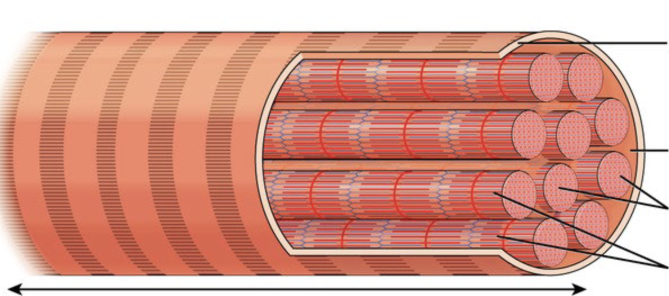
What is this image, and name the tags
Muscle Fiber, Sarcolemma, Sarcoplasm, Myofibrils, Striations.
What makes up myofibrils?
Myofilament
What are the types of myofilament?
Thin (mostly actin), Thick (myosin)
Where are Myofilaments found?
Compartments called sarcomeres
what is a sarcomere?
Basic functional unit of a myofibril, seperated by z discs, contains myofilaments.