Histology of the male/female reproductive system
1/20
There's no tags or description
Looks like no tags are added yet.
Name | Mastery | Learn | Test | Matching | Spaced |
|---|
No study sessions yet.
21 Terms
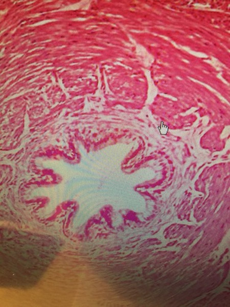
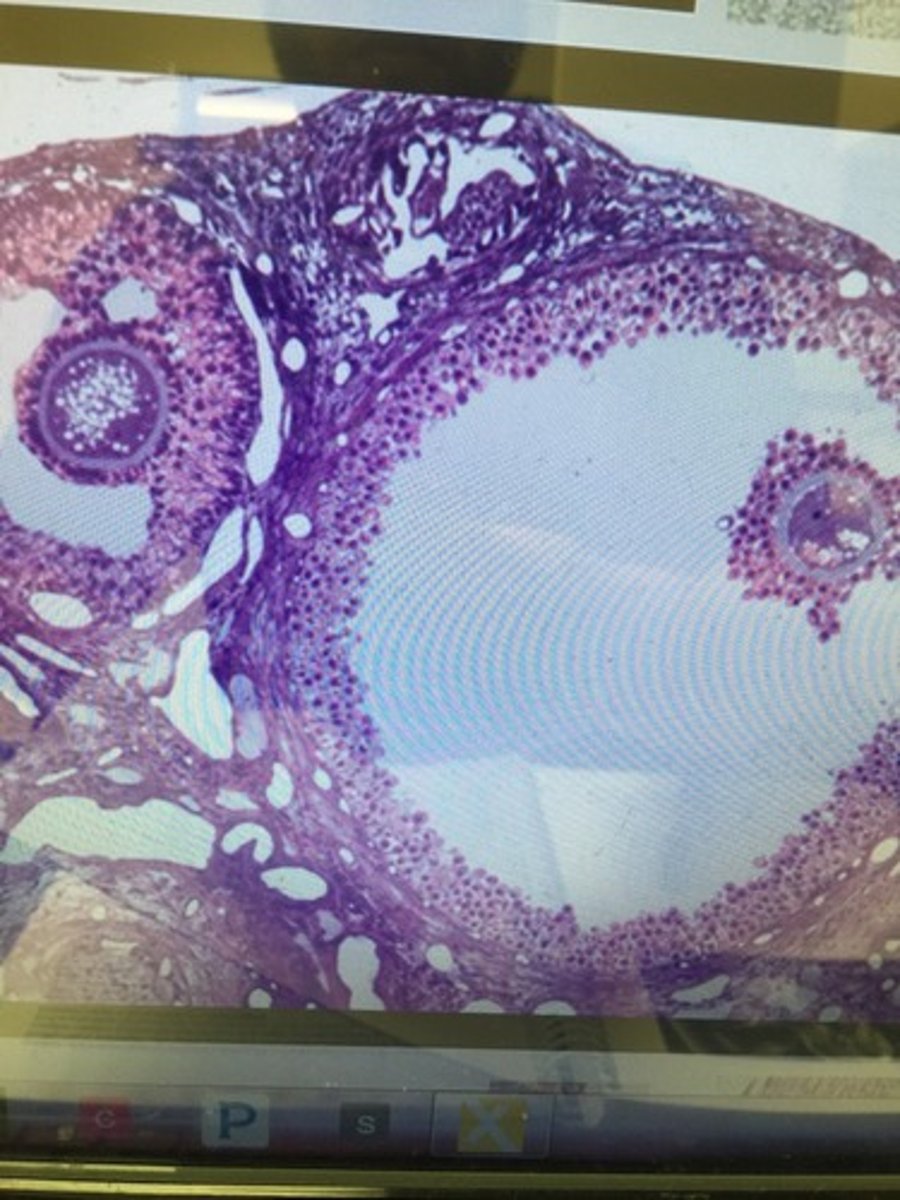
Ovary
What structure in the female reproductive is this slide?
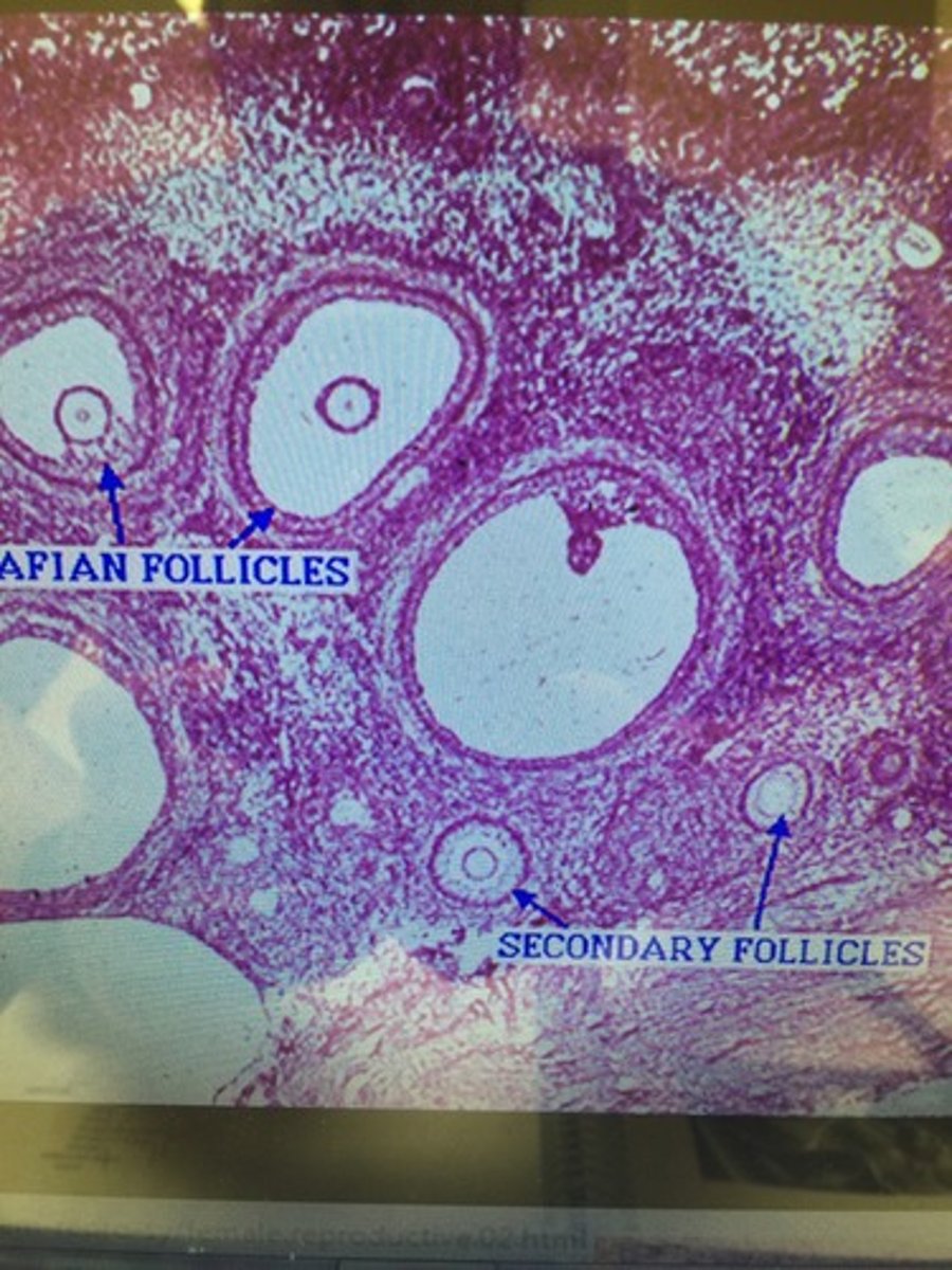
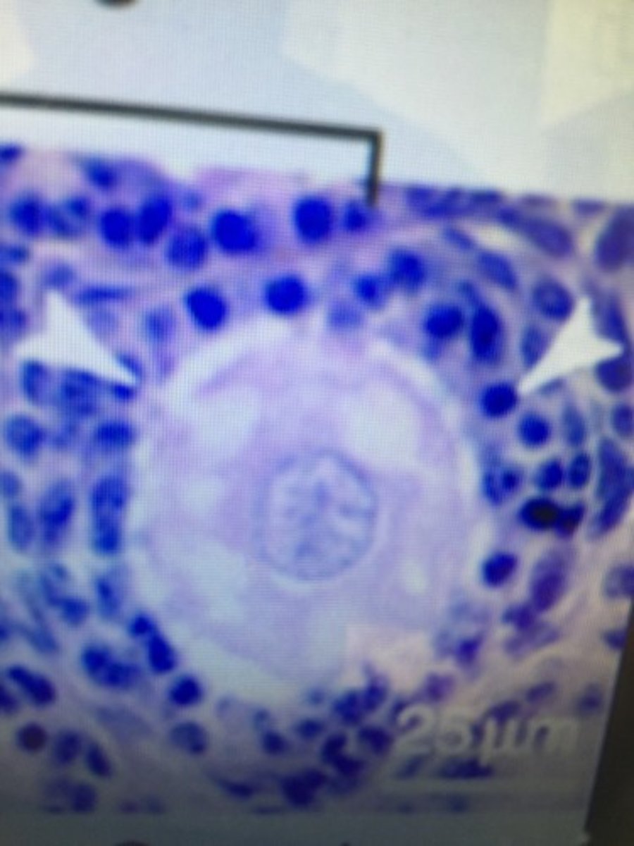
Primary Follicles
As follicular cells mature, they form these that pictured here...?
Secondary Follicle
As primary follicles continue to mature, they form these structures that are pictured here?
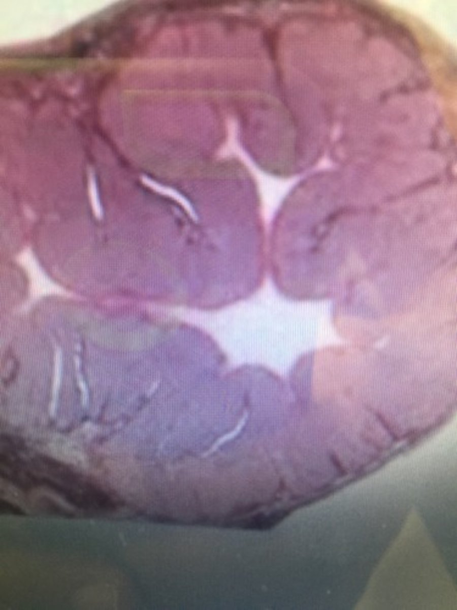
Tertiary Follicle
Every 28 days, a secondary follicle develops into one of these, which surrounds a secondary oocyte..?
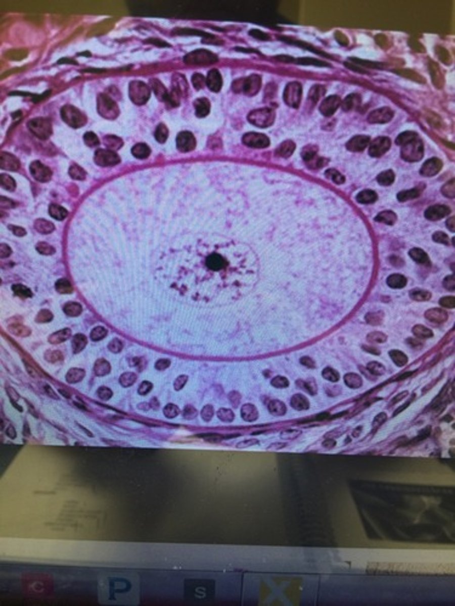
Granulosa cells
What smaller cells that form a layer around the primary follicles are pictured above?
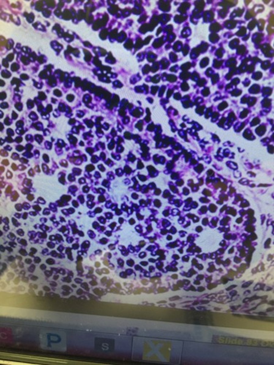
Antrum
Number four pictured above...central cavity that appears in a secondary follicle that is filled with fluid..?
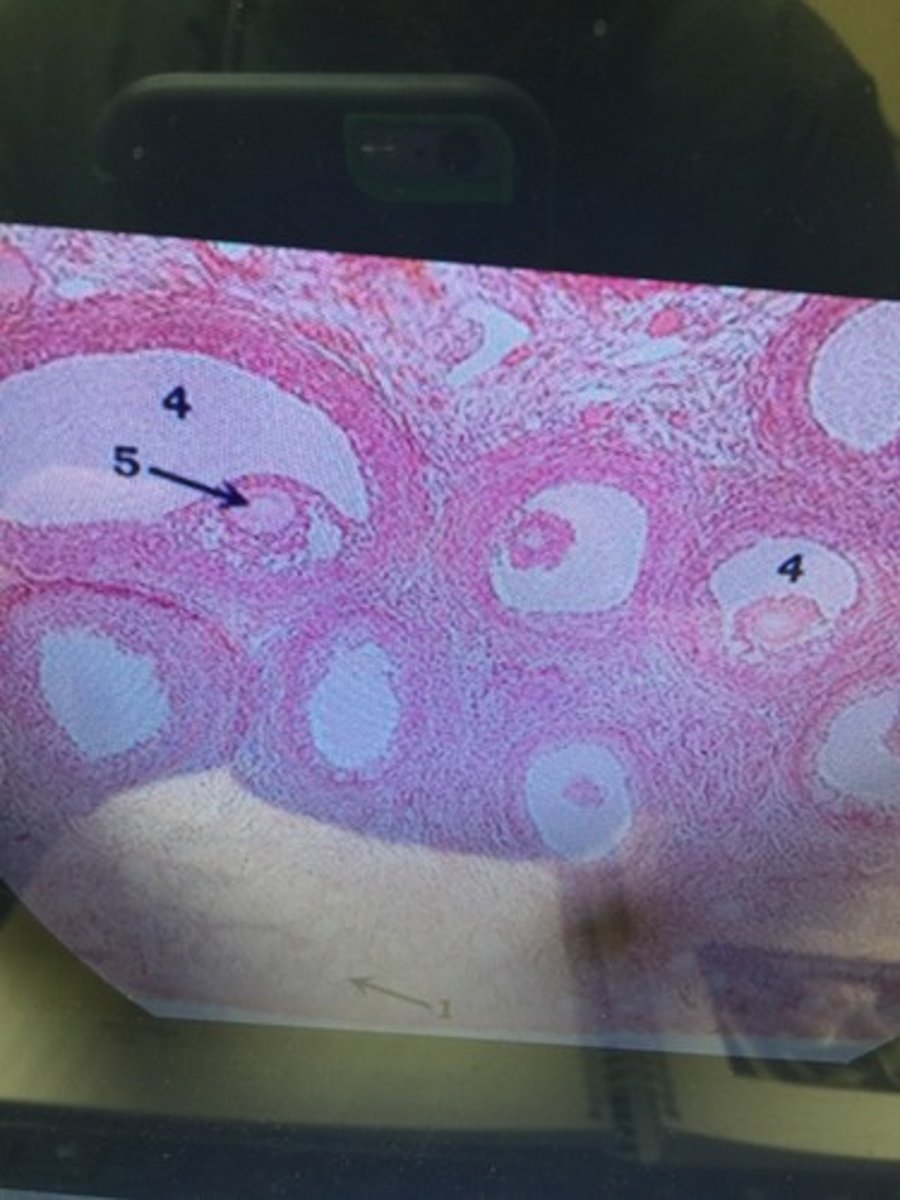
Oocyte
Number 5 pictured above... A cell in an ovary that may undergo meiosis division..?

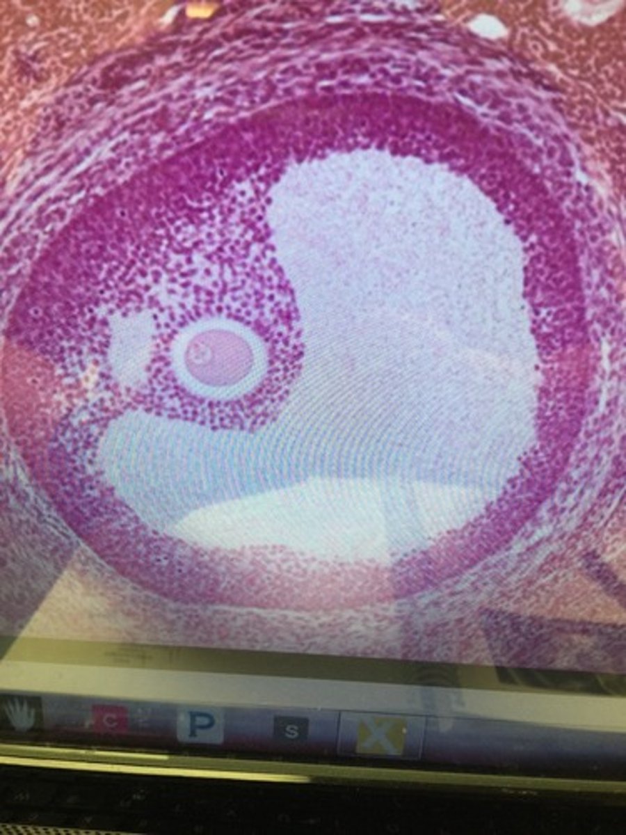
Primary Oocyte
The surviving germ cells following oogonia develop into these , which begin the first meiotic division before birth...?
Secondary Oocyte
The larger of the two haploid cells following meiosis I form this structure pictured above which is the start of Meiosis II
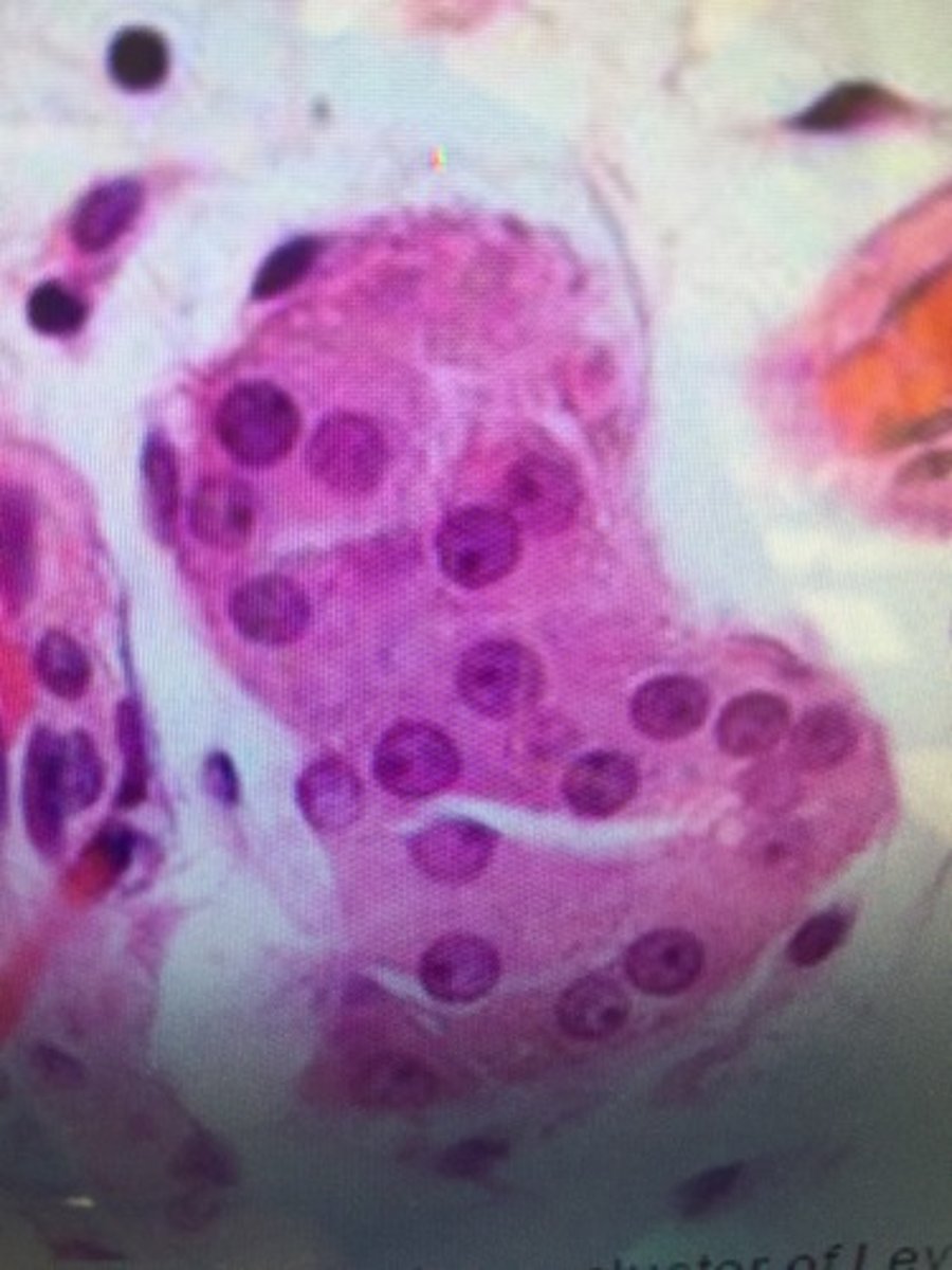
Corpus Luteum
After ovulation, the ruptured Graafian follicle differentiates to form this, which is a source of progesterone and estrogen..?
Endometrium
This layer of the uterus lines the uterine cavity and is a highly vascular and glandular mucosa...?
Myometrium
The thickest layer of the wall of the uterus.
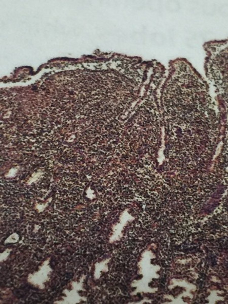
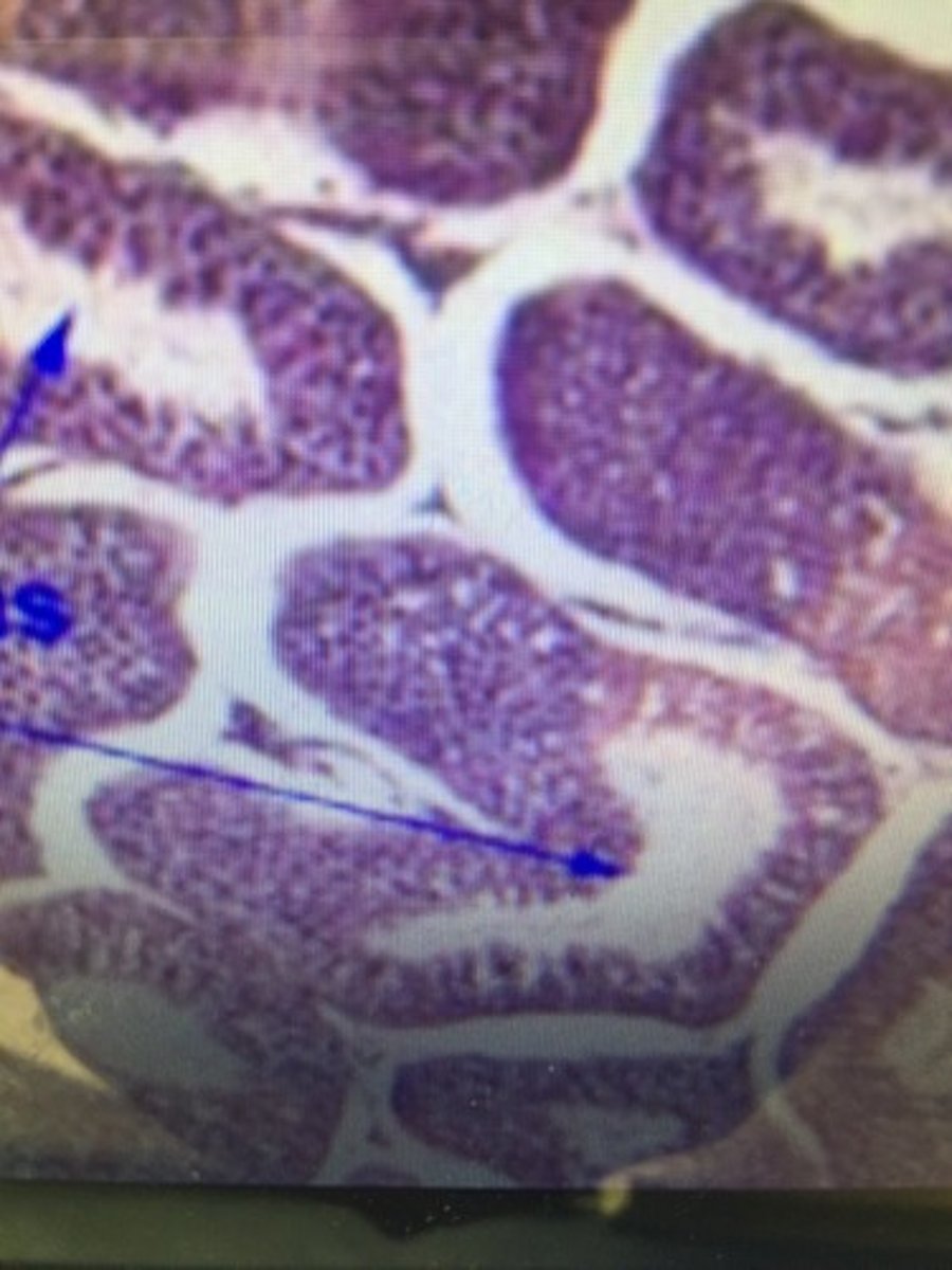
Seminiferous tubule
The site of germination, maturation, and transportation of the sperm cells within the male testes
Spermatogonia
A cell produced at an early stage in the formation of spermatozoa, formed in the wall of the seminiferous tubule.
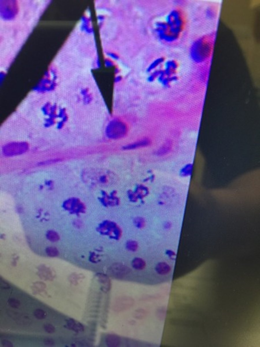
Spermatozoa
The mature motile make sex cells that typically have a compact head and one or more long flagella for swimming.
Lumen
Cavity that is connected to the epididymis and Spermatic duct.
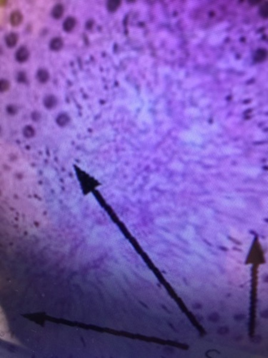
Nurse Cell
Also called a Sertoli cell, this is part of a seminiferous tubule and helps in the process of spermatogenesis.
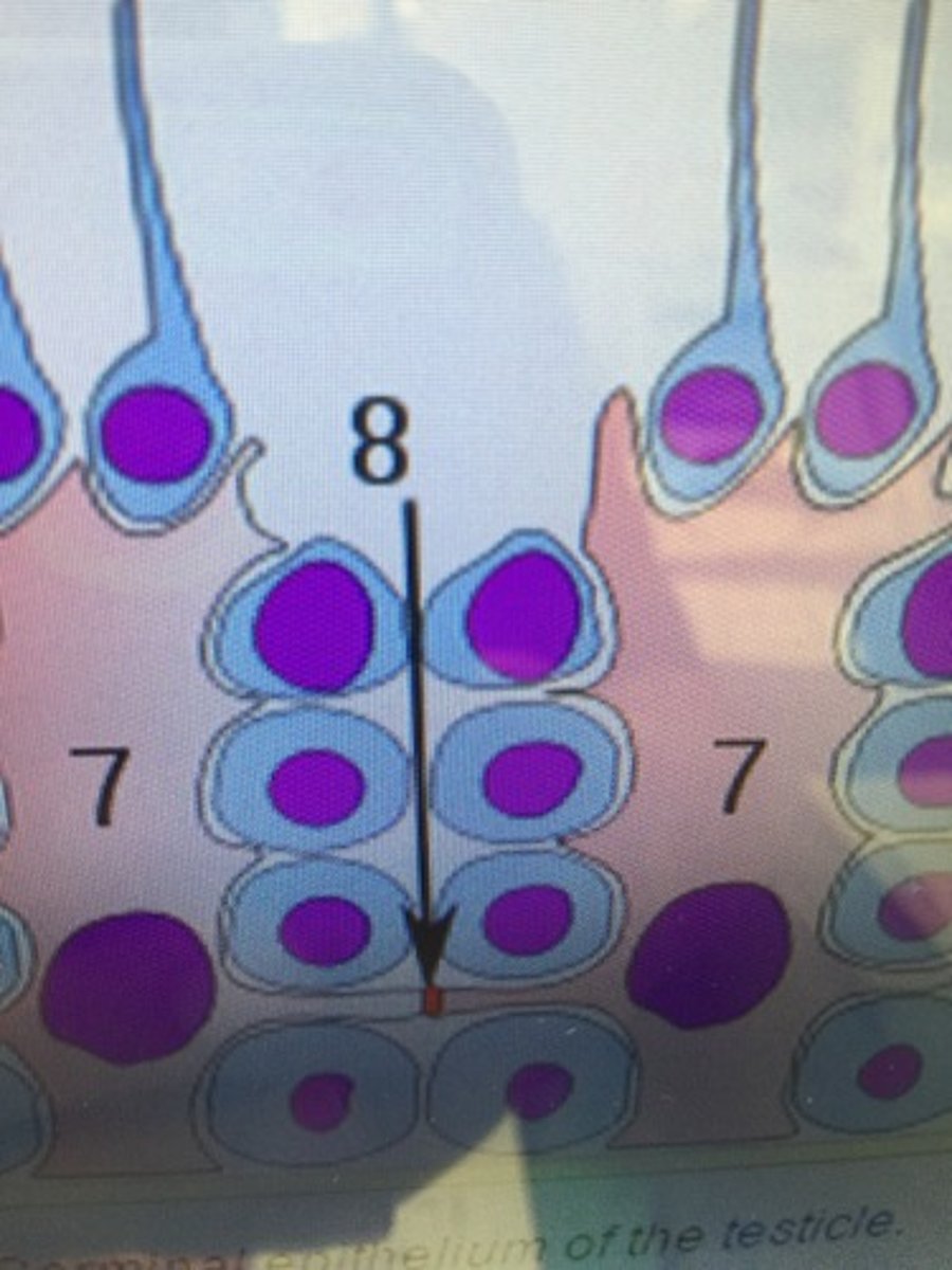
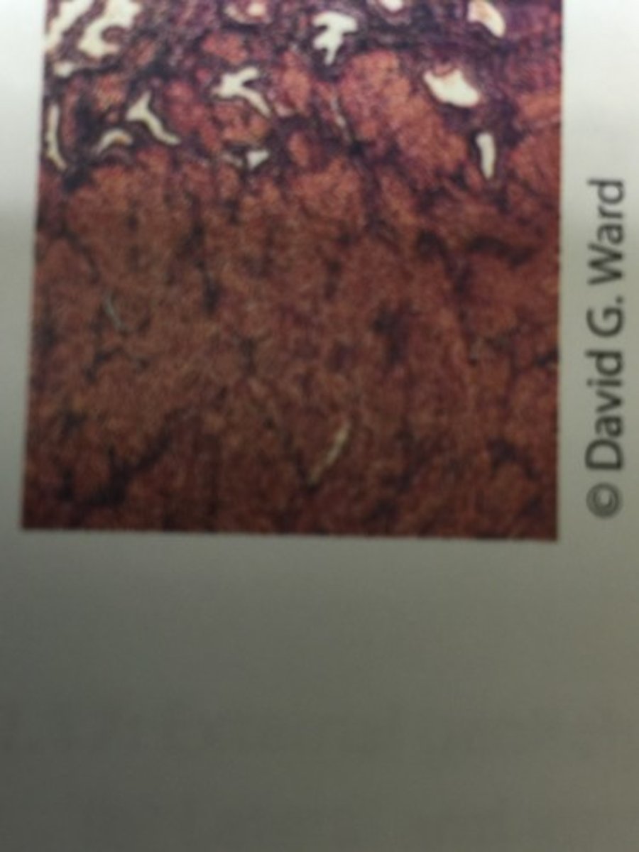
Interstitial cells
These are found adjacent to the seminiferous tubules in the testicle. They produce testosterone in the presence of luteinizing hormone (LH)
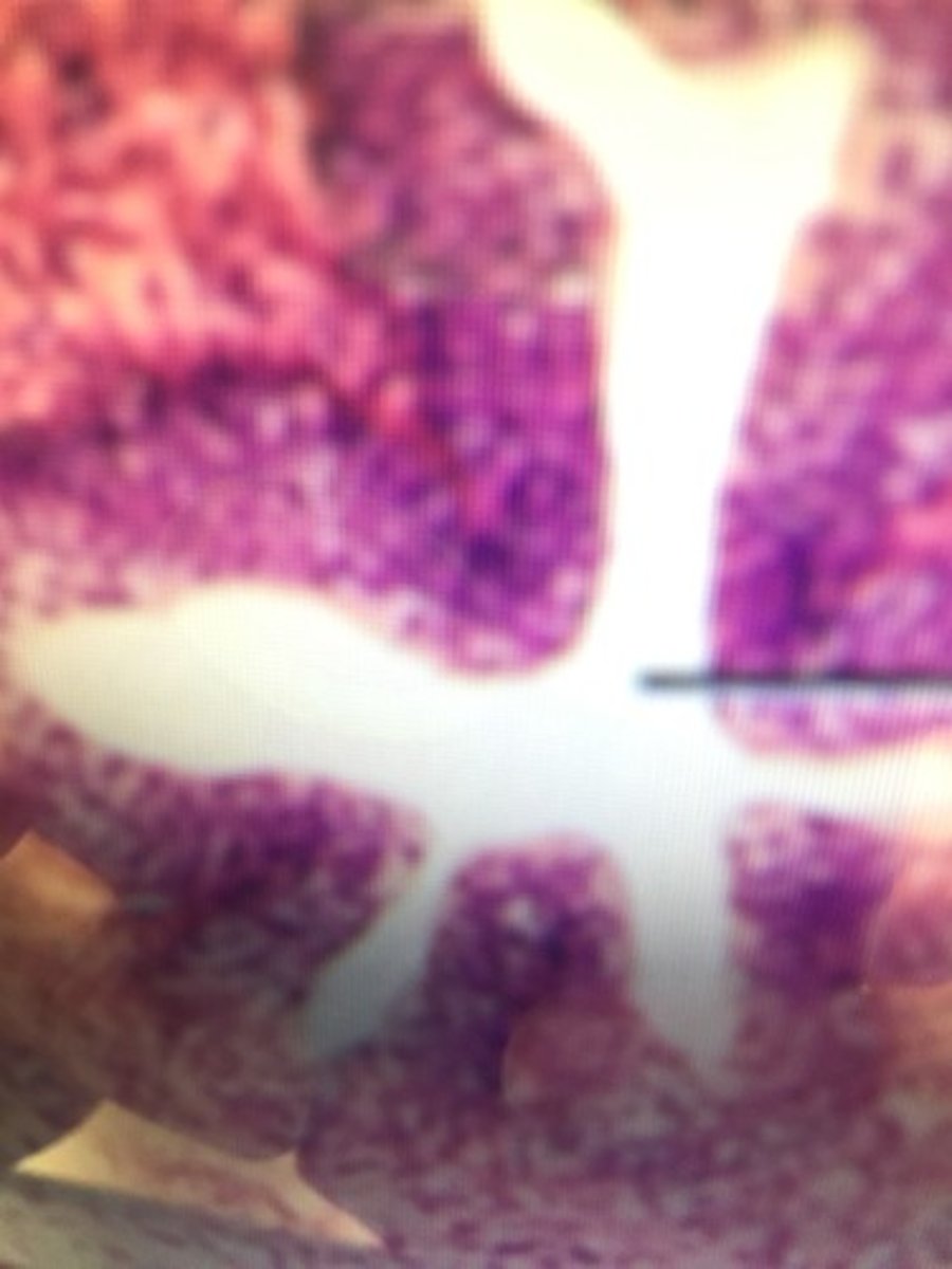
Ductus deferens
What histological structure is this?

Lumen
What is the center structure of this image called?

Pseudostratified ciliated columnar epithelium
What kind of tissue surround the lumen of the ductus deferens?