Retina
1/26
There's no tags or description
Looks like no tags are added yet.
Name | Mastery | Learn | Test | Matching | Spaced |
|---|
No study sessions yet.
27 Terms
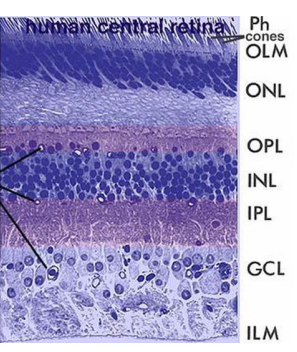
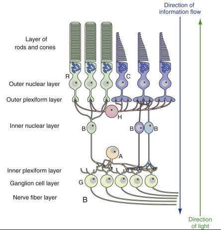
10 Layers of the Retina
Retinal pigment epithelium
single layer of pigmented cells; absorbs stray light and nutrient/waste exchange
Photoreceptor layer
Outer limiting membrane
fusion of photoreceptor cell membranes; basis of electrical charge of retina
Outer nuclear layer
cell bodies of photoreceptors
Outer plexiform layer
synapses between photoreceptors and bipolar/horizontal cells
Inner nuclear layer
cells bodies of bipolar cells, amacrine cells, horizontal cells
Inner plexiform layer
synapses between bipolar/amacrine and ganglion cells
Ganglion cell layer
cell bodies of ganglion cells; send axons out
Nerve fiber layer
axons of ganglion cells that exit the lamina cribrosa and form the optic nerve
Inner limiting membrane
footplate of Muller cells (glial cells), highly reflective in younger patients
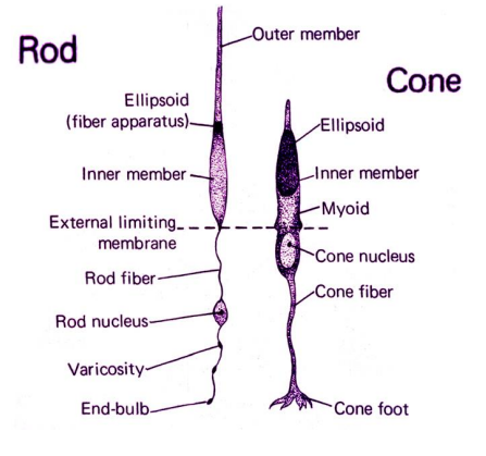
Photoreceptor Micro-Anatomy
Photoreceptors (rods and cones)
Common features across species
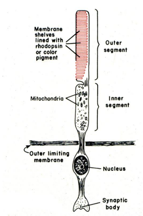
outer segment
Discs found in both rods and cones
discs are circular in shape when viewed from above
on sagittal section the disc has a double membrane with a high concentration of Na+ on the inside of the discs
discs made up of lipoprotein and visual pigment (50%) - 4 different pigments depending on the cone type (3) or rod (1)
inner segment (ellipsoid and myoid)
location of energy production
Ellipsoid: closer to outer segment; has mitochondria for energy production
Myoid: closer to cell body; has organelles like Golgi and ER for protein synthesis
connected to outer segment by cilium
contains a specialized cilia consisting of 9 double microtubules
transport from inner segment to outer segment
extend all the way to the discs and are involved in the regeneration of the outer segment
nucleus
synaptic termination
Differences: outer segments
cone outer segment is smaller than its inner segment
rod outer segment is the same size as its inner segment
Response time to stimulus: rods respond much slower due to larger disc space; cones detect temporal change better
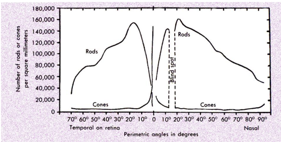
Photoreceptor Distribution varies with retinal location inspected
foveola (central 1.2 degrees) mostly cones and some glial cells
fovea (central 2.5 degrees) has both rods and cones
peak concentration of rods at 20 degrees from the fovea
Glial Cells of the Retina
Astrocytes – Type I, II, and III
star-shaped cells
protect blood vessels, maintain the blood-brain barrier
physically hold the blood vessels in place in the retina
Oligodendrocytes
myelination of the retinal ganglion cells occurs outside the retina after it passes through the lamina cribrosa
Muller cells - ependymal cells
transverse the retina from ILM to ELM
Guidance and physiological support
Finds nutrients, removes waste
Retina is part of CNS
Information Flow -- Vertical Connections CONES
cones ---> ON and OFF bipolar cells ---> ON and OFF ganglion cells ---> LGN

Information Flow -- Vertical Connections RODS (4 paths)
rod→ rod ON bipolar → rod A II Amacrine → cone ON bipolar (gap junction) → ON ganglion cell
rod→ rod bipolar → rod A II amacrine → OFF ganglion cell
rod→ cone (gap junction) → ON and OFF bipolar cell → ON and OFF ganglion cell
rod → OFF cone bipolar → OFF ganglion cell
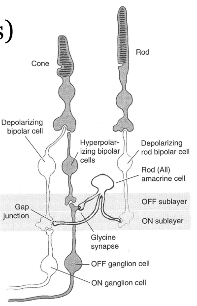
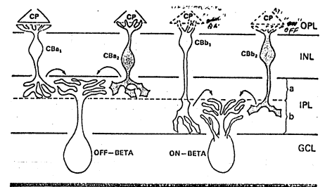
The sublamina of the IPL
Sublamina A - more distal or closer to bipolar cells
Sublamina B - more proximal or closer to ganglion cells
Two functionally different bipolar cells
‘ON’ bipolars - terminate in sublamina B
‘OFF’ bipolars - terminate in sublamina A
Biplexiform cell
a ganglion cell that receives direct input from the photoreceptors (unknown function and projection) - bypasses the bipolar cells
receives mostly rod input
recent studies (animals)
Projection: optic tectum
Function: visuomotor reflexes
Horizontal or Lateral Connections
Horizontal cells
Each horizontal cell contacts all cones within its dendritic field; thus, it is not wavelength selective
functionally produce the receptive field surround of the ganglion cells
Amacrine cells
30-40 different types based on morphology and cytochemistry
cell bodies located in the proximal INL and synapse in the IPL
connected with bipolar cells, ganglion cells and other amacrine cells
can provide direct input to ganglion cells
that is, midget ganglion cells receive as much input from amacrine cells as from bipolar cells
AII
mediates rod vision
contains glycine as its neurotransmitter
Functionally helps to shape the time course of the ganglion cell response and may be involved in the production of the surround of the ganglion cell RF
Interplexiform cells
carries information from amacrine cells back to bipolars, other amacrines, rods or cones
involved in a feedback loop in the retina
Mangel and Dowling (1987) suggested that these cells were involved with the changes that occur in the receptive fields of ganglion cells with dark adaptation
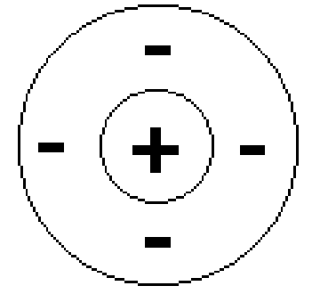
Receptive Fields
Central receptive field (inner circle) - on pathway
on pathway: rod → on bipolar cell → on ganglion cell
Surround receptive field (outer ring) - off pathway
off pathway: rod → horizontal cell → off bipolar cell → off ganglion cell
Area on the retina from which the neural discharge of a neuron can be influenced by light stimulation

Synapses at the outer plexiform layer
Cone pedicle
many invaginations in which bipolar and horizontal cells synapse (12 - 25 / pedicle)
commonly see a ribbon synapse, which has 2 horizontal cells and 1 bipolar cell synapsing
will also be noninvaginating synapses - typically with a flat bipolar (off synapses)
Rod spherule
typically more than 3 processes synapse (more than one central process)
common information flow is from rods to rod bipolar
no junction between the laterally placed horizontal cells (like that of cone pedicles)
one invagination per spherule
Gap Junction
Each adherent junction has 3 gap junctions
links the membrane of photoreceptor cells
Open channels between 2 cell membranes allow for communication, ion transport, electrical coupling
Synapses at the inner plexiform layer
Conventional dyad: most common in primates: 1 bipolar synapses with 1 ganglion and 1 amacrine
Amacrine-amacrine: second most common: 1 bipolar synapses with 2 amacrines
Reciprocal: information can flow both ways between a bipolar and an amacrine (feedback)
Serial: bipolar to amacrine to amacrine to ganglion cell: allows for informational integration
Types of Ganglion Cells
Cat ganglion cells
types seen with light microscopy and Nissl stain
Alpha cells (cell body > 21 microns) LARGEST
Beta cells (cell body 12 - 20 microns)
Gamma cells (cell body < 11 microns) SMALLEST
Diameter of dendritic spread
Alpha cells, 425 - 785 microns LARGEST
Beta cells, 90 - 370 microns SMALLEST
Gamma cells, 440 - 500 microns
Axon diameter
Alpha cells, 2.5 - 3.5 microns LARGEST
Beta cells, 0.8 - 1.5 microns
Gamma cells, 0.25 - 0.32 microns SMALLEST
Primate Ganglion Cells
P1: larger cells, larger axon diameter, larger dendritic spread
P2: smaller cells, smaller axon diameter, smaller dendritic spread
Bipolar Cell Categorization
Invaginating Midget Bipolar
Single cone at ribbon
Flat Midget Bipolar
Single cone at conventional synapse
Diffuse Flat Bipolar
Several cones conventional synapses .
Diffuse Invaginating Bipolar
Several cones ribbon synapse
Rod Bipolar
Rods only
Giant Bistratified Bipolar
Contacts many cones
S-cone Bipolar
Contacts only S-cones
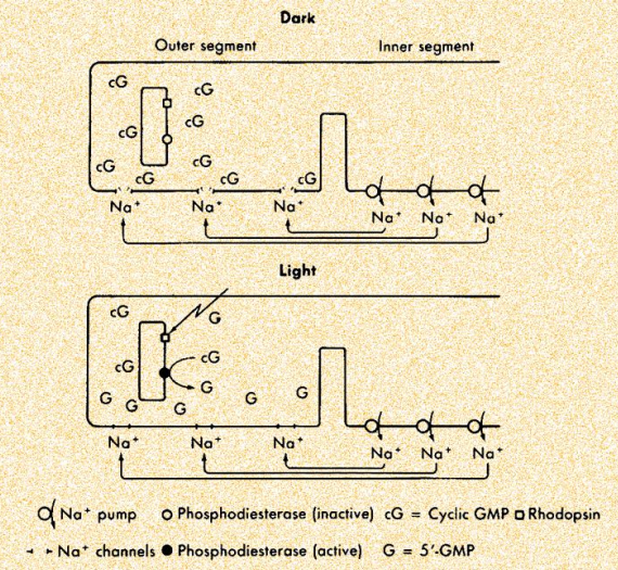
Photoreceptor Physiology: Transduction
photoreceptors convert light into a neuroelectrical response (act as a transducer)
unlike typical neural cells photoreceptors do not produce action potentials (only produce membrane potential changes)
unlike most CNS cells the photoreceptors do not depolarize when excited, they hyperpolarize
Dark current
in the dark the Na+ channels are open and Na+ flows into the outer segment and is pumped out the inner segment - a continuous cycle
light results in a closure of the Na+ channels so Na+ doesn't flow in but it is still pumped out - inside becomes more negative (hyperpolarizes)
in the dark the photoreceptors release neurotransmitter continually - glutamate
Effects of light
in the dark the inside of the photoreceptor is about 20 millivolts more negative than the outside
when light hits the photoreceptor there is a series of chemical reactions that results in the inside of the cell becoming more negative (-60 millivolts)
Chemical reactions
rhodopsin --> opsin + all-trans retinal
with light (photoreaction) - rhodopsin → bathorhodopsin
concentration of free cGMP decreases; cGMP needed to bind at Na+ channels to keep them open
decreasing the cGMP concentration closes some of the Na+ channels
results in the inside of the cell becoming more negative

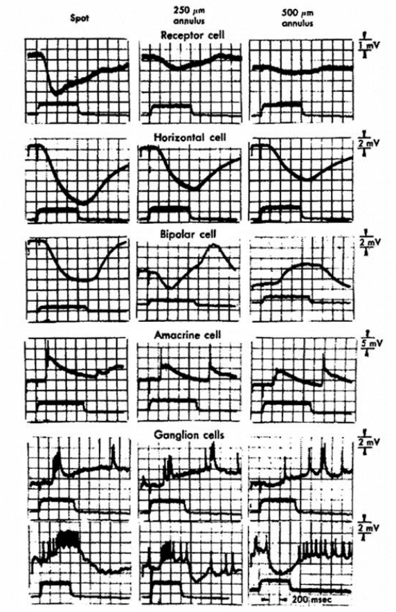
Effects of light on cells
Photoreceptor cell: hyperpolarization
Horizontal cell: hyperpolarization
Bipolar cell (off): hyperpolarization
Bipolar cell (on): depolarization
Amacrine cell (on): produces action potentials
Ganglion cell (on): produces action potentials