lesson 4 thorax
1/125
Earn XP
Description and Tags
human anatomy and embryology
Name | Mastery | Learn | Test | Matching | Spaced | Call with Kai |
|---|
No analytics yet
Send a link to your students to track their progress
126 Terms
what does the thorax consist of (4)
a wall
2 pleural cavities
the lungs
the mediastinum
functions of the thorax (4)
houses and protects the heart lungs and greater vessels
allows structures to pass between neck and abdomen
critical role in breathing
support to the upper limbs
what skeletal elements and muscles does the thorax contain POSTERIORLY
12 thoracic vertebra and their intervertebral discs
what skeletal elements and muscles does the thorax contain LATERALY
ribs (12 on each side) + muscles
what skeletal elements and muscles does the thorax contain ANTERIORLY
sternum (manubrium, body and xiphoid process)
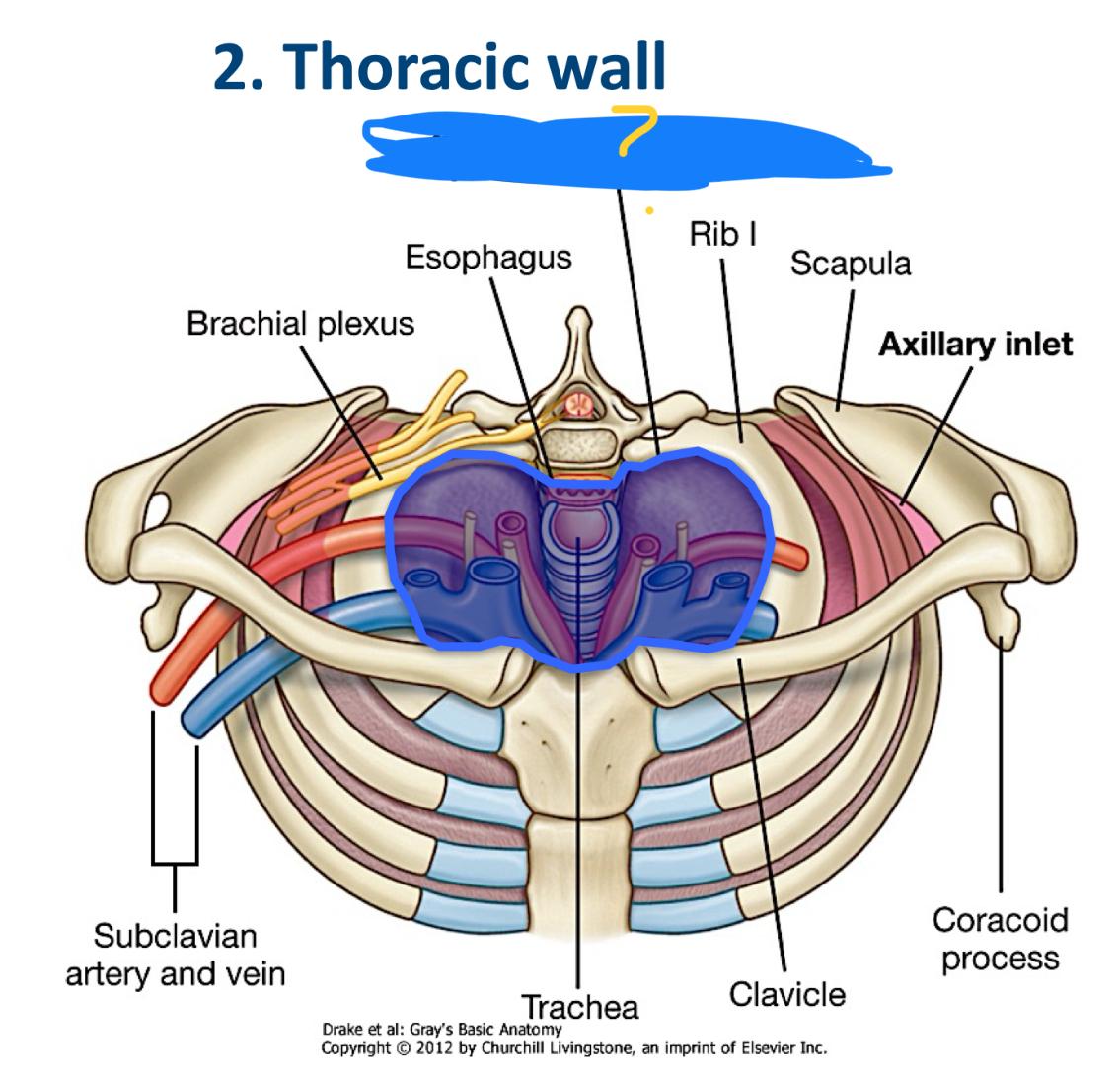
what is this
superior thoracic aperture
what is the superior thoracic aperture
body of the T1
the medial margin of rib 1
manubrium of the sternum
the inferior thoracic aperture is what part of the rib cage
the bottom
inferior thoracic aperture contains what
POSTERIOR (back rim)
the body of the TXII (main part of 12th thoracic vertebra)
Rib XII and distal end of XI (floating ribs at the very bottom and attach to that TXII)
inferior thoracic aperture contains what
ANTEROLATERAL
(sides and front )
Distal cartilaginous ends of ribs VII(7) to X(10)(fancy way of saying costal margins - the arch of cartilage)
Xiphoid process ( points but at the bottom of the sternum)
diaphragm
ribs Vii (7) and X (10) are
false ribs
what is the thoracic walls skeletal frame work
thoracic vertebrae
intervertebral discs
ribs
sternum
how many sites of articulation to ribs a typical thoracic vertebra have
3
what facets are located o a typical thoracic vertebra
2 demifacets located
oval facet
what are demi facets
half facets
2 demi facets types
superior costal face and inferior costal facet
what does the superior costal facet articulate with
part of the head of its own rib
what does the inferior costal facet articulate with
part of the head of the rib bellow
what is the oval facet
transverse costal facet
where is the oval facet and what does it articulate with
at the end of the transverse process and articulates with the tubercle of its own rib
…… has a vertebra with demi facets on the body and facets on the transverse process
thoracic vertebra
what are the atypical thoracic vertebrae
T1
TX
TXI
TXII
T1
complete superior costal facets on the body to articulate rib 1
TX
lacks inferior demi-facets and articulates only with its own ribs
TXI and TXII
lack transverse costal facet
articulate only with the head of their own ribs
how many pairs of ribs
12
how many pairs of true ribs
7
how many pairs of false ribs
5
what do the 7 true ribs articulate directly with
the sternum
what do the 5 pairs of false ribs articulate with
VII-X and XI and XII
ribs VII-X articulate with the costal cartaliges of the ribs above
ribs XI and XII have no anterior connection with other ribs or sternum (floating ribs)
which ribs are floating ribs
XI
XII
do the flashcards of the ribs
done
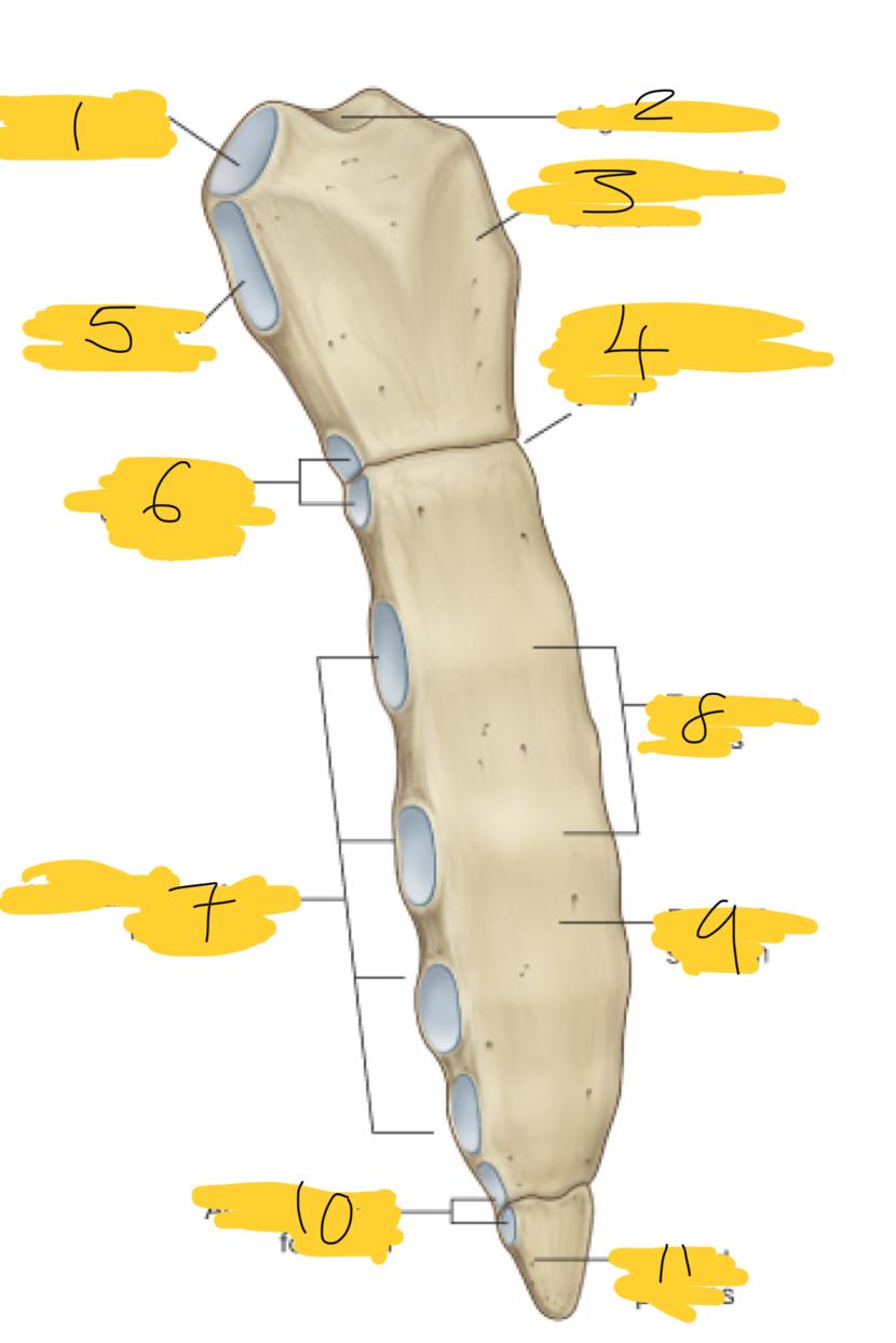
the posterior end articulates with …
the vertebral column and is characterised by a head neck and tubercle
the head of the rib has two articular facets (+size)
inferior costal facet (larger )
superior costal facet (smaller)
what does the inferior costal facet articulate with
the superior costal facet on the body of its own corresponding vertebra (rib 5 with T5)
what does the superior costal facet articulate with
the inferior costal facet on the vertebra above (rib 5 with T4)
what happens at the crest between these 2 facets on a rib
intra-articular ligaments attach - locking rib head into place
the neck of the rib
short region of bone that separates head from the tubercle
the tubercle of the rib consists of 2 regions
articular part
non-articular part
in the tubercle what is the articular part
is medial and has an oval facet for articulation with a corresponding facet on the transverse process of the associated vertebra
in the tubercle what is the non articular part
raised part
roughened by ligament attachments
shaft of the tubercle
has internal and external surfaces
superior margin?
superior margin = smooth and rounded
inferior margin?
inferior margin = sharp, marked by a distinct costal groove
the manubrium of the sternum has on its superior surface
jugular notch
the jugular notch on the manubrium has on either side
on either side there is a large oval fossa for articulation with the clavicle
the jugular notch on the manubrium has underneath
facet for the attachment of the first costal cartilage (rib

1
articular site for clavicle
2
jugular notch

3
manubrium of sterunm

4
sternal angle

5
attachment site of rib 1

6
articular demi facets for rib II

7
articular facets for rib III-VI

8
transverse ridges

9
body of sternum

10
articular facet for rib VII

11
xiphoid process
rib 1 articulates with sternum
directly to the manubrium (full facet)
rib 2 articulates with sternum
sternal angel (demifacets on both manubrium and body)
rib 3-6 articulates with the sternum
directly to the body ( full facets)
rib 7 articulates with the sternum
junction of Body and Xiphoid process (demi facets)
ribs 8-9 articulates with the sternum
false ribs - articulate with costal cartilage of the rib above not directly to the sternum
ribs 11-12 articulates with the sternum
floating ribs - No
what are sternocostal joints and where
articulation between costal cartilage of a rib and the sternum
upper 7 costa cartilages + sternum
what are the 2 sternocostal joints
Fibrocartilaginous joint
Synovial joint (2 compartments )
joint between rib 1 and sternum =
fibrocartilaginous joint
joints between rib II through VII and sternum =
synovial joints
why are synovial joints in 2 compartments
because of intraarticular ligaments
interchondral joints
are articulations between the costal cartilages of adjacent ribs
specially ribs 6-9 (sometimes 10)
what do the interchodral joints provide to the sternum
anchorage
what do interchondral joints form
inferior costal margin
what type of joint is an intercondral joint
synovial joint
Manubriosternal and xiphisternal joints are classified as
Symphysis (type of cartilaginous joint)
what is the manubriosternal joint
junction between the manubrium and the body of the sternum
what is the Xiphisternal joint
junction between the body of the sternum and the xyphoid process
what happends to Xiphisternal joints wit age
become ossified (fibrocartilage turns to bone - fusing the xiphoid process to the body of the sternum)
Manubriosternal and xiphisterna allow for
slight movement during respiration
the sternal angel passes through … ?
the intervertebral disc between TIV and TV
what is the intercostal space
space between 2 adjacent ribs (filled with muscles, nerves, and blood vessels)

1
serratus anterior muscle

2,3,4
external intercostal muscle
internal intercostal muscle
innermost intercostal muscle

5,6
skin
superficial fascia

7
lung

8,9,10
pleural cavity
visceral pleura
parietal pleura
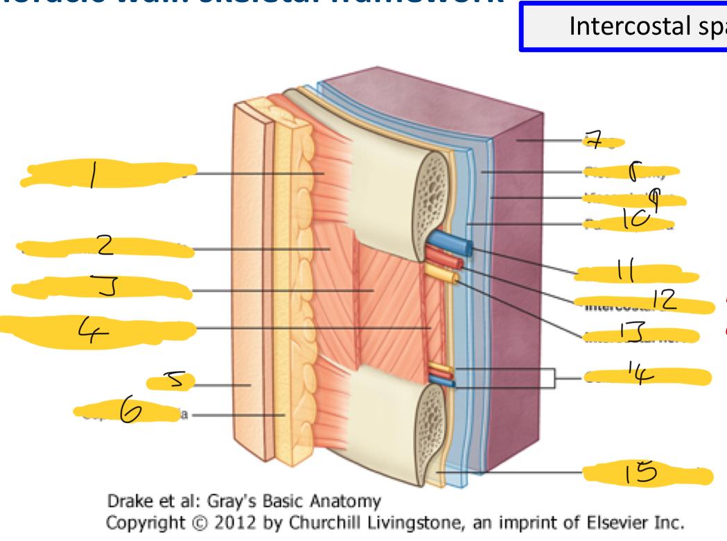
11,12,13
intercostal vein
intercostal artery
intercostal nerve

14
collateral branches

15
endothoracic fascia
where are intercostal muscles found
in each intercostal space (pass between adjacent ribs)
what do intercostal muscles do t the intercostal space
fill and support it
what are the types of intercostal muscles and how are they named
external (closest to the skin (superficial))
internal (in the middle (intermediate))
innermost (furthest away from skin (deep))
primary function of external intercostal muscles and how
inspiration (breathing in)
they contract
pulling ribcage up
increasing volume of the thorax
primary function of internal intercostal muscles and how
expiration (breathing out )
rib cage moved down when they contract
decreasing thoracic volume
primary function of innermost intercostal muscles
assists the internal intercostal with forced expiration
where are the subcostal muscles and transversus thoracis muscles in relation to the internal and innermost intercostal muscles
deeper
Transversus Thoracis muscle is located
inner surface of the anterior thoracic wall
(deep to the sternum and costal cartilages)
function of Transversus Thoracis muscle
depress costal cartilage
when is contracts - it pulls costal cartilage downwards
contributes to forced expiration
Subcostal muscle location
inner surface of the posterior thoracic wall
function of subcostal muscles
depress the ribs
contract it helps depress the ribs
during forced expiration
1
serratus posterior superior