Nursing Care of a Family When a Child Has a Cardiovascular Disorder
1/79
There's no tags or description
Looks like no tags are added yet.
Name | Mastery | Learn | Test | Matching | Spaced |
|---|
No study sessions yet.
80 Terms
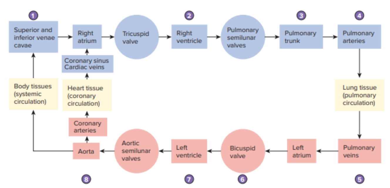
Right side of the heart
Pumps blood to the lungs where it will be oxygenated. (Pulmonary Circulation)
Left side of the heart
Pumps oxygenated blood to the peripheral tissues (Systemic Circulation)
Systole
Contraction of the heart chambers
Diastole
Relaxation of the heart chambers
Cardiac output
Volume of blood pumped by the ventricles each minute
Preload, contractility, afterload
Cardiac output is affected by three main factors
Preload
The volume of blood in the ventricles at the end of diastole or the point just before contraction
Contractility
The ability of the ventricles to stretch
afterload
The resistance against which the ventricles must pump
Right Atrium
Right Ventricle
Left Atrium
Left Ventricle
4 chambers of the heart
Superior vena cava and Inferior vena cava
The two vessels that connect to the Right Atrium
Knee-chest position or squatting
These positions trap blood in the lower extremities because of the sharp bend at the knee and hip, allowing the child to oxygenate the blood remaining in the upper body more fully and easily.
Cyanosis
occurs if a shunt is allowing deoxygenated blood to enter the arterial system.
Adult heart flow

Foramen ovale
Ductus arteriosus
Ductus venosus
Three fetal shunts
Fetal Heart Circulation
Highly oxygenated blood from the placenta returns via the umbilical vein, bypassing the liver through the ductus venosus and entering the IVC.
40%
About __ of this blood is shunted through the foramen ovale from the right atrium to the left atrium, then to the left ventricle and aorta, supplying the brain and upper body.
90%
Due to high pulmonary pressure, roughly __ of this blood bypasses the lungs via ductus arteriosus, flowing directly into the aorta.
Aortic
Pulmonic
Erb’s Point
Tricuspid
Mitral
Points of Maximal Impulse of the Heart
Apical impulse, thrills, lifts or heaves
In a physical examination, your place a hand on the left side of the chest to evaluate for?
Right 2nd intercoastal space
The Point of Maximal Impulse (PMI) of the heart for aortic is located?
Left 2nd intercoastal space
The Point of Maximal Impulse (PMI) of the heart for pulmonic is located?
(S1, S2) Left 3rd intercostal space
The Point of Maximal Impulse (PMI) of the heart for Erb’s point is located?
Lower left sternal border 4th intercoastal space
The Point of Maximal Impulse (PMI) of the heart for tricuspid is located?
Left 5th intercoastal, medial to midclavicular line
The Point of Maximal Impulse (PMI) of the heart for mitral is located?
Thrills, Lifts, Heaves
These are three abnormal sounds we can hear upon auscultation
Thrills
A vibration felt secondary to significant cardiac murmurs
Lifts
Forceful cardiac contraction that causes the hand to move up
Heaves
Forceful cardiac contraction that actually causes the hand to move up and laterally
S1
Produced by the closing of mitral and tricuspid valve.
Blood fills the ventricles, and as ventricular pressure exceeds atrial pressure due to the volume within, the mitral and tricuspid valve areas are forced to close.
What happens during the S1- First Heart Sound?
Systole
Aortic and pulmonic valves are forced open and the ventricles contract ejecting blood out of the aorta and pulmonary artery.
“Lub”
During the first heart sound, the contraction of the heart produces this sound.
S2
Produced by the closure of the aortic and pulmonic valves.
Diastole
The ventricles contract and empty, the aortic and pulmonic valves closure during the ventricular relaxation.
“Dub”
During the second heart sound, due to the ventricular relaxation it produces this sound
S3
Produced from the rapid filling of the ventricles in early diastole and is best heard with the bell of the stethoscope.
“Lub-Lub-Dub”
In the third heart sound, due to the extra contraction in the ventricles it produces this sound. Aka the ventricular gallop.
S4
Produced by atrial contraction in late diastole and is always pathologic. It is best heard with the bell of the stethoscope and produces a gallop rhythm.
Decreased ventricular compliance or Structural defects that cause obstruction to ventricular flow.
In the S4- Fourth Heart Sound an atrial gallop occurs, with this sound the following conditions are noted:
“Lub-Dub-Dub”
In the fourth heart sound, due to the atrial contraction in the late diastole, it produces this sound. Aka the atrial gallop.
Heart Murmurs
Turbulent flow through an abnormal valve, vessel, or chamber.
Innocent Murmurs
This type of heart murmurs are not associated with any intracardiac disease.
Increased metabolic states that occur with anemia, fever, pregnancy, or anxiety
The following can alter characteristics of an innocent murmur:
Pathologic murmurs
This type of heart murmurs are always associated with cardiac anomaly or altered cardiac function
Position in the cardiac cycle
Duration
Quality
Pitch
Intensity
Location
Presence of a thrill
Response of the murmur to exercise or change of position
(mnemonic na medyo bastos Please Don’t Quit Penetrating In Lusty Positions Roughly)
What to asses and document when noting a murmur?
Levine Grading Scale
What grading scale do you use for systolic murmurs?
Grade I
The characteristic of this murmur grade is at its lowest intensity and difficult to hear.
Grade II
The characteristic of this murmur grade is immediately audible but has lower intensity.
Grade III
The characteristic of this murmur grade is audible and louder than grade II.
Grade IV
The characteristic of this murmur grade is readily noted; has an associated palpable thrill.
Grade V
The characteristic of this murmur grade has a palpable thrill; murmur can be heard with stethoscope partially lifted off the chest.
Grade VI
The characteristic of this murmur grade has a palpable thrill; loudest intensity; murmur can be heard with stethoscope raised above the chest.
Systolic
The timing of an innocent heart murmur is?
Systolic or Diastolic
The timing of a pathologic heart murmur is?
Short
The duration of an innocent heart murmur is?
Longer
The duration of a pathologic heart murmur is?
Soft, musical, twangy
The quality of an innocent heart murmur is?
Harsh, blowing, to and fro
The quality of a pathologic heart murmur is?
Soft
The intensity of an innocent heart murmur is?
Loud
The intensity of a pathologic heart murmur is?
Supine position
The position in which innocent heart murmurs is heard?
All positions
The position in which pathologic heart murmurs is heard?
SA node
The heart’s electrical activity starts in the?
Right atrial wall near the entrance of the superior vena cava
SA node is found in the?
Bundle of His, Purkinje fibers
Fill in the blanks:
SA node >AV node >____________ >_____________> walls and septum of the ventricles
Asystole
Complete absence of electrical and mechanical activity in the heart, indicating no heartbeat.
Electrocardiogram
Is a written record of the electrical voltages generated by the contracting heart
P-wave
Represents atrial depolarization or atrial contraction
QRS complex
ventricular depolarization or contraction
T-wave
ventricular depolarization or relaxation
U-wave
An additional slow wave
PR interval
time it takes for an electrical impulse to travel from atria to ventricles, its normal time is 0.12 to 0.20 sec.
QRS interval
The time it takes for the ventricle to depolarize, which typically takes between 0.08 to 0.10 seconds.
ST segment
80 to 120 milliseconds
QT interval
the time it takes for the ventricles to depolarize and repolarize, and typically lasts 0.45 to 0.46
Acrocyanotic Heart Disease
Cyanotic Heart Disease
Classifications of Congenital Heart Diseases
Acrocyanotic Heart Disease
Involve heart or circulatory anomalies, either a stricture to the flow of blood or shunt that moves blood from arterial to the venous system (left to right shunts)
Cyanotic Heart Disease
Blood is shunted from the venous to the arterial system as a result of abnormal communication between two systems (right to left shunts)
Increased pulmonary blood flow
Obstruction to blood flow leaving the heart
Mixed blood flow (oxygenated and deoxygenated blood mixing in the heart or great vessels)
Decreased pulmonary blood flow
Second Classification System