Sensation and Perception Exam 1
1/101
There's no tags or description
Looks like no tags are added yet.
Name | Mastery | Learn | Test | Matching | Spaced |
|---|
No study sessions yet.
102 Terms
Sensation
Raw data processed by sensory receptors (eyes, nose, ears, etc.)
Perception
Sensations are processed by the brain (makes meaning out of perceptions)
Why study perception?
It is a multi disciplinary field that has broad applications and implications for science, technology, medicine, education, and life
Improving AI
Insights from human perceptions can inform the development of more human-like intelligence systems (bionic eyes and self-driving cars)
Enhancing Clinical Practices
Leads to better diagnosis and treatment of perceptual disorders (visual impairment, auditory processing, etc.)
Exploring Consciousness
Perception is closely linked to consciousness
Studying perception can shed light on the nature of conscious experience and the mechanisms underlying awareness
Cranial Nerves
Transmit neural activity from sensory organs to the central nervous system (i.e., the brain)
- Auditory nerve, optical nerve, etc.
Johannes Muller
Doctrine of Specific Nerve Energy
Perception is determined by which sensory nerve is stimulated, rather than the specific type of stimulus (e.g., light, sound, pressure) that triggers the nerve
Wilder Penfield
Montreal Neurological Institute
Electronically stimulated different portions of the cerebral cortex on awake (local anesthesia) patients to map different areas of the brain and understand their functions
How different regions of the cortex contribute to sensory perception, motor control, and various cognitive processes
MNI Brain - anatomical reference for brain structures
Santiago Ramon y Cajal
Made detailed drawings of nerve cells/ neurons and provided evidence that the nervous system was comprised of single cells
Otto Loewi
Showed that transmission between neurons at the synapse involves the release of chemicals called “neurotransmitters”
Brain
Contains specialized pathways for each of the senses
Some areas process info from only one sense (primary visual, auditory, somatosensory)
Some areas integrate info from multiple senses (thalamus (all except smell), superior temporal gyrus (auditory and vision)
Neurons
Fundamental building blocks of the nervous system. They are specialized cells responsible for transmitting information throughout the brain and body
Steps of the Perceptual Process
Environmental stimulus
Sensory receptors
Receptor process
Neural processing
Perception
Recognition
Action
Principle of Transformation
Stimuli and responses created by stimuli are transformed, or changed, between the distal stimulus (in the environment) and perception
Principle of Representation
Everything a person perceives is based not on direct contact with the stimuli but on representations of stimuli that are formed on the receptors and the resulting activity in the person’s nervous system
Transduction
Transformation of environmental energy (e.g., light, sound, energy) to electrical energy
Sensory Receptors
Cells specialized to respond to environmental energy
Top-Down Processing
Based on knowledge (previous experience)
Memory, expectations, knowledge about the nature of the stimulus
Select and examine features
Recognize stimulus
When a person has previous knowledge or experience and uses that to recognize a stimulus
Bottom-Up Processing
Based on incoming data that stimulates your response
Sensory receptors, detect specific feature of the stimulus
Combine features
Recognize stimulus
When receptors are stimulated by new data and using that data to create a recognizable stimulus without previous knowledge
Classical Methods to Measure Threshold (Gustave Fechner)
Method of limits
Method of Constant Stimuli
Method of Adjustment
Method of Limits
How is it done: the experimenter presents stimuli in either scending order (intensity is increased) or descending order (intensity decrease) - observer responds whether stimulus is perceived
Threshold: average of all crossover points
Advantage/Disadvantage: More efficient; has issues (subject anticipation)
Method of Constant Stimuli
How is it done: Different stimulus intensities are presented at one time, and the participant must respond whether they perceive it (same as method of limits), but the stimulus intensities are presented at random (rarely to almost always perceivable). The participant with respond yes/no or same/different
Threshold: the intensity that results in detection on 50% of trials
Advantage/Disadvantage: Most accurate (many observations and stimuli randomly presented); Time consuming
Method of Adjustments
How is it done: the participant adjusts the stimulus intensity continuously until they can just barely detect the stimulus
Threshold: taking the average setting
Advantages/Disadvantages: Few trials needed for observer to find their own threshold; faster; Subject anticipation
Above Threshold Measurement
Magnitude Estimates
Recognition Testing
Reaction Time
Phenomenological Report
Magnitude Estimation
Used to describe the relationship or mapping between the physical stimulus intensity and the magnitude of the perceptual experience
What is the procedure: the participant assigns values according to perceived magnitudes of stimulus
Response Compression: strength of the stimulus increases, so does the perception, but perception does not have a 1-to-1 relationship with stimulus increasing
Recognition Testing
Identifying or categorizing things
Used to assess brain damage and perceptual abilities
One thing we have learned from perception through recognition testing is that some things we perceive really fast but some details take longer
Reaction Time
Time between the perception of a stimulus and the person’s reaction to it
Phenomenological Report
This is our subjective experience of stimulus
Allows us to understand perceptions
Neuron Anatomy
KNOW THE LOCATIONS OF THE PARTS
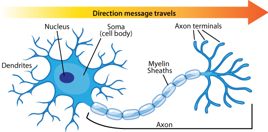
Soma
Cell nucleus (control center) integrates the information received from all other cells
Dendrites
Where the neuron receives the information from other neurons (a neuron can have many dendrite branches)
Axon
Long, thick fibers where the information that is being sent travels along
Terminal Buttons
Terminals at the end of the axon that sends the neurotransmitters to other cells’ dendrites when activated
Neurotransmitters
Chemical messengers are stored in synaptic vesicles (2 types: excitatory, inhibitory)
Synapse
Space between neurons
How do Researchers Measure APs?
Action/Membrane Potential: difference in electric potential between the interior and the exterior of the biological cell
Small electrodes are used to record from single neurons
Resting State
The difference between the recording electrode (inside nerve fiber) and reference (outside) is calculated
This negative charge of the neuron relative to its surroundings
Difference between inside and outside is -70 mV
Threshold for Firing an AP
Minimum potential difference threshold must be reached in order to fire an AP
- For most neurons in humans, this lies around ~ -50mV and -55mV
Depolarization
Electrically: The cell’s voltage becomes more positive (+)
Chemically: Influx of positive sodium ions
Repolarization
Electrically: Around +40, the voltage becomes more negative (-)
Chemically: the cells returns to its resting state as potassium comes in
Refractory Period
Electrically: A neuron can’t fire a new action potential
Chemically: Sodium channel gates close and potassium gates open
Synaptic Transmission
Excitatory Transmitters: causes depolarization
Neuron becomes more positive (+)
Increases the likelihood of an AP
Inhibitory Transmitters: causes hyperpolarization
Neuron becomes more negative (-)
Decreases the likelihood of an AP
Basic Properties of Action Potentials
Show propagated response: Once a response is triggered, it travels all the way down the axon without decreasing in size
All or nothing theory: an AP in a neuron will either occur fully or not at all, regardless of the strength of the stimulus
Increase in stimulus intensity results in an increase in rate of firing
Specificity Coding
A specialized neuron that responds only to one concept or one complex stimulus (also known as grandmother cells)
Quian Quiroga’s study provided evidence for this concept
Put 440 microelectrodes in 5 epilepsy patients
Recorded activity of single neurons
Major limitations of supporting research
Biologically limited: we only have so many cells, it would simply be impossible to have a cell for every concept
Research limitations: limited by the short recording time; with more time and materials, the neurons would likely respond to additional stimuli
Sparse Coding
A particular stimulus is represented by a pattern of firing of only a small group of neurons (most neurons remain silent)
Evidence that the code for representing objects, tones, and odors may involve a pattern of activity across a relatively small number of neurons
Advantages: Efficient - fewer neurons are active
Weaknesses: Not robust
Population Coding
Proposes that our experiences are represented by the pattern of firing across a large number of neurons
A large number of stimuli can be represented, because large groups of neurons can create a huge number of different patterns
Advantages: Robust: because information is distributed across many neurons, the system is less likely to lose information due tot damage or noise in individual neurons
Weaknesses: Takes up a lot of energy or capacity
Representations
Modulatory
Distributed
Connectivity
Modulatory or Modular Organization
The idea that specific brain areas (modules) are specialized to respond to specific types of stimuli or functions
One way to know the function of a brain region is to essentially turn it off and see what happens (Transcranial Magnetic Stimulation - TMS)
The Broca’s area is crucial for the production of speech and the Wernicke’s area is crucial for language comprehension
Distributed
The idea that the brain represents information in patterns distributed across the cortex, not just one brain area
This approach to representation focuses on the activity in multiple brain areas and the connections between those areas
Structural Connectivity
The anatomical organization of the brain through white matter tracts that connect cortical and subcortical regions (Taken with MRI - give structures)
Functional Connectivity
A measure of how brain regions interact with each other, even if they aren’t directly connected by white matter tracts (taken with fMRI - brain activity tracked with blood flow)
Resting State (Connectivity)
An fMRI measure when the brain is not involved in a specific task
How does measuring functional connectivity help us to understand perception?
It helps describe how different brain regions communicate and work together when making perceptions
Light
Electromagnetic radiation that travels in waves
Amplitude
Height of the wave
Wavelength
Distance between peaks
Eye Structure
KNOW THIS STUFF
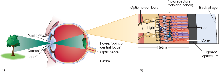
Pupil
Dark circular opening at the center of the iris in the eye, where light enters the eye
- Change size to allow more or less light; also change due to mental activity or drugs
Cornea
The transparent “window” into the eyeball also involved in focusing light on to the retina
Lens
The lens inside the eye, which focuses light onto the back of the eye
- Accommodation: process of lens changing size (object near - lens fat; object far - lens skinny)
Retina
A light sensitive membrane in the back of the eye that contains rods and cones (sensory receptors)
- The lends focuses an image on the retina, which then sends signals to the brain through the optic nerve (transduction occurs before sending signals)
Rods
Receptor better for vision in low light and peripheral vision (~120 million)
Cones
Vision in high light and color detailed vision (~6 million)
Fovea
Area that only contains cone receptors (point of central focus)
Peripheral Retina
Outside of the fovea; has both rods and cones (majority rods)
Optic Nerve
Nerve fiber transmitting impulses to the brain from the retina at the back of the eye (first to thalamus)
Optic Disc
Where the optic nerve exits the retina
Blind Spot
A small area at the back of the eye, where there are no visual receptors, so images that fall directly on the blind spot cannot be seen
Macular Degeneration
Damage of the macula (aka fovea), creating a central blind spot
Retinitis Pigmentosa
First damages the peripheral rod receptors (progressive genetic disorder)
Visual Transduction
When light energy is transformed into electrical signals
Dark Adaptation
Increasing sensitivity to the dark
Takes 30-40 minutes to adapt to the dark
Pupil size gets larger in short amount of time
The threshold decreases over time, meaning that the eye can detect progressively dimmer light as it adapts
How to Measure Dark Adaptation
Exposed to light = Light Adaptation - expose the subject to a bright light for a set period (5-10 minutes)
- Bleaches the photopigments in the retina, maximizing the contrast when moving to darkness
Darkness - Turn off the light source
Immediately begin recording the subjects’ ability to detect a dim light stimulus over time (presented at regular intervals)
Look at fixation point - Test light flashes to the side - peripheral retina with rods and cones
Methos of adjustment - adjusts the knob until it can be barely seen to measure threshold and sensitivity (when threshold is high then sensitivity is low)
Sensitivity
Low - reduced ability to detect light aka not great vision in dark
High - better ability to detect light aka good vision in dark
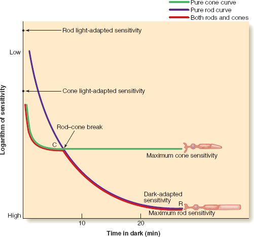
How can we measure the pure cone curve vs the pure rod curve?
Pure cone curve (test light only falls on the fovea)
Controls our vision in early stages, first 5 minutes or so
Pure rod curve
After 20-30 minutes rods reach their maximum sensitivity
Rod Monochromats
People with no cones (just rods)
Rod-Cone Break (2 stages)
Cones dominate the convo for about 5 minutes then petter out
Rods were there all along, but become more talkative at about 7 minutes
Opsin
Long/large protein
Retinal
A small molecule which fits into the binding site of a large protein called opsin
Isomerization
Changing of shape of the retinal when it absorbs photons of light
- The chemical basis of visual phototransduction - light detection
Visual Pigment Bleaching
Retinal molecules isomerize when they absorb light
It no longer fits into opsin, so they separate
Visual Pigment Regeneration
Retinal and opsin must recombine to respond to light
Cone pigment regenerates in 6 minutes
Rod pigment regenerates in over 30 minutes
How does Information from the Eye Flow to Various Parts of the Brain?
Visual receptors (rods and cones) in the retina trigger the firing of neural signals
Then sends impulses along the optic nerve (a collection of axons that connect the eye with the brain)
Optic Chiasm
The point at which axons from the inside half of each eye cross over and project to the opposite side
Lateral Geniculate Nucleus (LGN)
It sorts visual inputs and performs basic visual processing - it regulates neural information
Sends information to primary visual cortex and superior colliculus (important for controlling movement of the eyes)
Feedback Loops
Involve information flowing back from the visual cortex to the LGN
Allows for more complex, top-down processing where higher order brain regions influence earlier stages of visual processing
Primary Visual Cortex (V1)
First hits the primary visual cortex (v1) or the striate cortex in the occipital lobe
V1 has 6 layers that process depth perception, color, form, and motion
Hubel and Wiesel (1965)
Made substantial contributions to the study of receptive fields. They projected stimuli onto a screen and had anesthetized animals, usually a cat or monkey, look at the screen with glasses so the stimuli would be in focus on the back of the eye.
Receptive Field
A specific region of sensory space where a stimulus will elicit a response from a receptor (represents the area of input that the neuron is “sensitive” to
Types of Feature Detectors
Simple Cortical Cells
Complex Cortical Cells
End-Stopped Cortical Cells
Simple Cortical Cells
In the striate cortex (V1) receptive fields are side by side with excitatory (+) and inhibitory areas (-)
This cell likes horizontal bars, indicated by the high firing rate (an example)
These cells are tuned to bars in particular orientations
Finding the tuning of simple cells with orientation tuning curve - shows relationship between orientation and firing
Complex Cortical Cells
Like simple cells, respond best to particular orientation, BUT they only respond to a bar of light if it is moving across the receptive field
Often they will respond best to a particular Direction of Movement
End-Stopped Cortical Cells
Responds to corners, angles, or bars of a particular length moving in a particular direction
Selective Adaptation
Refers to when firing causes neurons to become fatigued (adapted)
Selective adaptation results in 2 physiological effects:
Firing rate decreases overtime
Firing rate is decreased when a particular stimulus immediately reappears
Selective Adaptation Procedure
Measure contrast threshold. Then view high contrast image (adapting stimulus) before measuring contrast threshold again
Selective Adaptation Results
The contrast in the repeated image should be more difficult to view than the non-repeated(in a different orientation)
Selective Adaptation - Why does this happen and what does it teach us about feature detectors?
The neurons become less sensitive making it difficult to perceive repeated stimuli in the same grating but doesn’t impact others as much
It teaches us that feature detectors adapting created a perceptual deficit
Selective Rearing
A procedure in which animals are raised in a special environment
Look at how the environment (IV) impacts behavior and brain functioning (DV)
Early environment exposure can shape neuronal functioning, like feature detectors (neural plasticity and experience-dependent plasticity)
Neural Plasticity
The brain changes and is adaptable. It forms new pathways depending on experience
Blackmore and Cooper (1970)
Raised cats in rooms with only vertical or horizontal stripes for 5 hours a day
Behavior
After 5months - visually impaired, the cats didn’t have grasping or startle reflexes
Vertical cats ignored horizontal things
Brain Scans
The horizontal brains didn’t respond to lines within 20 degrees of vertical line
Conclusions
Evidence of experience dependent plasticity, that the environment influenced the neurons
Connected feature detectors to perception
What/Where Pathway
What
Ventral Pathway
Object identification; helps with recognition of what things are
Where
Dorsal Pathway
Object spatial location; helps with guided behaviors
Both
Originate in retina
Have some interconnections
Receive feedback from higher brain areas
Ungerleider and Mishkin Experiment
Object discrimination problem - a monkey is trained to pick a particular object
Landmark discrimination problem - the monkey is trained to pick a location
Part of a monkey’s brain was removed
Removal of temporal lobe tissue → problems with the object discrimination task → what pathway
Removal of parietal lobe tissue → problems with the landmark discrimination task → where pathway