ch 19 – modulation of movement by cerebellum (motor control)
1/24
There's no tags or description
Looks like no tags are added yet.
Name | Mastery | Learn | Test | Matching | Spaced |
|---|
No study sessions yet.
25 Terms
upper
cerebellum modifies the activity of _____ motor neurons
laminated cortex and nucleu
two main gray matter structures:
laminated cortex
surface of gray matter
nuclei
deep in white matter
neurons are the main source of output of the cerebellum
detect and correct the error between intended and actual movements
primary function of cerebellum:
3 parts of the cerebellum
cerebrocerebellum
spinocerebellum
vestibulocerebellum

cerebrocerebellum
lateral hemispheres
largest in humans
receives input from cerebral cortex
controls highly skilled movements + speech
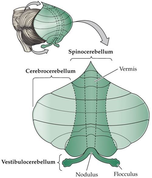
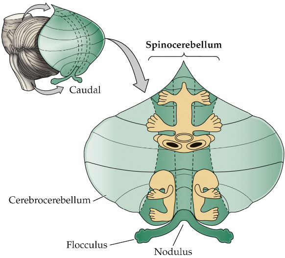
spinocerebellum
middle section
receives input from the spinal cord
vermis (center) → movements of proximal muscles
lateral areas → movements of distal muscles

vestibulocerebellum
receives inputs from brainstem
vestibulo-ocular reflex (VOR) → involuntary movement of the eyes when the head is moving
posture
equilibrium

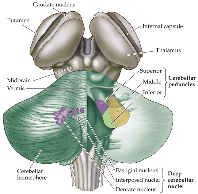
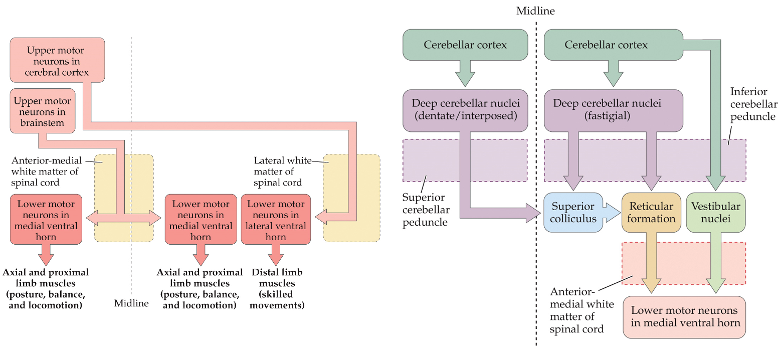
deep cerebellar nucleu
send outputs (efferents) to the thalamus, which projects to the motor cortex
cerebellum → thalamus → cortex
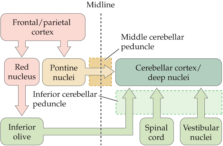
input projections TO the cerebellum
cerebral cortex axons project → pontine nuclei (brainstem) on the same side → pontine nuclei axons cross the midline → cerebellum
receives sensory info about body position and movement
spinocerebellar maps
spinocerebellum contains topographic maps for sensory inputs
fractured = areas of the body appear several times
inputs are ipsilateral
brain inputs are contralateral
output projections FROM the cerebellum
cerebellar cortex neurons → deep cerebellar nuclei → axons cross to other side of cerebral cortex
cerebral cortex neurons control contralateral musculature
each cerebellar hemisphere controls ipsilateral musculature

fastigial nuclei
neurons (deep cerebellar) send projections to brainstem
control medial tracts in spinal cord that regulate axial and proximal limb muscles
purkinje cells
inputs to the cerebellum target _______ _____
cerebellum circuit
cortex → pontine nuclei (brainstem) → cerebellum (contralateral)
mossy fibers
axons of pontine nuclei
connect to granule cells
granule cells
connected to mossy fibers
send parallel fibers to Purkinje cells = T branches
via excitatory synapses
50 billion
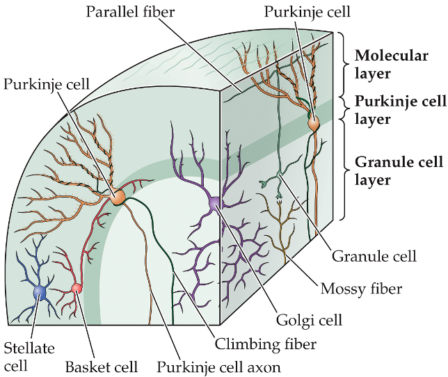
parallel fibers
at right angles to the plane of purkinje dendrites
connects many Purkinje cells
each Purkinje cell receives inputs from around 200,000
granule cells = wires - connecting to - Purkinje cells = poles
minimum contacts between wires
maximum number of contacts
spreading of information

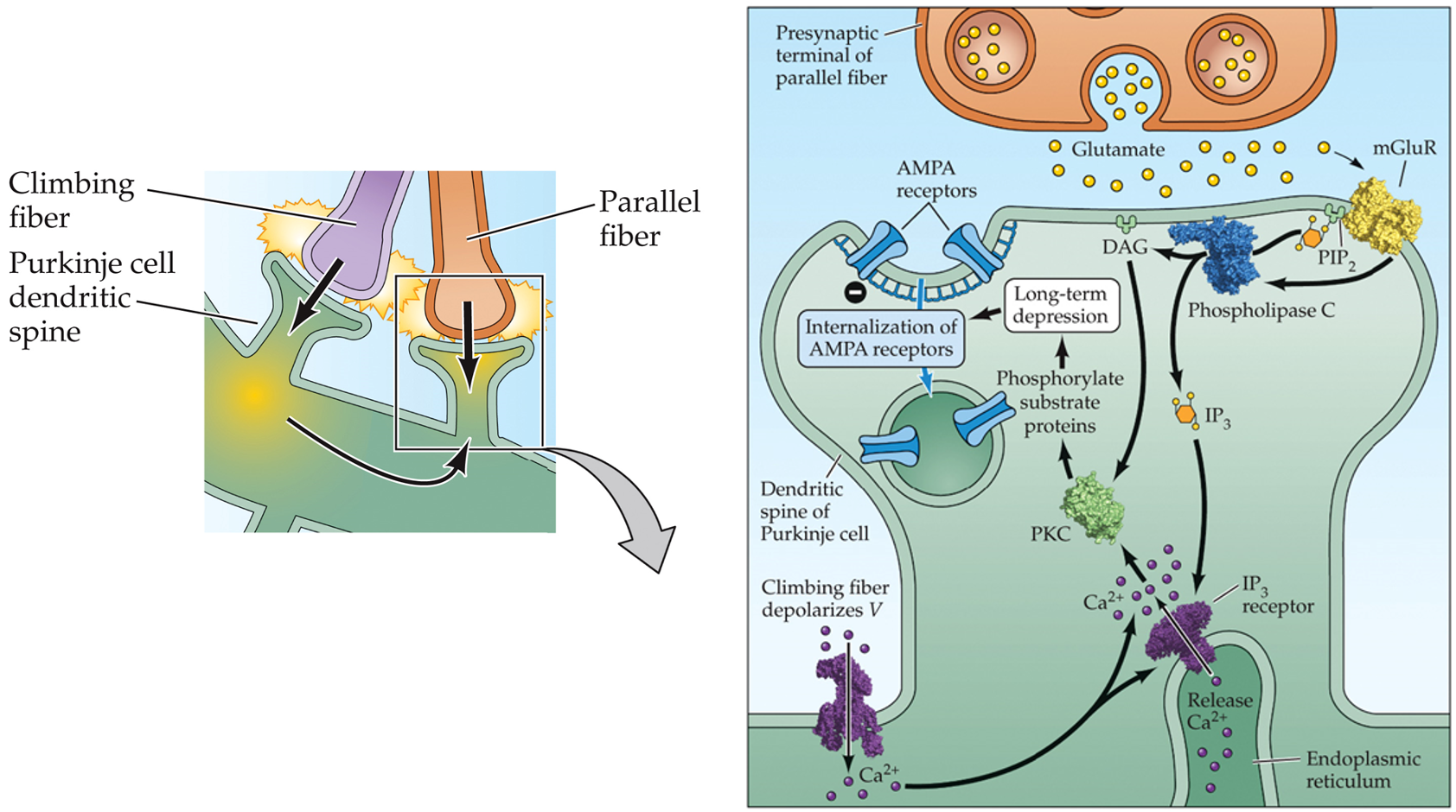
climbing fibers
connect Purkinje cell dendrites (wrapping around them) to form numerous connections
when fired, causes a Purkinje cell to fire
funneling of information
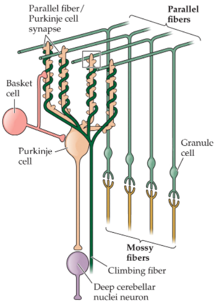
deep cerebellar nuclei neurons
receive excitatory inputs from mossy fibers and climbing fibers
Purkinje cells are GABAergic → modulate excitation via inhibition
Purkinje cells form a cortical inhibitory loop = can correct errors and modify movements

motor learning model
modulated by cerebellum
Purkinje cells compare climbing fiber input and parallel fiber input
if inputs arrive at the same time: depression of synapse with parallel fiber
via LTD = endocytosis of AMPAR
next time: similar input will have the Purkinje cells fire less → less inhibition from cerebellar loop → brain motor program controlled by that Purkinje cell will be more activated
movement control
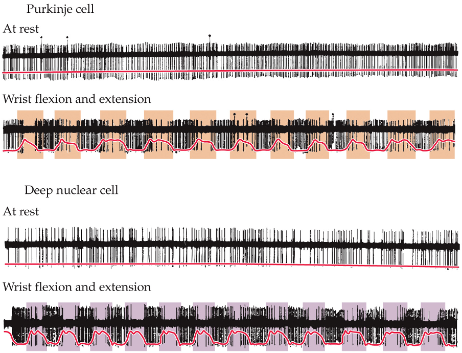
during movements, pattern of cerebellar activity in Purkinje cells and deep nuclei neurons changes continuously
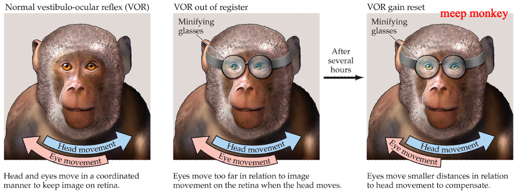
vestibulo-ocular reflex
when the head turns, the eyes move in the opposite direction to keep a stable image in the retina
if there is damage to the cerebellum, VOR can’t adapt to new conditions → cerebellar ataxia
minifying glasses:
glasses alter the size of the visual image
healthy cerebellum adapts VOR to compensate
damaged cerebellum prevents this adaptation
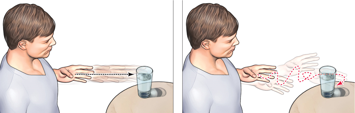
appendicular ataxia
irregular movements that typically overshoot or undershoot the visual target
require frequency corrective movements