spermatogenesis and semen
1/31
There's no tags or description
Looks like no tags are added yet.
Name | Mastery | Learn | Test | Matching | Spaced |
|---|
No study sessions yet.
32 Terms
3 goals of spermatogenesis
produce spermatozoa - cells capable of fertilization
replenish supply of primordial stem cells
create genetic diversity
3 phases of spermatogenesis
proliferation phase (mitosis) - spermatocytogeneis - begins with spermatogonia and finishes with spermatocytes
meiotic phase - spermatidogenesis - begins with primary spermatocytes and finish with haploid spermatids
differentiation phase - spermiogenesis - spherical shaped spermatid finishes with spermatozoa
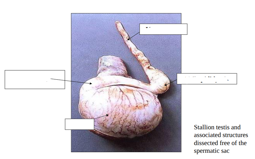
label testis - gross anatomy
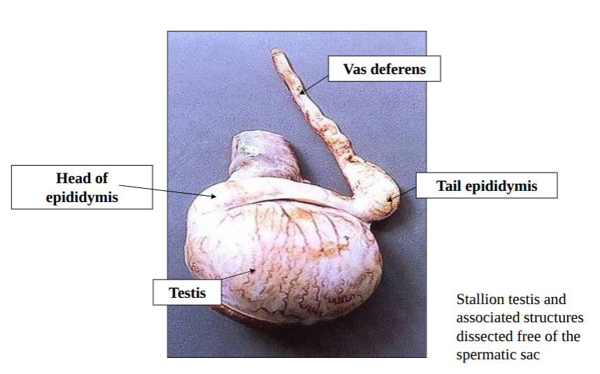
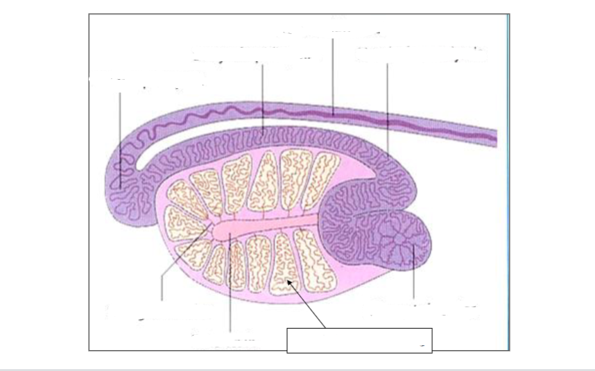
spermatizoa produced in seminiferous tubules of testes
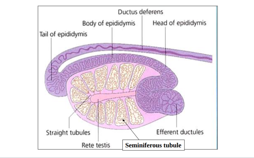
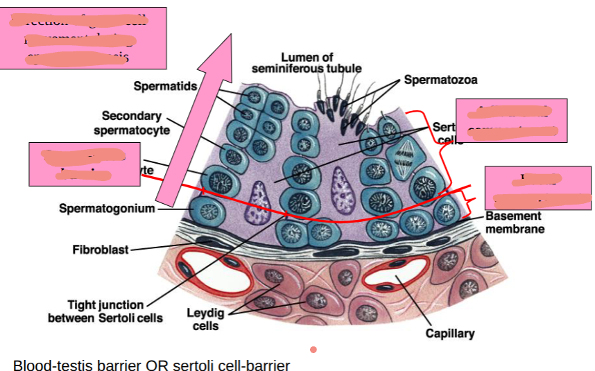
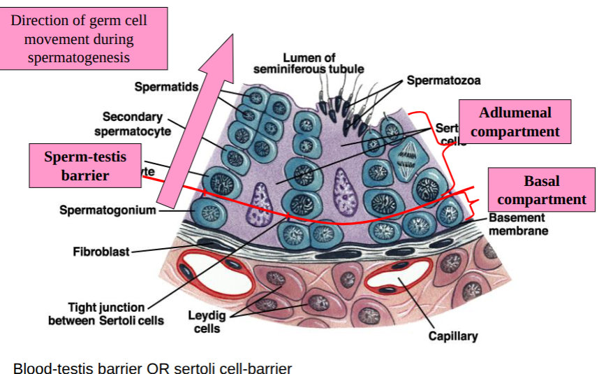
seminiferous tubules and spermatogenesis
occurs across the wall of tubules
sertoli cells - structurally support other cells involvedin producing spermatozoa - give them nutrition, produce fluid and mop things up, span from basement membrane to lumen, connect at tight junctions creating the sperm-testis barrier which created the basal compartment and adlumenal compartment
meiotic divisions
prophase of 1st division = relatively long process - lifespan of primary spermatocyte is longer than any other germ cell found on seminiferous epithelium
primary spermatocytes divide by meiosis to form secondary spematocytes in phase 1
in pahse 2 become spherical spermatids
lifespan of spermatocytes in bull
primary = 18-19 days
secondary = 1-1.7 days
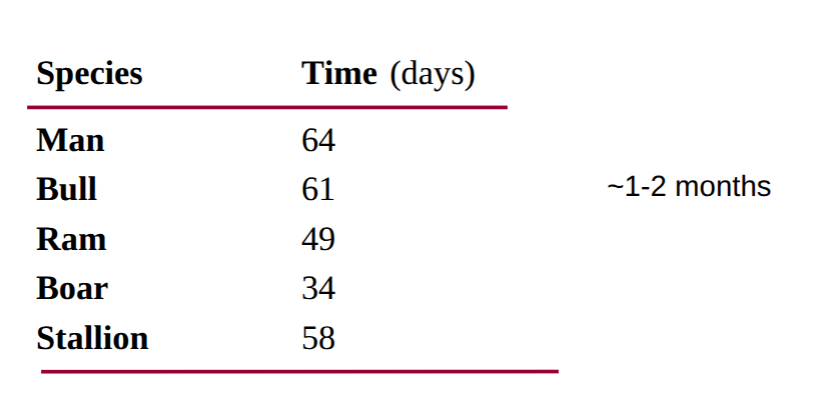
total duration of spermatogenesis = 61 days
differentiation phase
aims to produce a sophisticated self propelled package of enzymes and DNA
golgi phase, cap phase, acrosomal phase and maturation phase
each cell is genetically unique
then released into lumen = spermiation
head of spermatozoa
nucleus and acrosome and post nuclear cap
shape varies across species
acrosome contains hydrolytic enzymes required for penetration of zona pellucida
tail of spermatozoa
self powered flagellum
composed of - middle piece, principle piece and terminal piece
cycle of seminiferous epithelium
across species - length of cycle varies 4-9 weeks
spermatogenic wave - differences at any given instant in time along semiferous tubule
stage of cycle differs in adjacent regions of tubule
finite region of tubules releasing sperm at given time
time for completion of spermatogenesis
spermatozoa production
produced in testes
mature in head and body of epidiymis - move here via fluid produced by sertoli cells
stored in tail of epididymis until ejaculation
epididymis
transport through it takes 1-2 weeks
spermatozoa mature and develop motile capacity
epididyis stores 10-50 × 10^9 cells - enough for several ejaculations
copulation frequency - no effect on time spermatozoa in 1st part of epididymis - frequency copn → density of spermatozoa in epididymal tail and therefore decrease ejaculate
semen evalution
record volume, colour, appearance
NB EEJ sample cf. natural service = more dilute accessory gland secretion
live:dead ratio
mass motility/wave motion
density
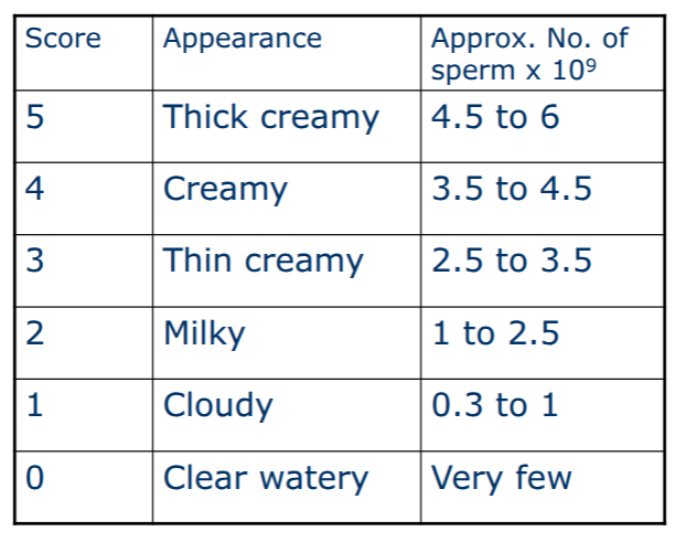
mass motility or wave motion
score 0-5
5 very good - billowing clouds
4 has clouds - good
3 fair
2 poor
1 very poor
0 dead
general appearance of ram semen
colour correlates to density
be careful with presence of infection as it will affect colour
head abnormalities
would have implication for ability to get in and fertilise egg
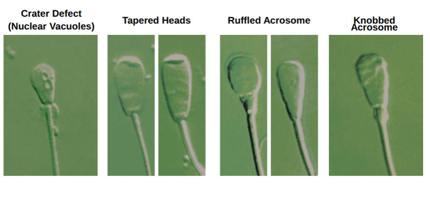

crater defect

tapered heads

ruffled acrosome

knobbed acrosome

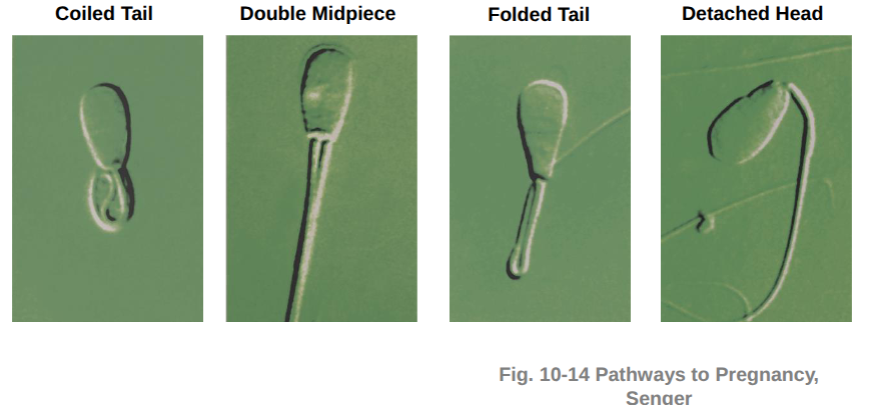
coiled tail

double midpiece

folded tail

detached head
tail abnormalities
would have difficulties of it getting places
spermatozoa sensitive to what
temp - keep sample, glassware, dilution and staioning fluids warm
water
bright light, blood, detergents, cigarette smoke, rubber bung in syringes
mitotic division
diploid
synchronised
occurs in basal compartment
do get cell nicrosis and death
endocrine regulation
hypothalamus produces GnRH and causes anterior pituitary to produce LH and FSH
LH acts on leydig cells which live between seminifeous tubules and produce testosterone which acts on sertoli cells and get -ve feedback at level of anteiror pituitary and hypothalamus
FSH acts on sertoli cells and produces inhibin and -ve feedback at level of anterior pituitary
sperm-testis barrier
immune system recognises spermatids as foreign divides semiferinous tubules into 2 compartments to prevent imune cells from breajing down spermatozoa - immune cells to large to fit through barrier
some viruses hide in testes and then can be passed to another animal in semen
morphology
primary vs secondary defects
compensable vs non-compensable defects