Cell death
1/14
There's no tags or description
Looks like no tags are added yet.
Name | Mastery | Learn | Test | Matching | Spaced | Call with Kai |
|---|
No study sessions yet.
15 Terms
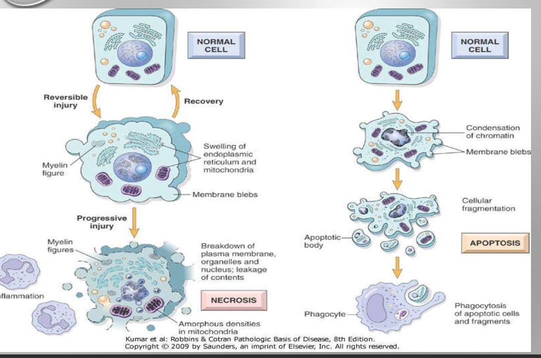
2 types of cell death
Necrosis
Apoptosis
Necrosis (definition + associated with , & results from)
Necrosis
Def: pathologic process where cells rupture and release content into extracellular space causing local inflammation
Associated with: Loss of memb integrity & cellular content leakage and dissolution of cells
Necrosis results from severe cell injury
Key features of necrosis
Membrane dissolution
cellular memb breakdown (plasma & lysosomal
Enzyme leakage:
Lysosomal enzyme escape into cytoplasm → digest cellular components worsening injury
Inflammatory response:
Cytokine release → induces inflammation
Immune activation
Phagocytosis of cell debris
Inflammatory cells release more proteolytic enzyme cause more tissue injury
Morphological changes of Necrosis (Cytoplasmic and nuclear)
Cytoplasmic: ↑Eosinophilia due to ↑binding of eosin to denatured cytoplasmic protein
Nuclear: Due to breakdown of DNA and chromatin
Pyknosis = Nuclear shrinkage & DNA condense into solid shrunken mass
Karyorrhexis = Pyknotic nucleus fragmentation
Karyolysis = Complete dissolution
Coagulative Necrosis: (Description,Mostly seen, Examples and describe the gross and microscopic image below)
Characteristic of hypoxia induced cell death most commonly by ischemia
Its called infarction seen mostly in solid organs like kidney and heart
Eg: Myocardial infarction, Renal infarction
A. Gross image = Kidney infarction (yellow) with outlines preserved
B Microscopic image = Normal kidney (N) and Necrotic cells of infarct (I). Necrotic cells show persevered outlines with loss of nuclei
Liquefactive Necrosis (Def, cause, Associated with, Example)
Def: Dead cells disintegrate and affected tissue is liquified
Causes: Dead cells digested by released enzymes in necrosis from bacterial, fungal infections & in brain ischemic infarcts (even if sterile)
Associated with: cellular destruction and pus formation
Eg: Cerebral infarction (ischemia in brain is liquefactive not coagulative)
Liquefactive necrosis in brain tissue
Pus formation
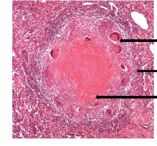
Caseous necrosis (Def, characteristic of, gross/ microscopically, peripheral collection)
Def: Dead tissue breaks down creating cheesy consistency on gross exam
Characteristic of : TB, some fungal infections (eg:histoplasmosis)
Microscopically: Collection of fragmented/lysed cells with amorphous granular pink (eosinophilic) appearance.
(Often) peripheral collection: macrophages forming granuloma
Gross: Large area of caseous necrosis containing yellow-white cheesy debris
Microscopically: Granuloma shows central necrosis surrounded by multiple Langerhans- giant cells, epitheliod cells and lymphocytes
Gangrenous necrosis (Def , Types + gross examination)
Def:
Death of soft tissue (applied for limb lost its blood supply)
Results from ischemia (eg: diabetic vasc disease affecting lower limbs)
Types
Dry gangrene : Dead tissue intact
Wet gangrene : Tissue liquifies (especially after bacterial infection)
Gross: Gangrenous necrosis is a dark/black colour
Fat necrosis (def, causes, clinical context, gross, microscope, types)
a. Def: Local areas of fat destruction
b. Causes: (usually) Activated pancreatic lipase release into pancreatic tissue and peritoneal cavity
c. Clinical context: Common in acute pancreatitis
d. Gross appearance:
Fatty acid released from necrotic cells + calcium → Fat saponification
Produces chalky white, visible lesions
e. Microscopic: Shadowy outlines of necrotic fat cells surrounded by basophilic Ca deposits. Also has inflammatory reaction.
f. Types: Enzymatic (fat necrosis by pancreatic enzymes), Traumatic (traumatic fat necrosis of breast)
Enzymatic Fat necrosis
Traumatic fat necrosis of breast
Fibrinoid necrosis (Def , Causes/Mech)
Def: Microscopic pattern of necrosis seen mostly commonly in cell mediated reactions
Causes/Mechanism: Deposition of immune complexes (antigen+ antibodies) & extravasated plasma protein in blood vessel walls.
Laboratory diagnosis of necrosis
Lab diagnosis of necrosis relies on detecting ↑ intracellular protein serum levels that leak out due to cell memb damage.
Eg of diagnostic markers:
Troponin → diagnose myocardial infarction
Transaminases (ALT,AST) → Diagnose liver disease
Pancreatic enzyme (amylase, lipase…) → Diagnose pancreatitis
Apoptosis (Def, Purpose, Key feature, Physiological and Pathological example)
Def: Apoptosis is form of programmed cell death (cellular suicide)
Purpose: Eliminate (/remove) cells no longer needed or damaged beyond repair
Key feature: Occurs without eliciting (harmful) inflammatory response
Physiological and Pathological apoptosis examples
Physiological = eg: During development for removal of excess cells during embryogenesis
Pathological = eg: Eliminate cells with DNA damage by radiation, cytotoxic agents etc
Apoptosis mech involves activation of caspases (enzyme)
Activated caspases → activation of nucleases degrade DNA & other enzymes that destroy necleoproteins and cytoskeletal proteins.
2 distinct pathways involved on caspase activation
Mitochondrial pathway
Death receptor pathway
Mitochondrial pathway = Cell injury → Bcl 2 family effectors (Bax, Bak) → Mitochondria → cytochrome c → initiator caspases
Death receptor pathway = Receptor ligand interactions → Adapter proteins → initiator caspases
Morphology of apoptosis
Cell shrinkage
Condensation of nuclear chromatin
Formed apoptotic bodies by fragmentation of cells and nuclei
Fragments are memb bound and contain cell organelles
Phagocytosis of apoptopic bodies by phagocytes
Necrosis vs Apoptosis ( Cell size , Nucleus, Plasma memb, Cell contents, Adjacent inflammation, Physiological or pathological)
Feature (N = necrosis , A = Apoptosis
Cell Size :
N = Enlarged swelling
A = Reduced (shrinkage)
Nucleus
N = Pyknosis → Karryohexis → Karyolysis ,
A = Fragmentation → Nuclosome sized fragment
Plasma membrane
N = Disrupted
A = Intact (altered struct especially lipid orientation)
Cellular contents
N = Enzymatic digestion (may leak out of cell)
A = Intact (may be released in apoptotic bodies)
Adjacent inflammation
N = Frequent
A = No
Physiological/ Pathological role
N = Pathological (culmination of irreversible cell injury)
A = Often Physiological (eliminate unwanted cells) may be pathological after some forms of cell injury especially DNA and protein damage