Kalat - Exam Test Bank Questions 14th Ed
1/172
There's no tags or description
Looks like no tags are added yet.
Name | Mastery | Learn | Test | Matching | Spaced | Call with Kai |
|---|
No analytics yet
Send a link to your students to track their progress
173 Terms
The two kinds of cells in the nervous system are ___, which receive and transmit information to other cells, and ___ which do not transmit information.
neurons; glial cells
The outer surface of a cell is called the ___ and the fluid inside the cell is the ___.
membrane; cytoplasm
Which part of a neuron contains the nucleus?
Cell body (AKA soma)
Neurons have one ___, but can have any number of ___.
Axon, dendrites.
As a general rule, axons convey information ___.
Away from the cell body.
What is an interneuron?
A neuron that receives all its information from other neurons and conveys impulses only to other neurons - it just relays.
A neuron that conveys information toward the hippocampus is considered a (an) ___ cell, with regard to the hippocampus.
Afferent.
A neuron that conveys information away from the hippocampus is considered a (an) ___ cell, with regard to the hippocampus.
efferent
In the human brain, glia cells are ___.
More numerous than neurons.
What do glia do?
Remove waste materials.
Build myelin sheaths.
Guide the growth of axons and dendrites.
They do NOT transmit information.
The difference in voltage between the inside and the outside of a neuron that typically exists is called the ___.
Resting potential.
What is meant by the term ‘concentration gradient’?
Potassium ions are more concentrated inside the cell and sodium ions are more concentrated outside.
The sodium potassium pump pumps sodium ions ___ and potassium ions ___.
Out of the cell, into the cell
The sodium potassium pump makes which of the following features of a neuron possible?
Resting potential.
When the neuron is at rest, which of the following forces tends to move potassium ions out of the cell?
Concentration gradient.
If a stimulus shifts the potential inside a neuron from the resting potential to a more negative potential, the result is ___.
Hyperpolarization.
If a stimulus shifts the potential inside a neuron from the resting potential to a potential slightly closer to zero, the result is known as ___.
Depolarization.
A membrane produces an action potential whenever the potential across it reaches ___.
The threshold
According to the all-or-none law, ___.
The size of the action potential is independent of the strength of the stimulus that initiated it.
For a given neuron, the resting potential is 70 mV and the threshold is 55 mV. Stimulus A depolarizes the membrane to exactly 55 mV. Stimulus B depolarizes the membrane to 40 mV. What can we expect to happen?
Stimulus A and stimulus B will produce action potentials of the same size.
During the entire course of events from the start of an action potential until the membrane returns to its resting potential, the net movement of ions is ___.
Sodium in, potassium out.
What is the refractory period of a neuron?
A period in which a usually adequate stimulus cannot produce an action potential.
Most action potentials begin ___.
At the axon hillock.
The velocity of an action potential is ___.
1100 m/sec.
The function of a myelin sheath is to ___.
Increase the velocity of transmission along an axon.
What are the nodes of Ranvier?
Interruptions in the myelin sheath.
Where does the abbreviation EPSP stand for?
Excitatory post synaptic potential.
What is an EPSP?
Graded depolarization.
Spatial summation refers to ___.
Adding two stimuli from different sources at the same time.
What is an IPSP?
Temporary hyperpolarization.
The synthesis of neurotransmitter molecules takes place ___.
In either the cell body or the presynaptic terminal, depending on the particular neurotransmitter.
When an action potential reaches the end of an axon, the depolarization causes what ionic movement?
Calcium into the cell.
What is the synaptic cleft?
The gap between the presynaptic neuron and the postsynaptic neuron.
The occipital lobe is at the ___ of the brain.
Posterior (back)
What isn’t part of the subcortical areas?
Hypothalamus
What is the function of the sympathetic nervous system (part of the autonomic NS)?
It prepares the organs for activity & expends energy (as opposed to rest and digest - parasympathetic nervous system).
The cerebellum has different functions. What are some of these?
It plays a role in movement.
It plays a role in balance and coordination.
The prefrontal cortex ___.
Is important for working memory.
How does fMRI work?
It shows an image of the brain by measuring blood flow with magnetic fields.
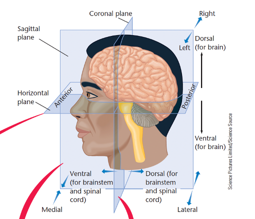
What are the anatomical directions?
· Dorsal – toward the back (think dorsal fin on a shark)
· Ventral – towards the stomach (think Ventriloquist ‘stomach talker’)
· Anterior – towards the front (e.g. the genitals)
· Posterior – towards the back (e.g. the butt)
· Superior – above another part (the head is superior to the torso)
· Inferior – below another part (the torso is inferior to the head)
· Lateral – towards the side, away from the midline (the arms are lateral to the midline)
· Medial – toward the midline, away from the side (the kidneys are medial to the midline)
· Proximal – close to the point of origin or attachment (the finger is proximal to the hand)
· Distal – more distant from the point of origin or attachment (the finger is distal to the elbow)
· Ipsilateral – on the same side of the body (e.g. the right arm and right leg)
· Contralateral – on the opposite side of the body (e.g. the left arm and right leg)
What is the central nervous system made up of?
The brain and spinal cord.
What is the peripheral nervous system made up of?
· Nerves outside the brain and spinal cord, connects to the rest of the body.
Somatic NS – controls voluntary muscles and conveys sensory information to the CNS.
Autonomic NS – controls the heart, intestine and other organs.
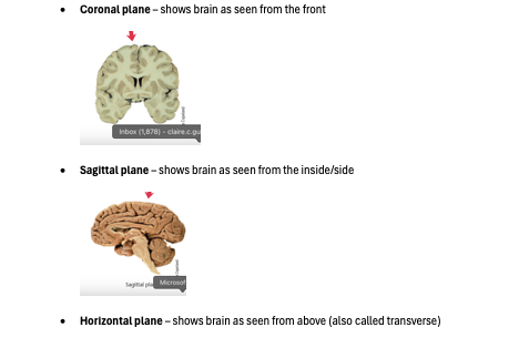
What are the main planes?
· Coronal plane – shows brain as seen from the front
· Sagittal plane – shows brain as seen from the inside/side
· Horizontal plane – shows brain as seen from above (also called transverse)
What does the hindbrain consist of?
Medulla, pons, and cerebellum. Controls vital reflexes such as: breathing, heart rate, vomiting, salivation, coughing, sneezing.
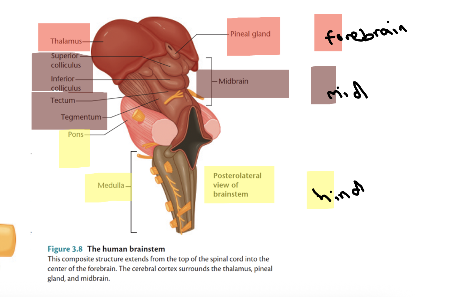
What does the midbrain consist of?
· Tectum – roof of the midbrain.
· Superior / inferior colliculus: sensory processing.
o Inferior – hearing.
o Superior – vision.
· Tegmentum – intermediate level of the midbrain.
Substantia nigra – dopamine-containing

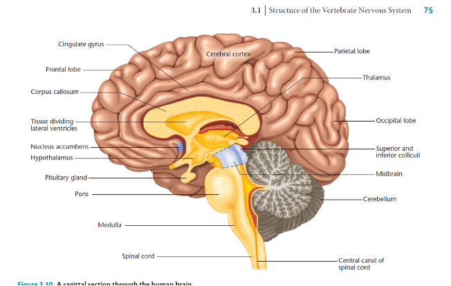
What does the forebrain consist of?
· Cerebral cortex – (think of bark of tree).
· Under the cerebral cortex are the thalamus, basal ganglia, and limbic system (olfactory bulb, hypothalamus, amygdala, cingulate gyrus).
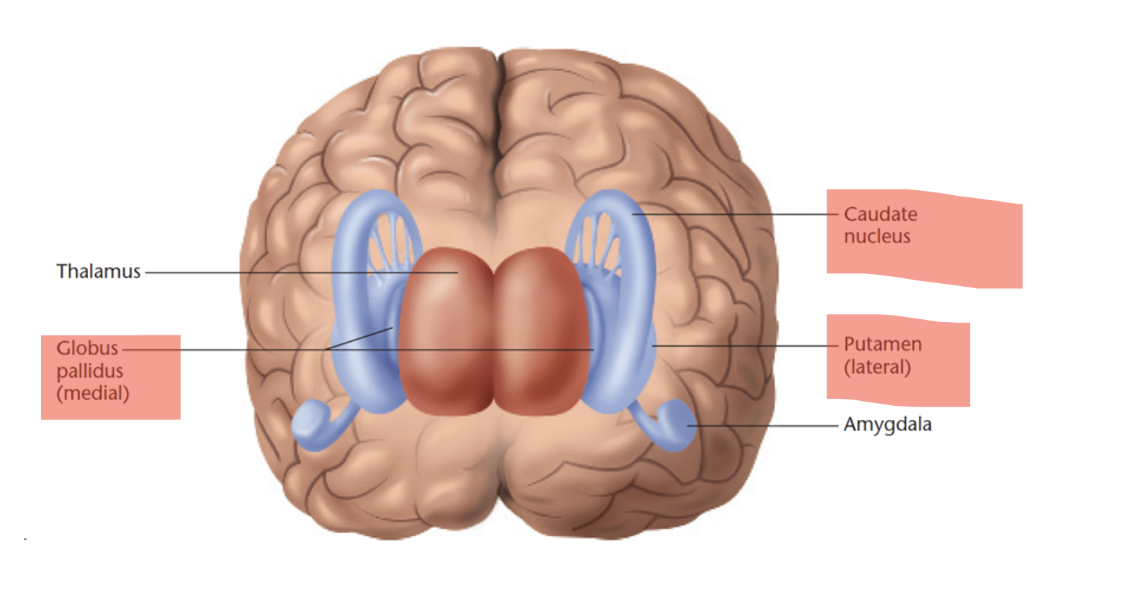
What are the basal ganglia (part of the forebrain)?
A group of subcortical structures lateral to the thalamus, including:
- Caudate nucleus
- Putamen
- Globus pallidus
Associated with movement, motivation/emotional vigour - CPG
What does the limbic system contain (part of the forebrain)

What do the hypothalamus and pituitary gland do (part of the forebrain)
o Motivated behaviours – feeding, drinking, temperature, sex, fighting, or activity level.
o Pituitary gland – synthesises hormones > blood > organs throughout body.
What does the basal forebrain contain (part of the forebrain)
o Nucleus basalis – receives input from hypothalamus
o Basal ganglia sends axons to release acetylcholine to widespread areas in the cerebral cortex.
o Key for arousal, wakefulness, and attention.
What does the hippocampus do (part of the forebrain)
o Memories – esp. individual events/declarative (explicit) memories.
o Monitoring where you are and where you are going.
Latin for seahorse.
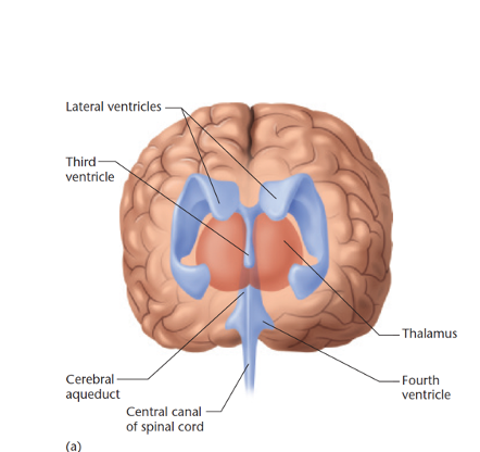
What are the ventricles and what do they do?
In fetal development, the neural tube becomes the central canal of the spinal cord and 4 fluid-filled ventricles.
Cushions brain against mechanical shock, buoyancy etc.
Also has hormones and nutrition for brain and spinal cord.
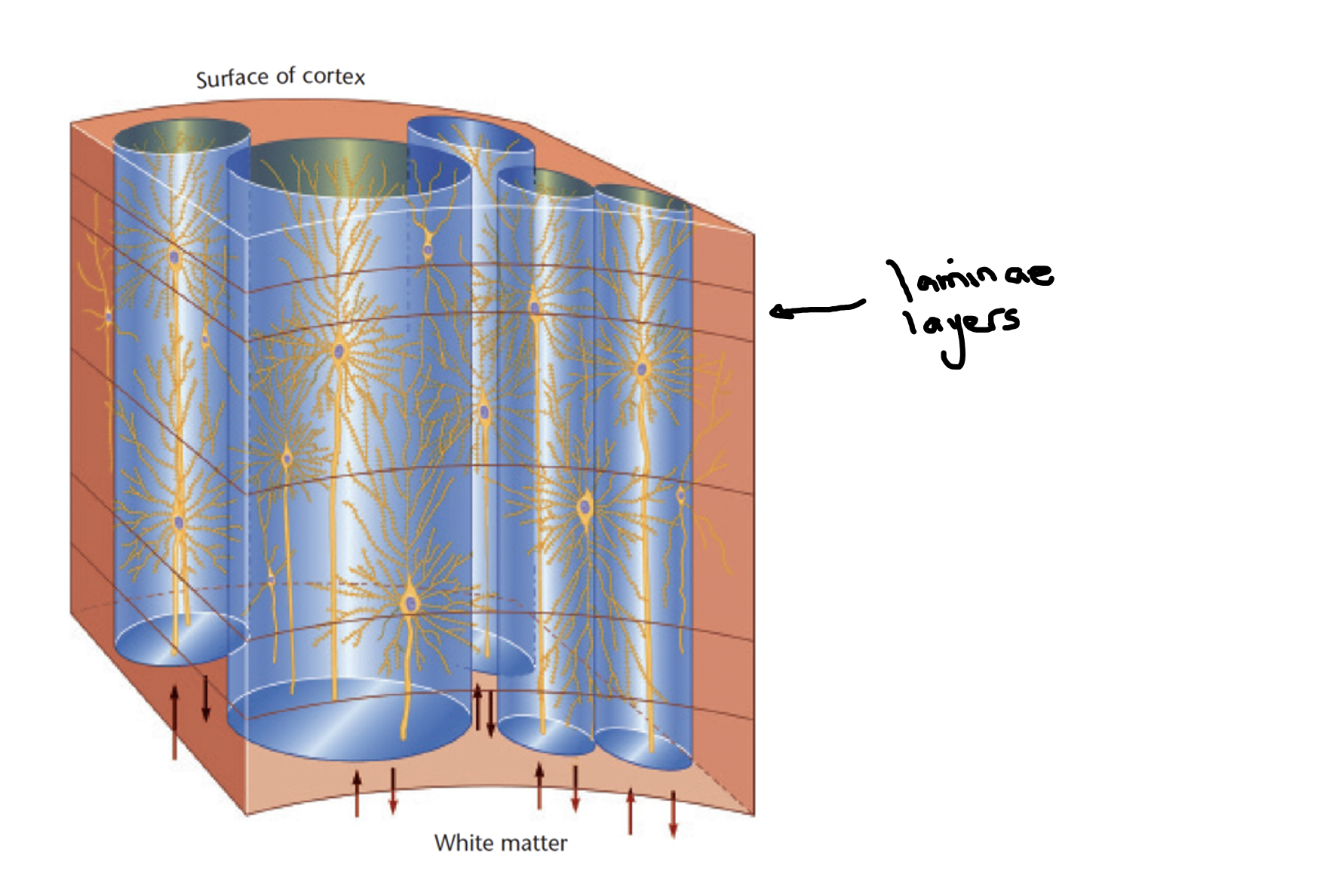
re: cerebral cortex, what are laminae and columns?
Cells on outer surface are grey matter, axons extending inward are white matter.
· Laminae – layers of cell bodies parallel to the surface of cortex,
· Columns: organised cells, perpendicular to the laminae.
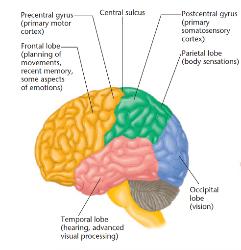
What are the main lobes of the cerebral cortex?
The main lobes of the cerebral cortex include the frontal, parietal, temporal, and occipital lobes, each responsible for different functions such as executive functions, sensory integration, auditory processing, and visual processing, respectively.
What is the binding problem?
The mystery of how the brain can perceive two things happening at the same time, in approx. the same space. AKA large-scale integration problem.
In many ways the eye is analogous to a camera. The light sensitive surface in the back of the eye that would correspond to the film in a camera is the ___.
Retina.
Where are the rods and cones of the eye located?
Retina.
The fovea is the part of the retina ___.
With the greatest perception of detail.
If you want to see something in fine detail, you should focus the light on which part of your retina?
fovea
Why is the blind spot of the retina blind?
It is the point where the optic nerve leaves the retina and there are no rods or cones.
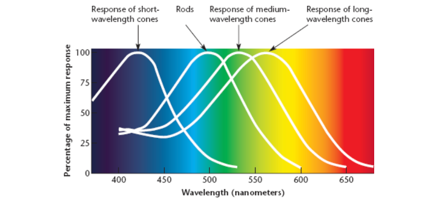
The perception of colour depends on ___.
Cones.
In comparison to the cones, the rods are ___.
More sensitive to dim light.
According to the Young Helmholtz theory, colour vision is based on ___.
Three kinds of receptors.
In the most common form of colour blindness people have difficulty distinguishing between what two colours?
Red and green.
Males are ___ likely to be colour blind compared to females.
More
Lateral inhibition refers to ___.
The reduction of activity in one neuron by activity in a neighbouring neuron, enhancing contrast and perception of edges.
In which layer of the retina is visual information coded in series of action potentials?
In the layer of the ganglion cells.
The function of the horizontal cells in the retina is related to ___.
lateral inhibition and contrast enhancement.
Where is the receptive field of a lateral geniculate cell located?
In the retina, corresponding to a specific area of visual space.
The three types of cells in the primary visual cortex are known as ___.
Simple, complex, and hypercomplex.
What are the colours corresponding with which wavelengths?
Blue = short
Green/yellow = medium
Red = long.
According to the law of specific nerve energies, ___.
Electrical stimulation of the auditory nerve is perceived as sound.
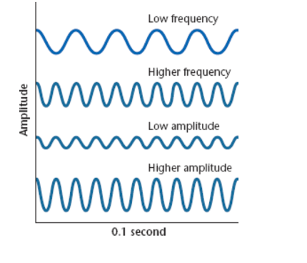
The intensity of a sound wave is its ___; the perception of that intensity is its ___. Frequency is_____
amplitude; loudness; how many waves occur per second
Suppose the highest pitch you can hear is about 20,000 Hz. Under what circumstances will that limit decrease?
It drops naturally as you grow older.
Three small bones connect the tympanic membrane to the oval window of the inner ear. The function of those bones is to ___.
Convert air waves into waves of greater pressure.
The cochlea is part of which sensory system?
Auditory
How are the receptor cells of the auditory system known?
Hair cells in the cochlea.
"Every sound causes one location along the basilar membrane to resonate, and thereby excites neurons in that area." This is one way to state which theory about pitch perception?
Place theory of pitch perception.
Damage to the cochlea, hair cells, or auditory nerve can produce ___.
Nerve deafness
Touch, pain, and other body sensations are known as ___ senses.
Mechnical
Which two sensory systems are based on the responses of hair cells?
Hearing and vestibular sensation.
What is the pathway for vision?
1.Retina
Photoreceptors (rods/cones)
Bipolar cells
Gangion cells
Optic nerve
Thalamus
V1 (aka primary visual cortex)
V2
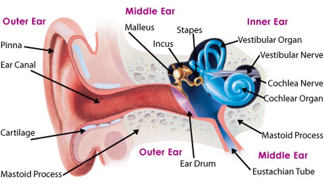
What is the pathway for sound/hearing?
Outer ear - pinna
Middle ear - eardrum (aka tympanic membrane) and hammer, anvil, stirrup (malleus, incus, stapes)
Inner ear - Cochlea
Auditory nerve
Brainstem
Thalamus (relay station)
A1 for processing (aka primary auditory cortex)
In vision, what is the dorsal and ventral streams?
The dorsal stream at the top of the brain is the "how pathway," processes spatial awareness and action. The ventral stream towards the bottom, known as the "what pathway," is involved in object recognition, while
What is visual agnosia?
An inability to recognize objects despite otherwise satisfactory vision
What is prosopagnosia?
An impaired ability to recognize faces, often referred to as face blindness, even when vision remains intact. Caused by damage to the fusiform gyrus.
What causes motion blindness?
Damage to the medial temporal cortex (MT) and MST (medial superior temporal cortex).
SAQ: Damage to the primary visual cortex (area V1) leaves someone blind, however damage to the primary auditory cortex does not produce deafness. Why doesn't damage to the primary auditory cortex produce deafness?
The primary auditory cortex (A1) is responsible for processing auditory information, but it is not the only area involved in hearing. Unlike damage to V1, damage to the primary auditory cortex does not produce complete deafness because auditory processing begins in the ear and continues through various pathways in the brain before reaching the primary auditory cortex. Therefore, other parts of the auditory pathway can still process sound to some extent, allowing for the perception of auditory stimuli.
Where are olfactory receptors located?
olfactory epithelium (back of nasal passages).
· Receptors detect chemicals in:
- Air we inhale (orthonasal olfaction).
- Food we eat (retronasal olfaction).
What is the basic olfactory pathway?
Odorant signals in clusters from the olfactory receptors in the nasal epithelium > olfactory bulb (glomeruli) > sends signals to the primary olfactory cortex in the temporal lobe and other brain regions for processing.
Which muscle is ‘antagonistic’ to a flexor muscle in the left leg?
An extensor muscle in the left leg.
Which disorder is commonly treated with LDOPA?
Parkinson's disease.
Which would be especially important (i.e. used much more than normal) when you run up a flight of stairs at full speed?
Fast-twitch muscles.
What statement is valid regarding Parkinson's disease?
The basal ganglia are affected, expressed in a tremor during rest.
The cerebellum is most important for ___.
Learned motor programs of ballistic movements.
People who have suffered damage to the cerebellum ___.
Have to plan their movements one at a time, not as a smooth sequence.
Tests for alcoholic intoxication resemble the tests for damage to the ___.
Cerebellum.
The caudate nucleus, putamen, globus pallidus, substantia nigra, and subthalamic nucleus make up the ___.
Basal ganglia.
Following damage to the basal ganglia, people ___.
Have difficulty initiating movements.