Fine Needle Smear Preservation
1/20
There's no tags or description
Looks like no tags are added yet.
Name | Mastery | Learn | Test | Matching | Spaced | Call with Kai |
|---|
No study sessions yet.
21 Terms
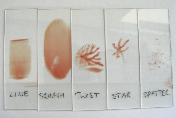
There are several methods for smear preparation. There is no scientific evidence to suggest one technique is better over another, often the choice is down to personal preference. Usefulness can depend on preference and the content/viscosity of the sample. These include ..
The squash technique - most effective
Push/pull technique - most effective
Line concentration technique
Star
Twist
When producing a smear what much be produced?
A monolayer to allow the slide to be appropriately examined
To avoid degradation of cells, the collected sample should be ..
Rapidly transferred from the needle to a microscope slide
A delay in transfer from the needle to the slide can lead to ..
Clotting within the needle shaft, trapping the sample in the needle shaft
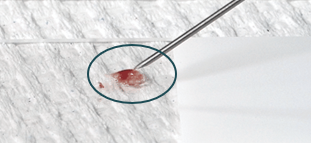
Placing the sample onto the slide
Rest the needle, bevel down, onto the labelled slide and depress the plunger briskly, resulting in a droplet on the slide
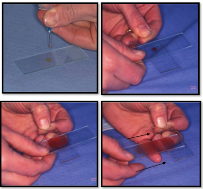
Squash technique
Simple and often preferred method
Excellent for semisolid, mucoid and viscous materials and small volume samples
The sample is expelled from the needle, in the middle of the slide
A second slide is placed at right angles to the first slide, on top of the sample, with an overlap either side
Second slide is then pulled along the lower slide, spreading the sample
Contact must be maintained, and the slides must be parallel to one another
Downwards pressure should be avoided to prevent cell damage
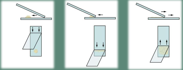
Push/pull technique
This technique is slower than other, more preferred techniques
Cell degradation due to desiccation of the sample
Used for samples with suspected low cellularity e.g. fluid washes
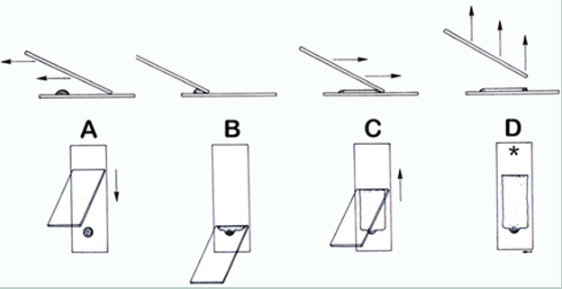
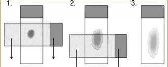
Line concentration technique
Excellent for semisolid material and small volume samples
Used for samples with suspected low cellularity, e.g. fluid washes
Same technique as push/pull but spreader slide lifted from the sample slide when approx. 2/3rd along the slide
The end line formed by using this technique will contain a concentrated quantity of cells
Can cause damage and lysis of cells due to shearing action of the slide
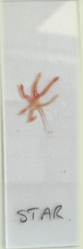
Star technique
The sample is placed in the centre of the slide, and the aspirate needle is used to drag the material into radiating streaks from the central droplet
Minimally traumatic to cells but is a relatively ineffective method of spreading the sample thinly
Often too thick for examination, but can be used for samples with suspected low cellularity, e.g. fluid washes
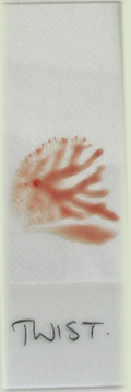
Twisting technique
The sample is expelled in the middle of the slide. A second slide is applied over the sample and the slides are twisted at 90 degrees counter to each other before being lifted off
The slides stick together which can cause cell damage
Smear is often too thick to assess, but can be used for samples with suspected low cellularity, e.g. fluid washes
Smears should be rapidly air dried to avoid ..
Shrinking of cells (drying artefact)
When transferring to the slide, keep the sample in a single droplet form, this is to avoid ..
The sample drying out before smearing is possible
Why is it best to use pencil when labeling the slide as opposed to pen?
Ink may be washed off during staining whereas pencil won’t
Ensure smears are thin and no blood is present within the sample, this will allow ..
Full visualisation of cells within the sample
Do not apply pressure to the sample as this will ..
Damage the cells
Ensure the sample is central and away from slide edges - why?
May not stain effectively, finger contamination, difficult to access under microscope
DO NOT exposure smear slides to formalin (including fumes), when shipping the smear should be ..
Packaged separately if shipping other samples preserved in formalin to prevent exposure of the slide
Slide preparation tips
Transfers and make the smear as quickly as possible
Minimise handling of slide and ensure it is clean prior to sample preparation
Label slide with patient ID and date
Prepare at least two slides from each sample collection
Repeated sample and sample preparation is recommended if poor initial exfoliation or sample preparation
RVN roles
Sampling techniques that enter a body cavity cannot be performed by an RVN/SVN - All these procedures are outside the remit of schedule 3
Most, but not all, fluid samples harvested will be prepared and preserved in a similar way ..
Using a sterile EDTA tube and plain blood tube - to inhibit bacterial growth and reduce cell lysis
Fresh smears can be a useful cell preservation technique, but this should not be used for CSF cells, due to ..
The fragility of cells