Overview of the Peripheral Nervous System and Sensory Pathways
1/97
There's no tags or description
Looks like no tags are added yet.
Name | Mastery | Learn | Test | Matching | Spaced |
|---|
No study sessions yet.
98 Terms
Facial Nerve
Has five major branches (and two minor ones) that innervate muscles of facial expression and other structures.
Temporal Branch
Innervates forehead muscles (e.g., raises eyebrows).
Zygomatic Branch
Innervates orbital muscles (e.g., closes eyelids).
Buccal Branch
Innervates cheek muscles (e.g., smiling).
Mandibular Branch
Innervates lower lip and chin (e.g., pouting).
Cervical Branch
Innervates platysma (neck muscle).
PNS
Provides links from and to world outside our body.
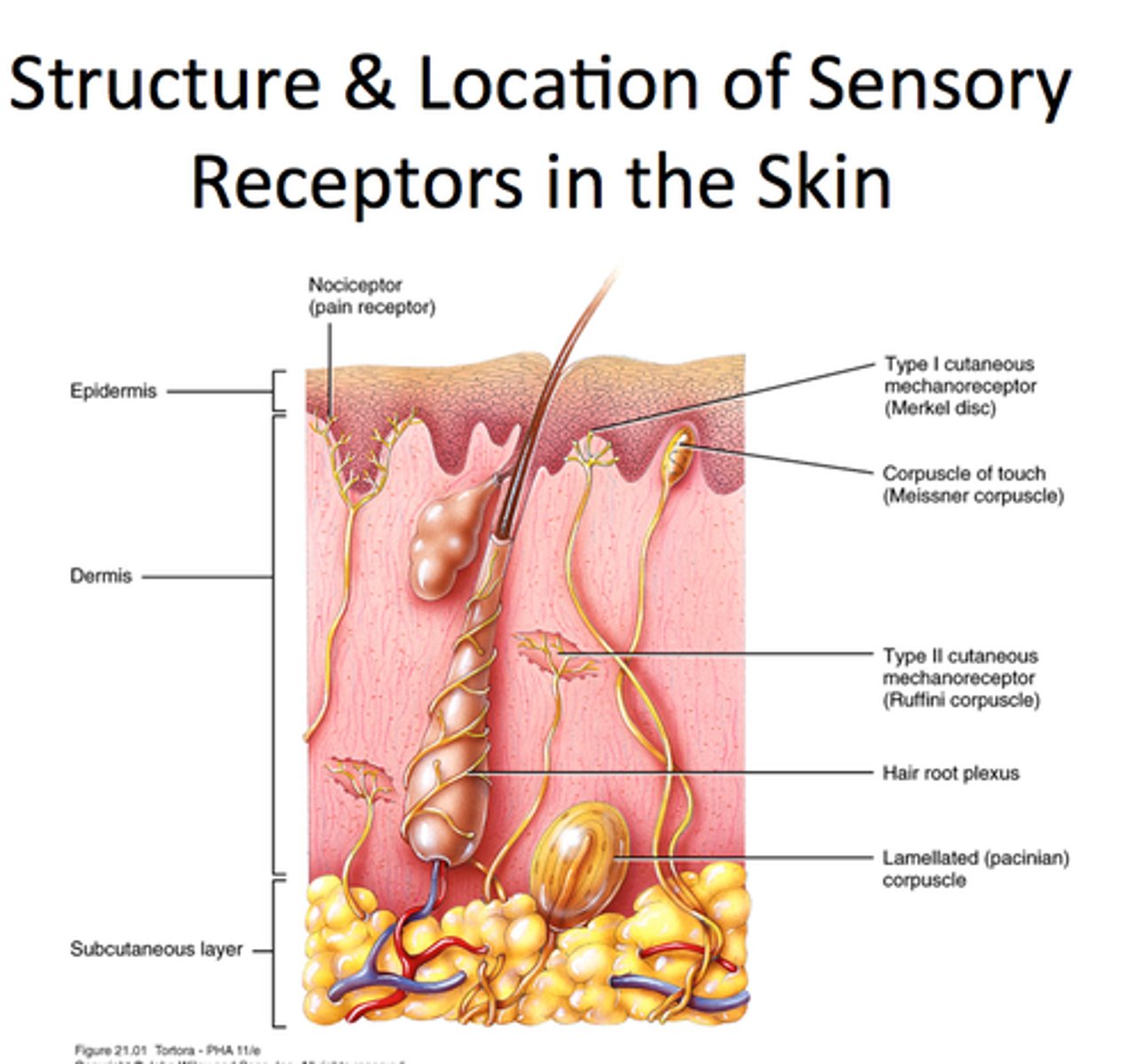
Sensory Receptors
Specialized to respond to changes in environment (stimuli).
Graded Potentials
Activation of sensory receptors results in graded potentials that trigger nerve impulses.
Sensation
Awareness of stimulus occurs in the brain.
Perception
Interpretation of meaning of stimulus occurs in the brain.
Classification of Receptors
Three ways to classify receptors: by type of stimulus, body location, and structural complexity.
Mechanoreceptors
Respond to touch, pressure, vibration, and stretch.
Thermoreceptors
Sensitive to changes in temperature.
Photoreceptors
Respond to light energy (example: retina).
Chemoreceptors
Respond to chemicals (examples: smell, taste, changes in blood chemistry).
Nociceptors
Sensitive to pain-causing stimuli (examples: extreme heat or cold, excessive pressure, inflammatory chemicals).
Exteroceptors
Respond to stimuli arising outside body, including receptors in skin for touch, pressure, pain, and temperature.
Interoceptors
Respond to stimuli arising in internal viscera and blood vessels, sensitive to chemical changes, tissue stretch, and temperature changes.
Proprioceptors
Respond to stretch in skeletal muscles, tendons, joints, ligaments, and connective tissue coverings of bones and muscles.
Simple Receptors
Modified dendritic endings of sensory neurons found throughout the body that monitor various sensations.
Special Sense Receptors
Housed in complex organs (e.g., retina for vision, cochlea for hearing) and highly specialized for one type of stimulus.
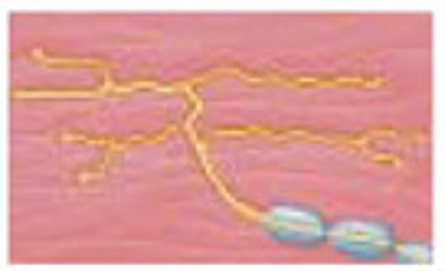
Nonencapsulated Nerve Endings
Bare dendritic endings (no connective tissue wrapping) abundant in epithelia and connective tissues.; Mostly unmyelinated C fibers (slow, dull pain/temperature) or lightly myelinated Aδ fibers (sharp, fast pain).
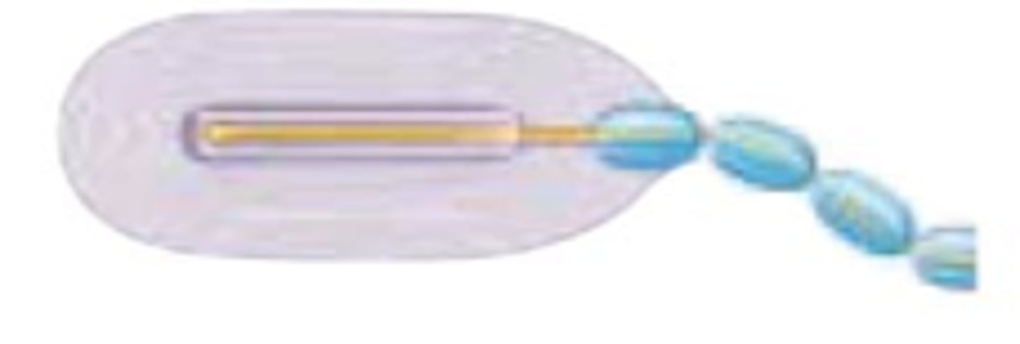
Transmission Lines
Nerves are bundles of axons (myelinated/unmyelinated) in connective tissue that can regenerate if damaged.
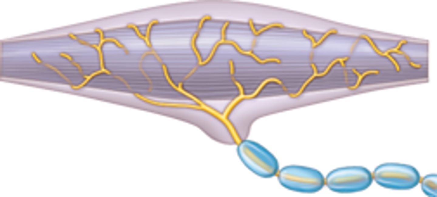
Somatic Motor Neurons
Innervate skeletal muscles (voluntary).
Autonomic Motor Neurons
Innervate smooth/cardiac muscles and glands (involuntary).
Reflex Activity
Rapid, involuntary responses (e.g., pulling hand from heat).
Cold receptors
Active at 10-40°C (superficial dermis).
Heat receptors
Active at 32-48°C (deeper dermis).
Vanilloid receptor (TRPV1)
Ion channel activated by heat (>43°C), acid (low pH), or capsaicin (why chili peppers feel 'hot').
Itch receptors
Stimulated by chemicals like histamine (allergic reactions).
Tactile (Merkel) discs
Light touch receptors in epidermis (detect steady pressure/texture).
Hair follicle receptors
Detect hair movement (e.g., mosquito landing).
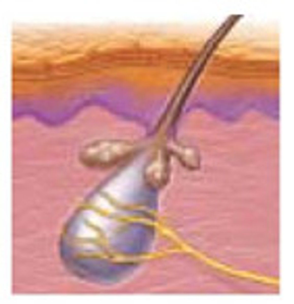
Encapsulated Nerve Endings
Structure: Dendrites wrapped in connective tissue (enhances sensitivity/specificity).
Meissner's corpuscles
Light touch/vibration in hairless skin (e.g., fingertips).
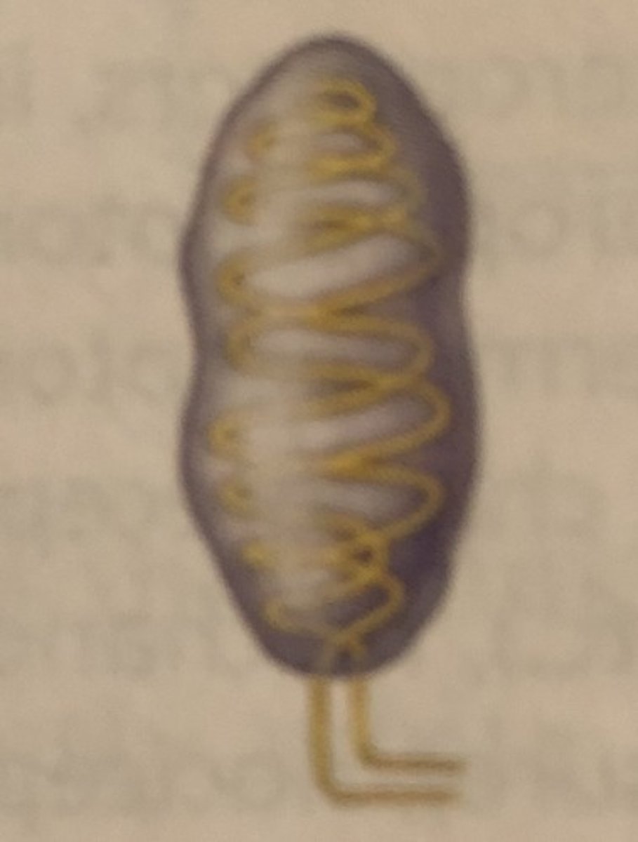
Pacinian corpuscles
Deep pressure/vibration (e.g., subcutaneous tissue).
Ruffini endings
Stretch/persistent pressure (e.g., joints, ligaments).
Somatosensory system
Part of sensory system serving body wall and limbs; Receives inputs from: Exteroceptors, proprioceptors, and interoceptors.
Receptor level
Sensory receptors.
Circuit level
Processing in ascending pathways.
Perceptual level
Processing in cortical sensory areas.
Adaptation
Change in sensitivity in presence of constant stimulus.
Phasic receptors
(fast-adapting) send signals at beginning or end of stimulus.
Tonic receptors
Adapt slowly or not at all.
Spinal Cord/Brainstem
Initiates reflex arcs (e.g., withdrawing a hand from heat before feeling pain).
Thalamus
Acts as the 'gateway' to the cortex, filtering irrelevant inputs (e.g., ignoring constant clothing pressure).
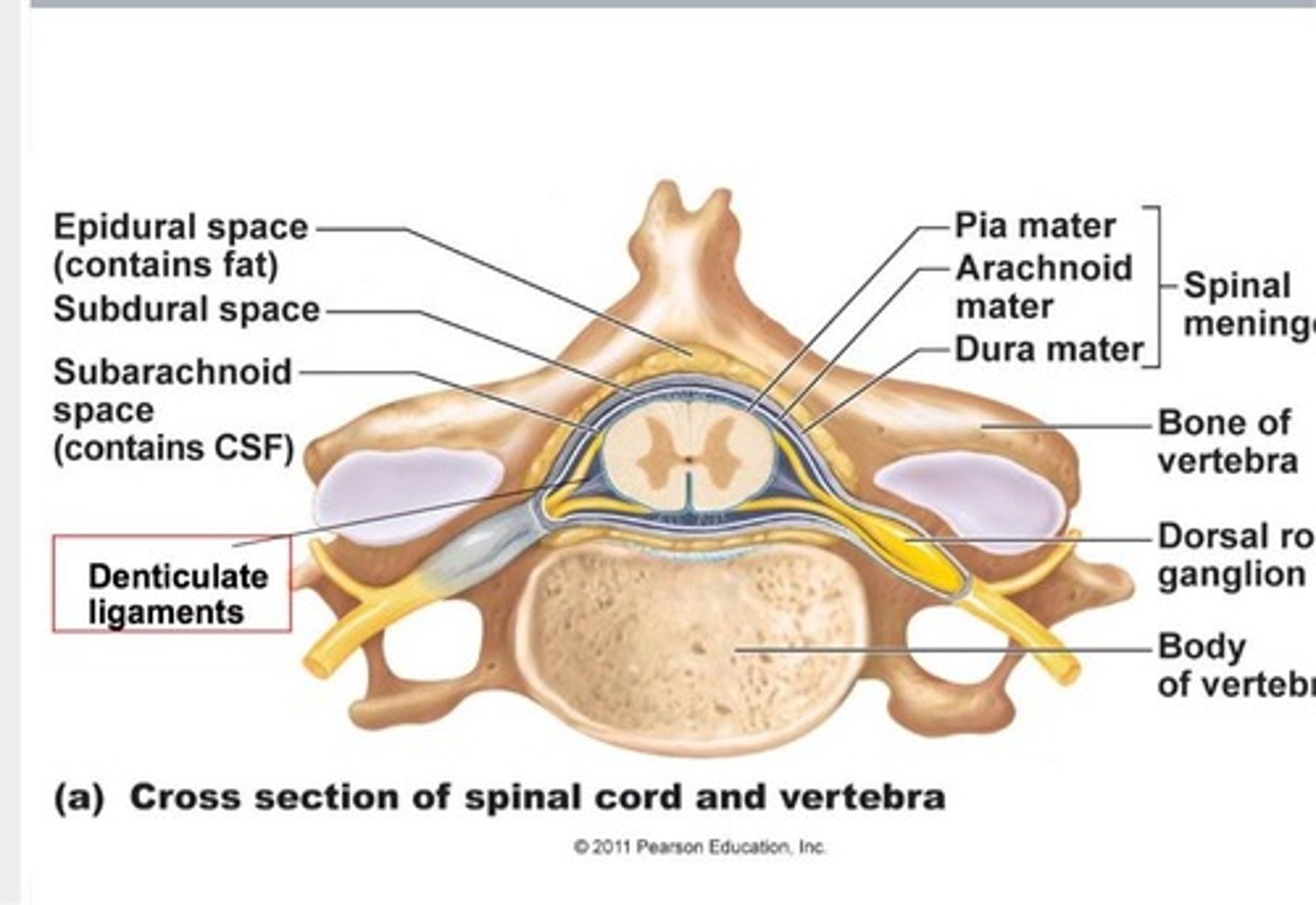
Spinal Cord
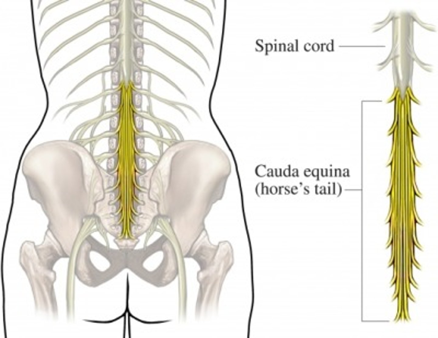
Enclosed in vertebral column, begins at the foramen magnum and ends at L1 or L2 vertebra; provides two-way communication to and from brain and body.
Major Reflex Center
Reflexes are initiated and completed at spinal cord.
Spinal Dura Mater
Is one layer thick; does not attach to vertebrae.
Epidural Space
Cushion of fat and network of veins in space between vertebrae and spinal dura mater.
CSF
Fills subarachnoid space between arachnoid and pia maters.
Meninges and CSF
Create a protected physical environment for sensory pathways to transmit signals uninterrupted.
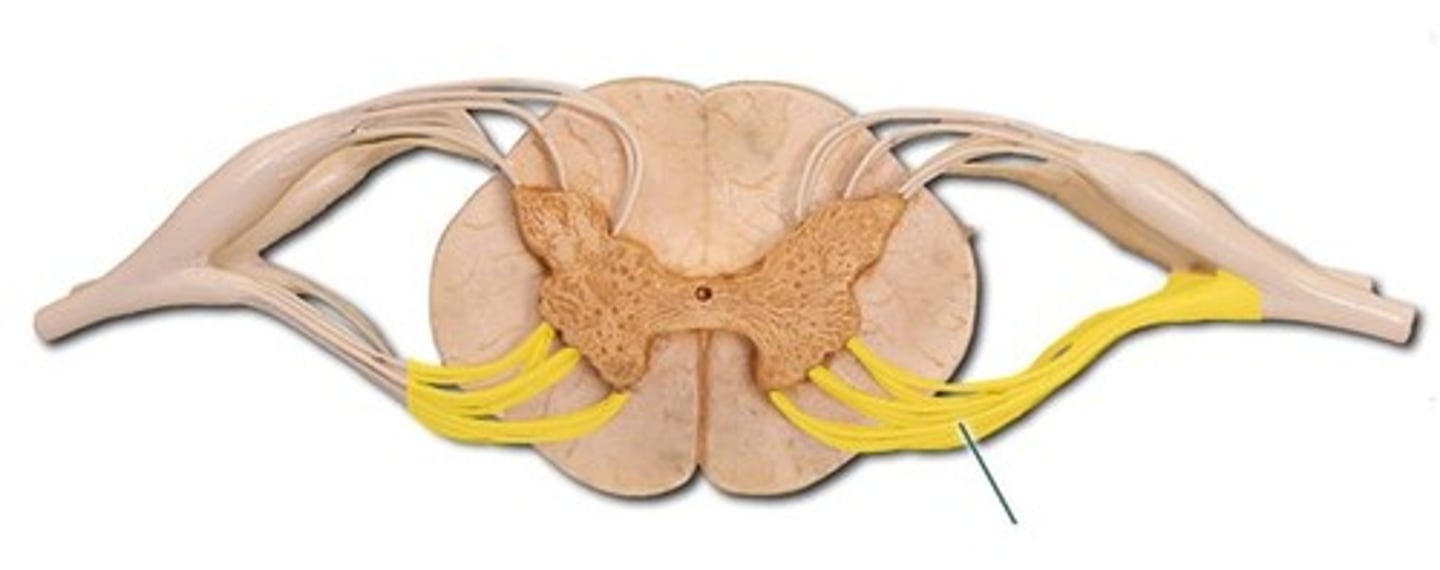
Spinal Nerves
Part of PNS; attach to spinal cord by 31 paired roots.
Cervical and Lumbosacral Enlargements
Nerves serving upper and lower limbs emerge here.
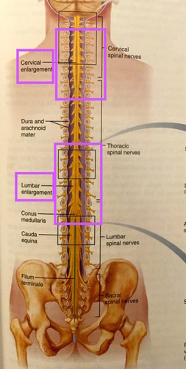
Cauda Equina
Bundle of nerve roots below L1/L2; the cord itself ends here.
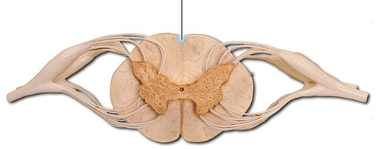
Ventral Median Fissure
Deep groove dividing the spinal cord.
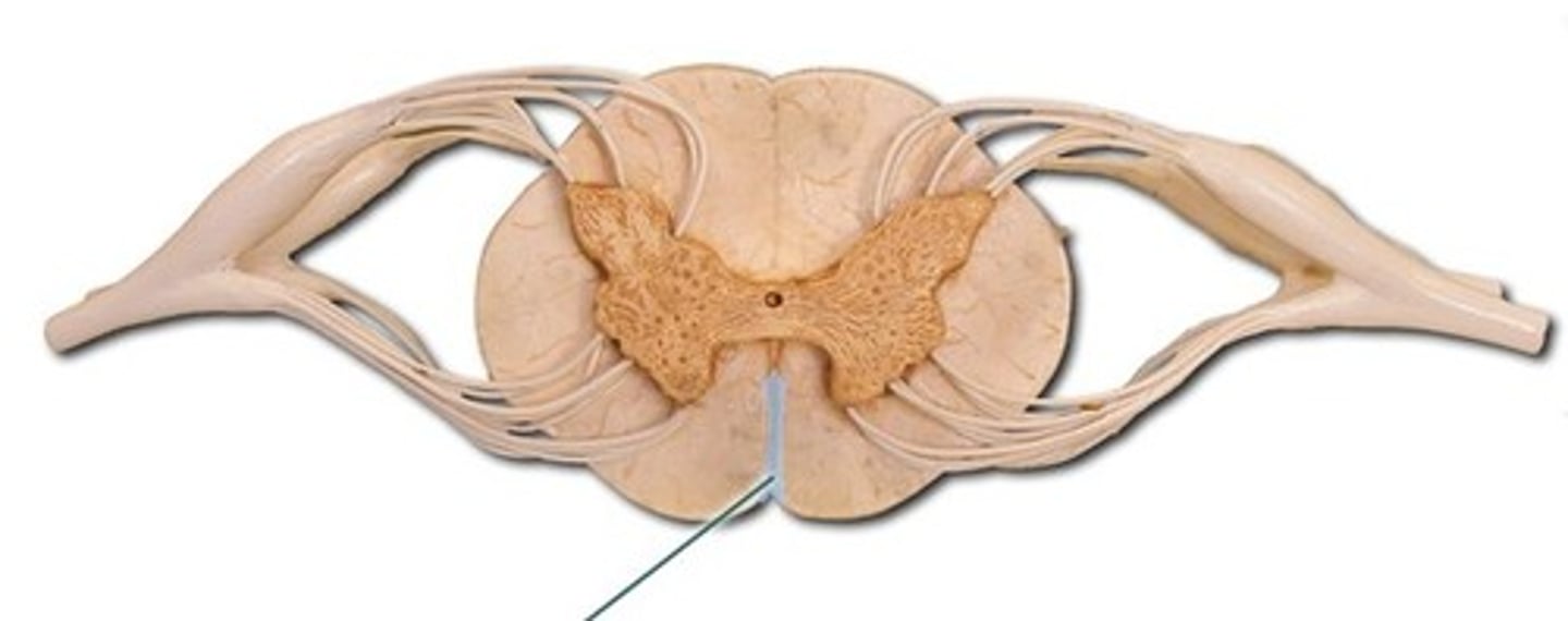
Dorsal Median Sulcus
Shallow groove dividing the spinal cord posteriorly.
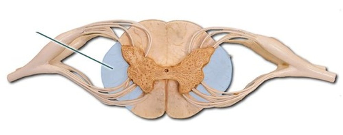
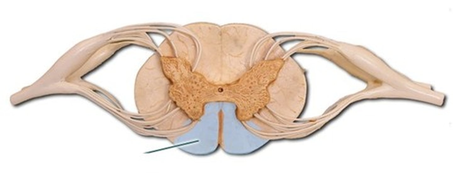
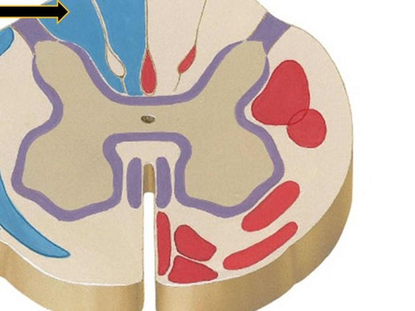
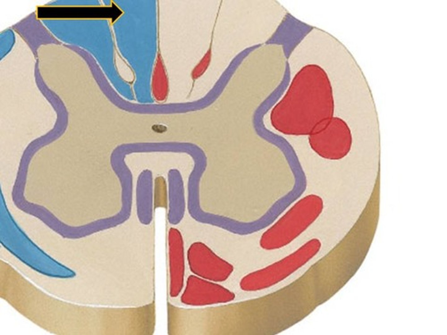
Gray Matter
H-shaped core of spinal cord containing neuron cell bodies, synapses, and interneurons.
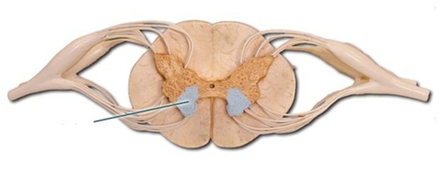
Dorsal Horns
Receive sensory input (somatic + visceral).
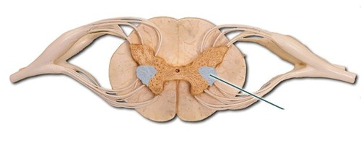
Ventral Horns
Send motor output (somatic muscles).
Lateral Horns
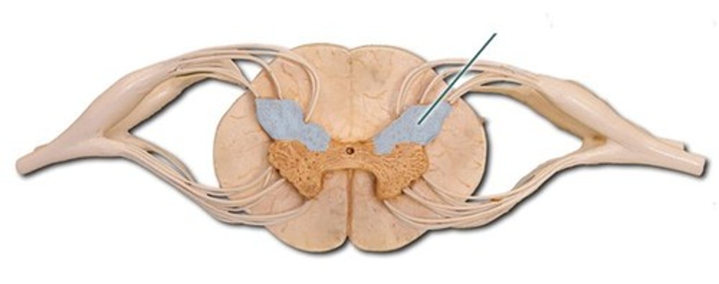
House sympathetic neurons (visceral motor) and are present only in T1-L2.
Gray Commissure
Connects left/right sides; surrounds the central canal (filled with CSF).
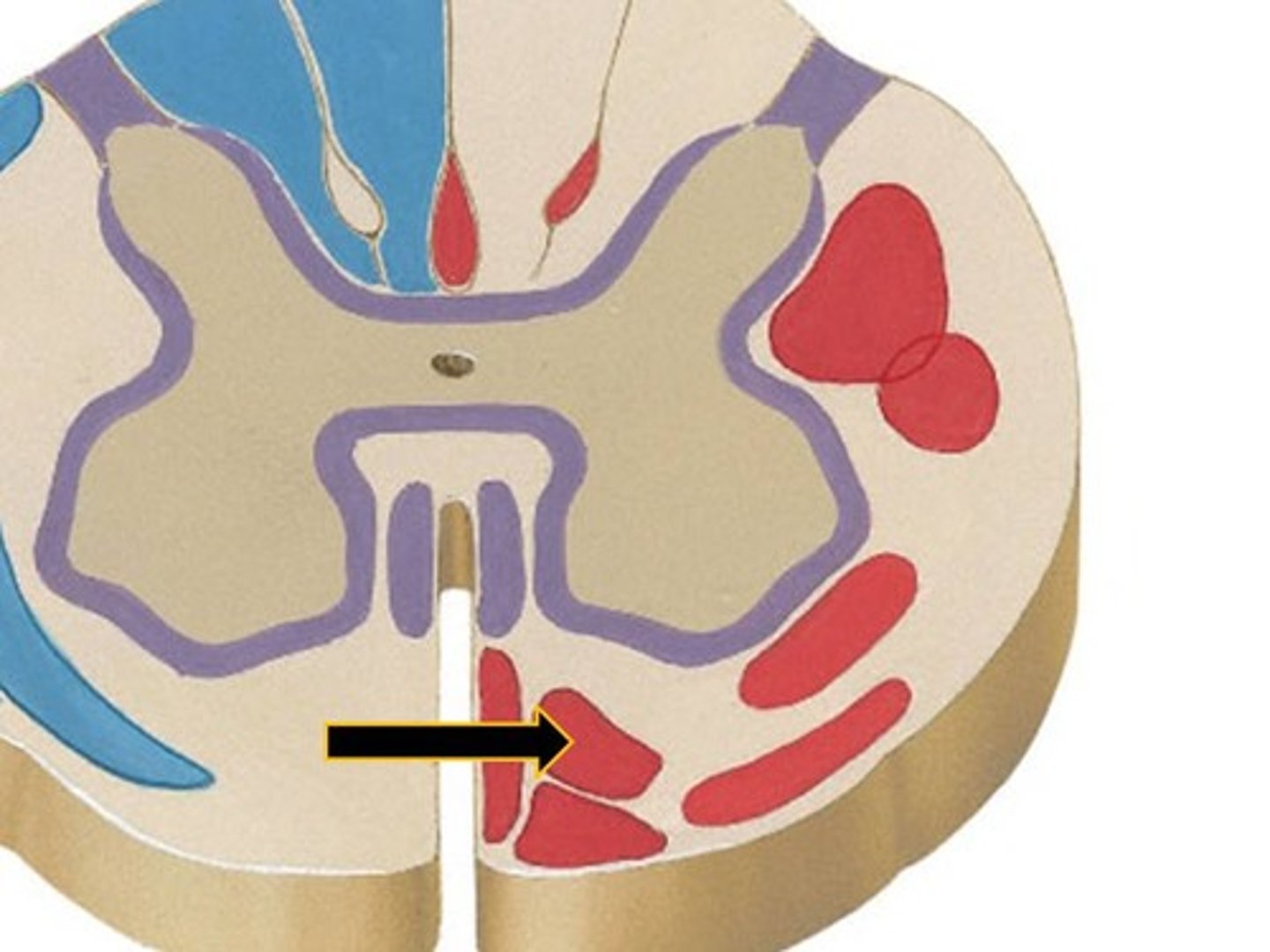
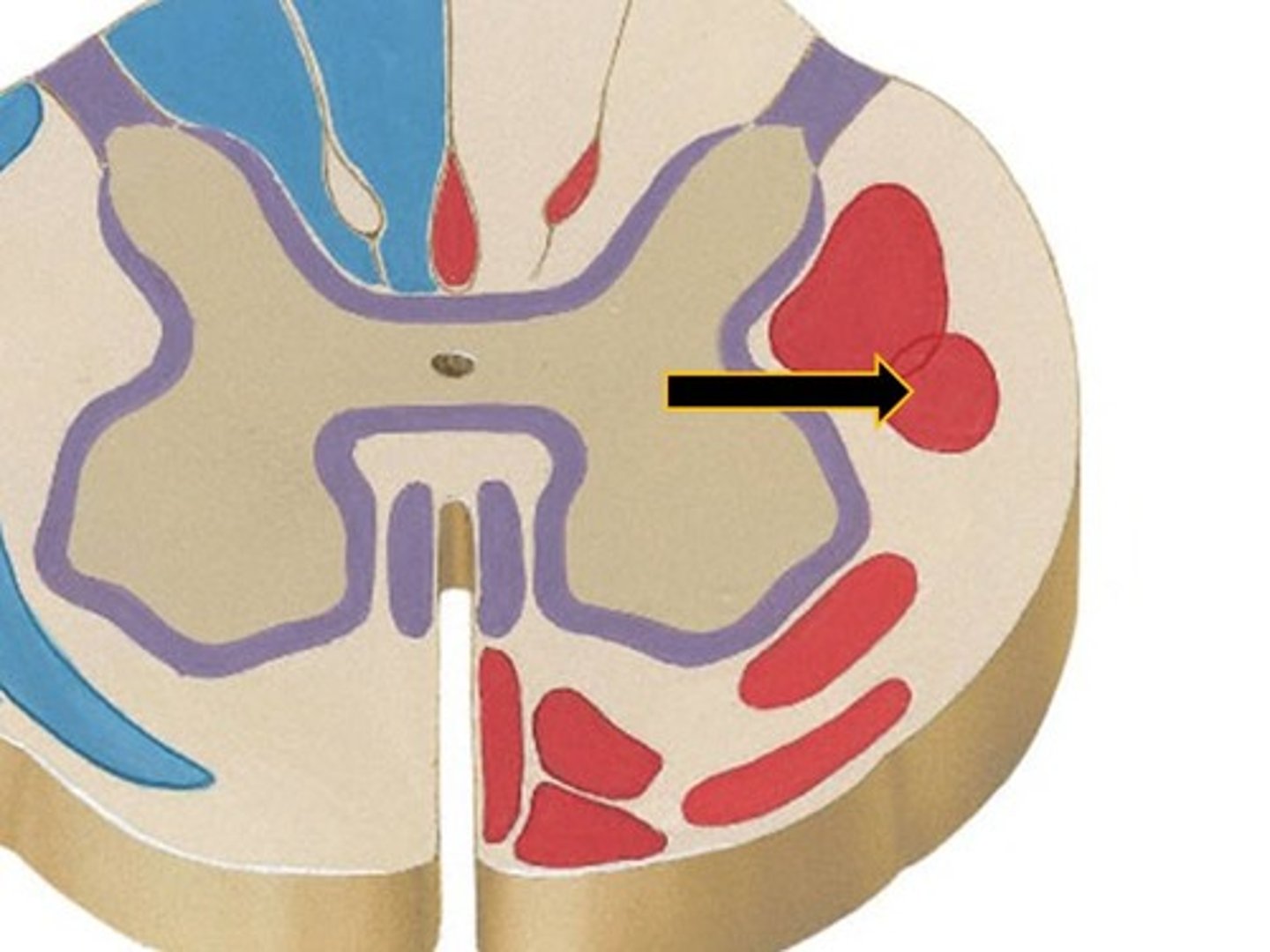
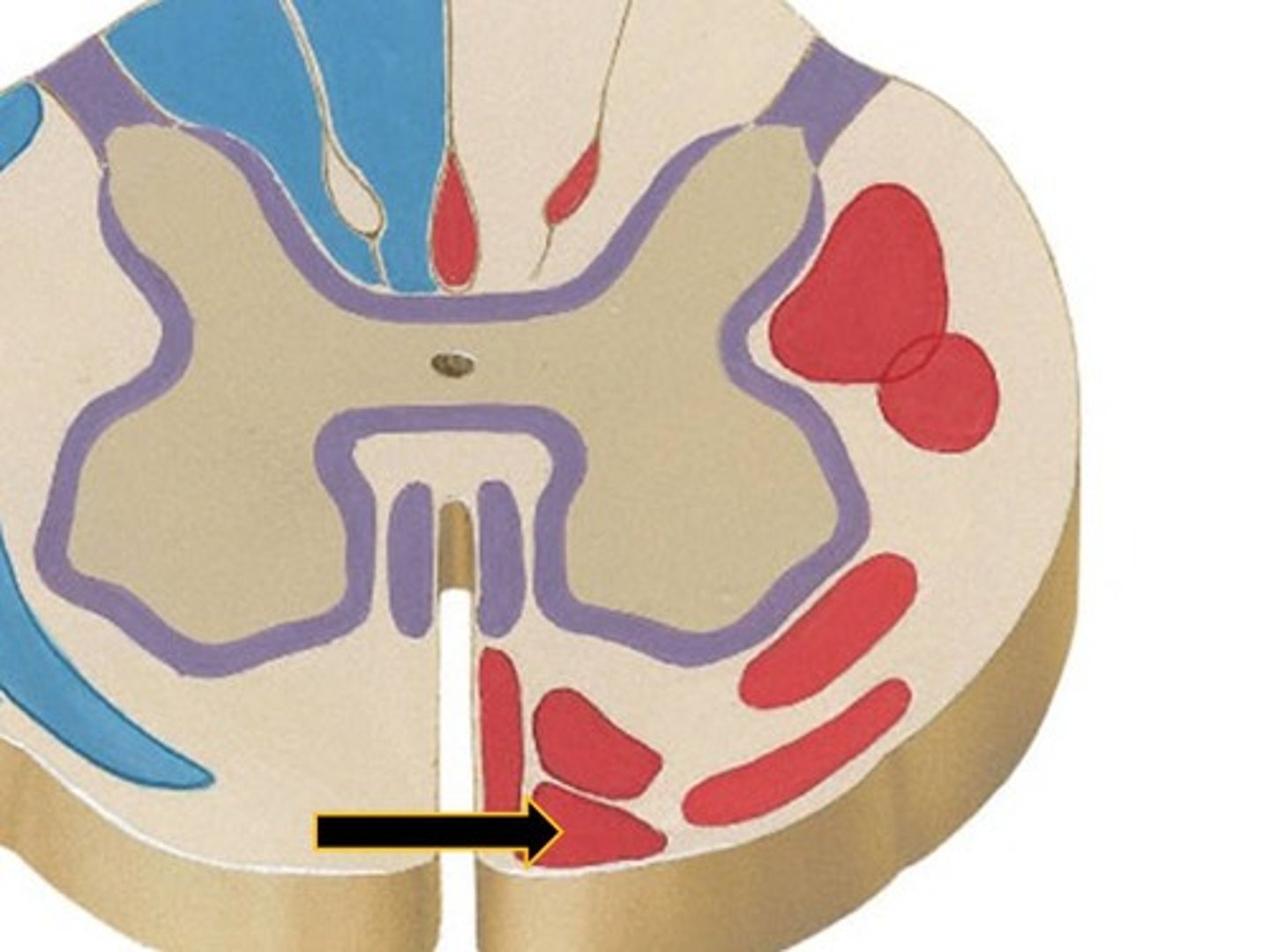
White Matter
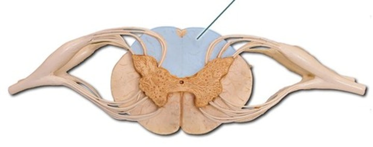
Outer region of spinal cord containing myelinated axons for rapid signal transmission.
Dorsal Funiculus
Ascending sensory tracts (e.g., touch).
Lateral Funiculus
Contains both ascending (e.g., pain) and descending (e.g., motor) tracts.
Ventral Funiculus
Mostly descending motor tracts.
Dorsal Roots
Carry sensory input into the cord.
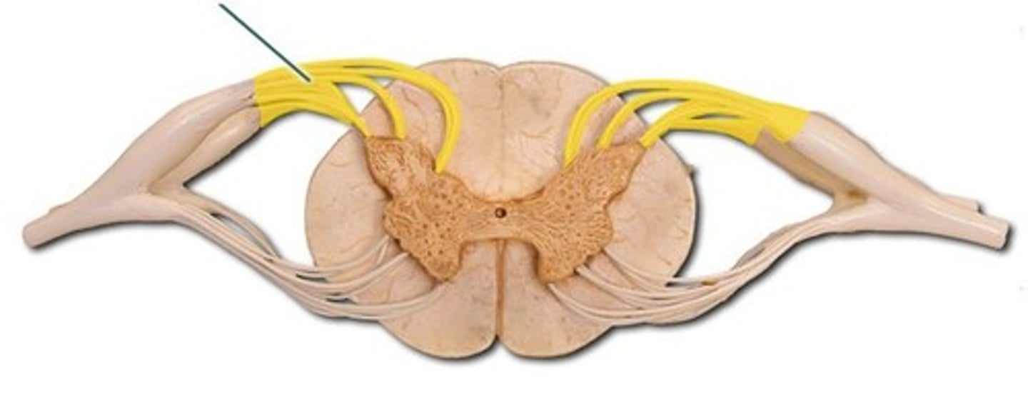
Dorsal Root Ganglia
Swellings containing sensory neuron cell bodies.
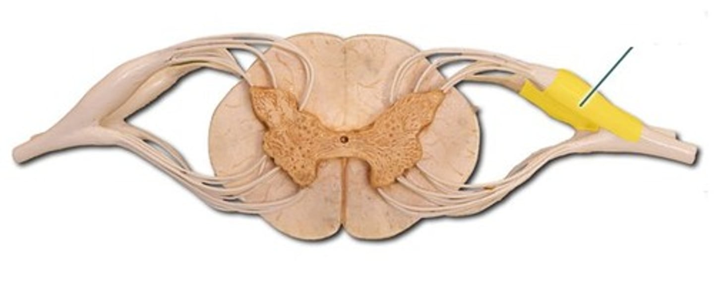
Ventral Roots
Carry motor output away from the cord.
Decussation
Most pathways cross from one side of CNS to other at some point.
Relay
Consist of chain of two or three neurons.
Somatotopy
Precise spatial relationship in CNS correspond to spatial relationship in body.
Symmetry
Pathways are paired symmetrically (right and left).
Dorsal column-medial lemniscal pathways
Transmit input to somatosensory cortex for discriminative touch and vibrations.
Fasciculus cuneatus
Paired structure in spinal cord part of dorsal column-medial lemniscal pathways.
Fasciculus gracilis
Paired structure in spinal cord part of dorsal column-medial lemniscal pathways.
Medial lemniscus
Pathway in brain (medulla to thalamus) for dorsal column-medial lemniscal pathways.
Spinothalamic pathways
Transmit pain, temperature, coarse touch, and pressure impulses.
Lateral spinothalamic tract
Part of spinothalamic pathways that transmits pain and temperature.
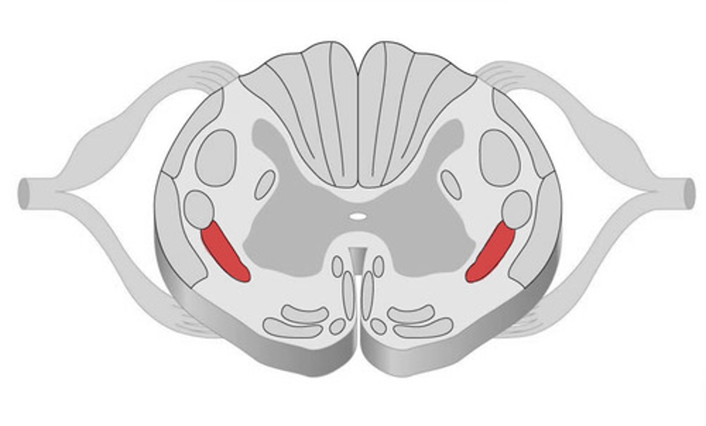
Ventral spinothalamic tract
Part of spinothalamic pathways that transmits coarse touch and pressure impulses.
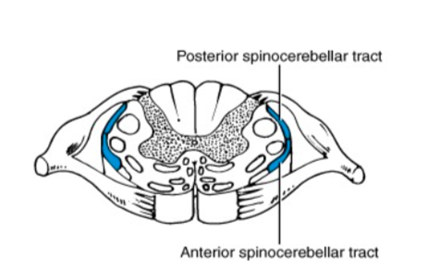
Spinocerebellar tracts
Convey information about muscle or tendon stretch to cerebellum.
First-order neuron
Conducts impulses from cutaneous receptors and proprioceptors.
Second-order neuron
Interneuron with cell body in dorsal horn of spinal cord or medullary nuclei.
Third-order neuron
Interneuron with cell bodies in thalamus, extending to somatosensory cortex.
Descending pathways
Deliver efferent impulses from brain to spinal cord.
Direct pathways
Pyramidal tracts in primary motor cortex for precise, voluntary movements.
Indirect pathways
All other pathways originating in brainstem nuclei for automatic movements.
Upper motor neurons
Pyramidal cells in primary motor cortex that initiate voluntary movement.
Lower motor neurons
Ventral horn motor neurons that innervate skeletal muscles.
Reticulospinal tracts
Maintain balance by varying tone of postural muscles.
Rubrospinal tracts
Control flexor muscles.
Tectospinal tracts
Originate from superior colliculi and mediate head movements in response to visual stimuli.
Perceptual detection
Ability to detect a stimulus (requires summation of impulses).
Magnitude estimation
Intensity coded in frequency of impulses.
Spatial discrimination
Identifying site or pattern of stimulus (studied by two-point discrimination test).
Feature abstraction
Identification of more complex aspects and several stimulus properties.
Quality discrimination
Ability to identify submodalities of a sensation (e.g., sweet or sour tastes).
Pattern recognition
Recognition of familiar or significant patterns in stimuli (e.g., melody in piece of music).