Week 10 Lab Assignment
1/5
There's no tags or description
Looks like no tags are added yet.
Name | Mastery | Learn | Test | Matching | Spaced | Call with Kai |
|---|
No analytics yet
Send a link to your students to track their progress
6 Terms
During what part of the cardiac cycle should the LVOT be measured?
mid systole
During what part of the cardiac cycle should M-Mode Aortic Cusp Separation be measured?
early systole
When measuring the LA volume in Apical 4CH, what structure must we be careful not to include?
pulmonary vein
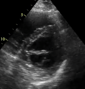
1. What view is this?
2. What is wrong with this image?
3. What would we do to correct this image? Be specific, how would we manipulate the transducer?
1. Parasternal short axis mitral valve level.
2. is more of an egg shape instead of a circle.
3. would rotate the transducer counter clockwise until it became the shape of a circle.
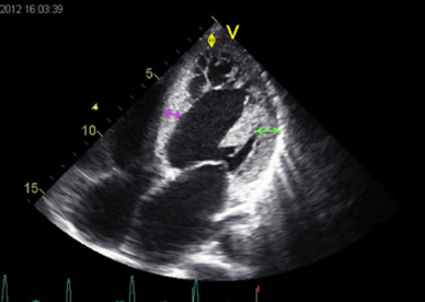
1. What view is this?
2. What is wrong with this image? (Do not worry about pathology)
3. What would we do to correct this image? Be specific, how would we manipulate the transducer?
1. Apical 4 chamber
2. Not centered, not enough depth, foreshortened and is off axis
3. rotate clockwise to bring in right side of heart. swing the transducer tail lateral and pull the transducer face down and point superiorly to get the ventricle more bullet shaped. increase depth a notch
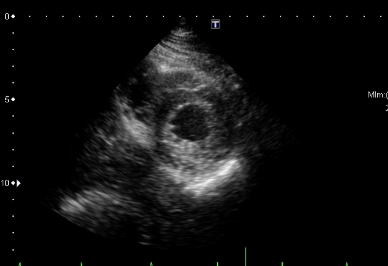
1. What view are we looking at?
2. What four wall segments can we see in this view?
1. Parasternal short axis apex level.
2. we can see the septal, anterior, lateral and inferior segments.