CH. 3 Histology: The Study of Tissues
1/128
There's no tags or description
Looks like no tags are added yet.
Name | Mastery | Learn | Test | Matching | Spaced | Call with Kai |
|---|
No study sessions yet.
129 Terms
Tissues
groups of cells located in a distinct region of the body that works together to carry out a specific function.
A. Histology- the study of tissues
B. Tissues differ from each other based on the types of cells that they contain and the specific functions of those cells.
C. Tissues are most effectively observed using microscopes. In most cases, small tissues sections are removed from the body, stained, and prepared for observation on a microscope slide.
D. Tissues are often sectioned and stained for easy observation under the microscope.
What are the 3 sections for observation that tissue can be cut along?
1) A longitudinal Section
2) A Cross Section (Transverse Section)
3) An oblique Section
Types of Tissues in the Human Body
A) Epithelial Tissue
B) Connective Tissue
C) Muscle Tissue
D) Nerve Tissue
Epithelial Tissue (Epithelium-singular)
occurs has a sheet of cells that covers or lines surfaces and cavities in the body.
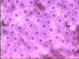
General Features of Epithelial Tissue
1) The cells of epithelium are arranged in sheets and are packed closely together.
2) It is avascular (does not contain blood vessels). Epithelial cells are nourished by nutrients that diffuse from blood vessels in the underlying connective tissue.
3) Epithelium has a nerve supply
4) Epithelium has a high regeneration rate. Cell division occurs rapidly in epithelial cells. This allows epithelial tissue to heal quickly.
5) Epithelial tissue has several major functions in the body including: protection, absorption, filtration, excretion, secretion and sensory reception.
6) Nearly all substances received or given off by the body must pass through a layer of epithelial tissue.
Structure of Epithelial Tissue
1) The Apical Surface
2) All epithelial tissues rest upon and are supported by connective tissue.
The Apical Surface
upper, free surface of epithelial tissue that is exposed to the exterior or to a body cavity.
This layer may contain fingerlike projections called microvilli or hair like structures known as cilia.
These referred to as a brush border and they aid in increasing the surface area of the tissue.

Basement membrane
a layer of attachment between epithelial and connective tissue
Functions by holding epithelial tissue in place.
A key role in regulating the movement of materials between the epithelium and the lower connective tissue

2 Layers of the Basement Membrane
A) Basal lamina
B) Reticular lamina

Basal lamina
secreted by the epithelial tissue.
Acts as a filter that determines which molecules will diffuse from underlying connective tissue.
Composed primarily of proteins and sugars, it is acellular.
Referred to as the basal surface.
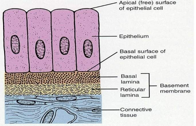
Reticular lamina
secreted by connective tissue.
functions by providing support to the overlying epithelial tissue.

Arrangement of Cells in Epithelial Tissue- an organization of cells in epithelial tissue
1) Simple epithelium
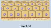
2) Stratified epithelium
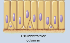
3) Pseudostratified epithelium
Simple epithelium
is only one cell layer thick
found in areas where absorption, filtration, and osmosis occurs.
Stratified epithelium
is composed of two or more cell layers.
found in areas where protection is important.
Pseudostratified epithelium
has the appearance of being several cell layers thick
only one cell layer thick
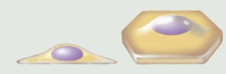
Cells Shapes in Covering and Lining Epithelium
A) Squamous cells
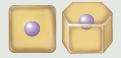
B) Cuboidal cells
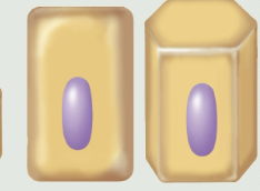
C) Columnar cells
D) Transitional cells
Squamous cells
are flat and scalelike
Cuboidal cells
are boxlike, almost as tall as they are wide
Columnar cells
are column or rectangular in shape.
Transitional cells
have the ability to change shape.
Two Broad Categories of Epithelial Tissue (both are further subdivided into specific types)
1) Simple Epithelium
2) Stratified Epithelium
Specific Types of Simple Epithelium
1) Simple Squamous epithelium
2) Simple cuboidal epithelium
3) Simple columnar epithelium
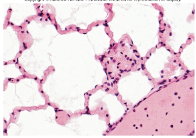
Simple squamous epithelium
composed of a single layer of flat cells
a) is a very thin tissue, so it is highly adapted for diffusion and filtration.
b) Forms endothelium, which lines blood vessels and many of the hollow organs of the body.
c) Mesothelium- serous membrane that covers the ventral body cavity
D) the most abundant epithelial tissue in the body
E) Specific functions of this tissue include allowing for rapid diffusion and secreting serous fluid.
Exfoliation
the loss of simple squamous epithelium from the surface
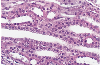
Simple cuboidal epithelium
lines glands and is involved in secretion and absorption
found in the kidney
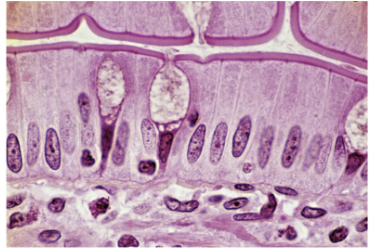
Simple columnar epithelium
A) can be nonciliated (as in the gallbladder) or ciliated (as in the Fallopian tubes- the cilia here move eggs to the uterus)
B) is often covered by microvilli
C) lines the digestive system from the mouth to the anus
D) Contains goblet cells which produce mucus.
Specific Types of Stratified Epithelium
1) Stratified Squamous Epithelium
2) Stratified Cuboidal Epithelium
3) Urothelium
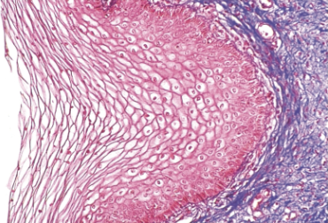
Stratified Squamous Epithelium
A) can be keratinized (covered by the thick protein keratin or nonkeratinized)
B) Keratinized squamous epithelium makes up the bulk of the epidermis while nonkeratinized squamous epithelium forms a covering over the tongue.
C) Keratinized stratified squamous epithelium resists abrasions and water loss. Nonkeratinized stratified squamous epithelium protects the body from pathogenic organisms.
Keratinized squamous epithelium
Makes up the bulk of the epidermis
resists abrasions and water loss
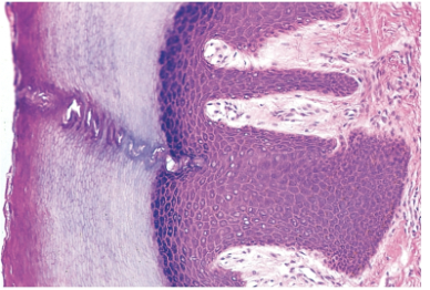
Nonkeratinized stratified squamous epithelium
forms the covering of the tongue
protects the body from pathogenic organisms.
Stratified Cuboidal Epithelium
lines the sweat glands and a portion of the ovaries.
Capable of secreting numerous hormones in the body.
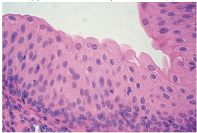
Urothelium
is composed of cuboidal cells that often bulge above the apical surface.
located in the kidney and ureter
Has an ability to stretch beyond its normal size.
Thick and often serves as a protective tissue over body surfaces.
Pseudostratified epithelium
looks thick bit is only one cell layer thick.
Pseudostratified columnar epithelium
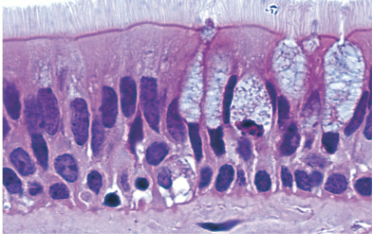
Pseudostratified columnar epithelium
located in the trachea and nasal cavity.
Contains:
Goblet cells
Cilia
Connective Tissue
the most abundant type of tissue in the human body.
Functions of Connective Tissue
1. Binds structures together
2. Provides support
3. Protects, provides immunity
4. Insulates, pads
5. Transports materials through the body
6. Movement
7. Storage
8. Some heat production
General Features of Connective Tissue
1) Has an extensive nerve supply
2) Most types are highly vascular
Structure and Organization of Connective Tissue
connective tissues is composed of 3 basic elements
a) cells
b) Fibers
c) matrix
d) Mesenchyme
Cells
make up the bulk of most connective tissues.
1) Prefix blast- refers to unspecialized, immature types of connective tissue embryonic cells.
Typically, blasts become cytes.
Ex. fibroblasts
2) Suffix -cyte refers to mature types of connective tissue cells
Fibers
provides support and strength to connective tissue
Collagen elastic fibers and reticular fibers are 3 major types of fibers found in connective tissue.
composed of various proteins
1) Collagen fibers
2) Reticular fibers
3) Elastic fibers
Collagen fibers
are extremely strong and resist stretching
These fibers are abundant in the skin, bones, and cartilage of the body
Reticular fibers
form an internal framework for several organs
the spleen and lymph nodes
Elastic fibers
composed of the protein elastin and have the ability to stretch as needed.
abundant in the connective tissue associated with the lungs and arteries of the body
Matrix
the material that surround connective tissue cells.
noncellular
It can be fluid, gelatinous or solid
Ground substance- fills the space around the connective tissue cells
Ground substance
fills the spaces around the connective tissue cells
composed of matrix and fibers
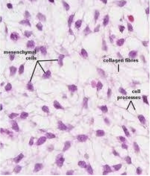
Mesenchyme
embryonic connective tissue
develops early in embryonic development and specializes into the different connective tissues that make up the body
some remains in the human body throughout our lives
Types of Connective Tissue in the Body
1) Areolar Connective Tissue
2)Adipose Tissue
3) Reticular Tissue
4) Dense Regular Connective Tissue
5) Dense Irregular Connective Tissue
6) Cartilage
7) Bone (Osseous) Tissue
8) Blood
Areolar Connective Tissue
contains all 3 types of connective tissue fibers
provides strength, support, and elasticity to body structures
Found beneath epithelial tissue
Plays a role in anchoring the epithelium in place
Serves as a packaging tissue in the body
if inflamed, areolar tissue soaks up the excess fluid to prevent edema
classified as a type of loose connective tissue
Can be stronger than steel
Adipose Tissue
fat tissue
a) Adipocytes- mature cells in adipose tissue. Specializes for fat storage. Fat is stored in a fat vacuole
b) functions as an insulator, a source of stored energy and as a padding around body storage
c) classified as a loose type of connective tissue
d) Subcutaneous layer- composed of areolar and adipose tissue. Attaches the skin to the underlying tissues and muscles.
e) white fat- typical fat in an adult
f) brown fat tissue- found primarily in a developing fetus and remains in the infant after birth for a long period of time. Get it color from the abundant supply of blood vessels found in the tissue itself. Brown fat generators heat that warms the fetus and infant
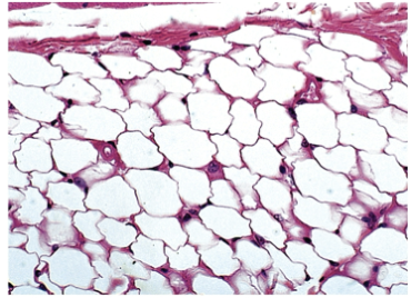
Adipocytes
mature cells in adipose tissue
specialized for fat storage
The fat is stored in a fat vacuole
Subcutaneous layer
composed of areolar and adipose tissue
attaches the skin to the underlying tissues and muscles
White fat tissue
typical fat in adults
Brown Fat Tissue
found primarily in a developing fetus and remains in the infant after birth for a period of time
gets it color from the abundant supply of blood vessels in the tissue itself
generates heat that warms the fetus and infant
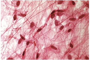
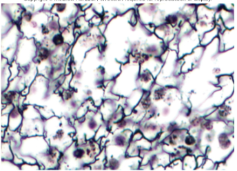
Reticular Tissue
contains only reticular fibers and forms a loose connective tissue.
a) very abundant in many lymphatic structures; like the lymph nodes, spleen, and bone marrow.
hold structures together and it can form a framework for blood cells to attach to
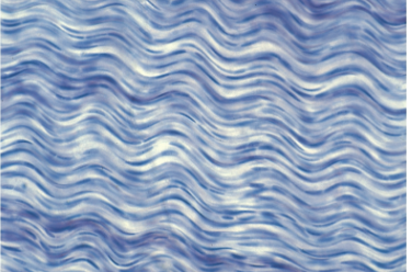
Dense Regular Connective Tissue
fibers in this tissue are pack tightly together.
Contain collagen fibers; offers a great deal of strength
provides strength and supply to body structure
tendons- composed of dense regular connective tissue
connective tissue cords that attach muscles to bone
ligaments- dense regular connective cords that attach bone to bone
Tendons
dense regular connective cord that attaches muscles to bone
Ligaments
dense regular connective cord that attaches bone to bone
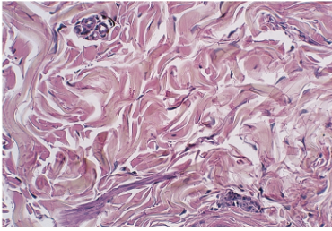
Dense Irregular Connective Tissue
fibers have an irregular arrangement
forms sheets that cover and protect organs and structures within the body
found in heart valves and in the periosteum (membrane that covers bones)
forms protective, fibrous coverings over some organs
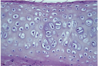
Cartilage
composed of fibers that are packaged tightly together
can withstand a great deal of stress
A) Chondrocytes- mature cartilage cells.
located in open spaces known as lacunae
Matrix surrounds the lacunae
Perichondrium- the membrane that surround cartilage tissue
Cartilage- greatly reduced blood supply and nerve supply
Very slow growing and healing
B) Chondroblasts- secrete new cartilage matrix
develop into chondrocytes
Chondrocyte
mature cartilage cells
located in open space known as lacunae
Matrix surrounds the lacunae
Perichondrium- the membrane that surrounds cartilage tissue
Chondroblasts
secrete new cartilage matrix
develop into chondrocytes
Types of Cartilage Tissue
1) Hyaline Cartilage
2) Elastic Cartilage
3) Fibrocartilage
Hyaline Cartilage
most abundant type of cartilage in the body
located at the ends of long bones
reduces friction and absorbs shock at the end of bones
make up the bulk of the embryonic skeleton
forms the epiphyseal plates of bones
referred to a articular cartilage
Elastic Cartilage
similar to hyaline cartilage in structure
form the bulk of the ears, nose, glottis (voice box), and epiglottis (flap that covers the trachea)
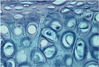
Fibrocartilage
forms pads in the human body
can withstand extreme pressure
Ex. intervertebral discs and the public symphysis
Bone (Osseous) Tissue
due to its hardness, bone has the ability to support and protect body structures
stores calcium and phosphorous for body
forms the skeleton of the body
2 Types of Bone Tissue
1) Compact Bone
2) Spongy Bone
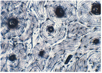
Compact Bone
forms the external covering over all human bones
composed of repeating units Haversian System (Ostenons)
Components of Haversian Systems
1) Osteocytes
2) Lamella
3) Canaliculi
4) Haversian (Central) Canals
Osteocytes
mature bone cells
located in lacunae
Lamella
rings of matrix
composed of mineral salts
Can be calcium carbonate or calcium phosphate
Canaliculi
small canals that extend from lacunae, through the lamellae of bone
nutrients and wastes pass to/ from osteocytes through these small canals
Haversian (Central) Canals
holes in the center of each Haversian system.
openings contain blood vessels and nerves
Spongy Bone
is not composed of Haversian systems
composed of thin plates- Trabeculae
Trabeculae are composed of osteocytes and lamellae
Bood
has a liquid matrix
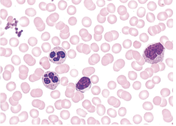
Components of Blood
1) Plasma
2) Formed elements
Plasma
the liquid portion of blood
the matrix of blood
Contains a variety of of dissolved compounds
Formed Elements
cells and cell fragments in blood
formed elements in the body include:
Erythrocytes (Red Blood Cells)
Leukocytes (White Blood Cells)
Thrombocytes (Platelets)
Erythrocytes (Red Blood Cells)
involved in carrying oxygen to the body cells and carbon dioxide to the lungs
lack a nucleus and cannot divide.
contain the red pigment hemoglobin
Leukocytes (White Blood Cells)
function in fighting infection and in providing immunity to the body
have a distinct nucleus and can divide
Thrombocytes
involved in blood clotting
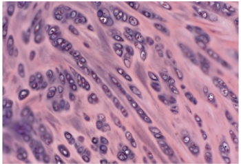
Muscle Tissue
composed of fibers that are capable of generating force from contraction
A) Function to move bones, provide body support and protection and thermogenesis (heat production)
B) compose of numerous cells- MUSCLE FIBERS
Types of Muscle Tissue in the Human Body
1) Skeletal Muscle Tissue
2) Cardiac Muscle Tissue
3) Smooth Muscle Tissue
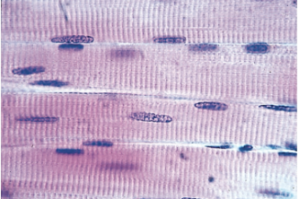
Skeletal Muscle Tissue
attaches to and moves bones
classified as being striated and voluntary
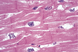
Cardiac Muscle Tissue
located in the wall of the heart
is striated and involuntary
contains intercalated discs- swelling of the cell membrane of cardiac muscle fibers
Hold cardiac muscle fibers together during muscle contraction
responsible for pumping blood throughout the body
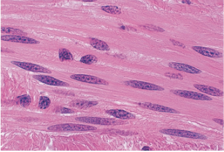
Smooth Muscle Tissue
located in the internal organs and blood vessels of the body
Is nonstriated and involuntary
regulates blood flow in and out of organs
regulates movement within the digestive system
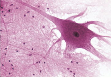
Nervous Tissue
makes up the brain, spinal cord and nerves of the nervous system
2 Types of Cell in Nervous System
1) Neuroglia (Support Cells)
2) Neurons
Neuroglia (Support Cells)
support, insulate and protect the nervous system
Neurons
the primary type of nerve cells
capable of generating and conducting impulses (electric signals) throughout the body.
Cannot divide
function by responding to stimuli via impulse formation and conduction
Tissue Repair
the process by which tissues replace dead or damaged cells
A) Tissue repair can occur by regeneration and fibrosis
B) Epithelial and connective tissue have a high capacity for continuous renewal
C) Muscle tissue repair can occur, it is generally a slow process
D) Nerve tissue does not have much capacity for renewal
What are the 2 major ways tissue repair can occur?
1) Regeneration
2) Fibrosis
Regeneration
the replacement of destroyed tissue with the same kind of tissue
Fibrosis
the formation of fibrosis connective tissue (Scar tissue) over injured tissue
Conditions that Influence Tissue Repair
1) Tissue type
2) Nutrition- nutrients needed to direct healing of tissue include vitamins A, C, E and K
3) Proper Blood Circulation- carried oxygen, nutrients to the injury site
4) Age- young tissue generally repairs faster than older tissues
Tissue Growth
A) Hyperplasia
B) Hypertrophy
C) Neoplasia
D) Metaplasia
Hyperplasia
occurs as cells increase their number
Epithelium grows in this fashion
Hypertrophy
occurs as preexisting cells enlarge in size
adipose and muscle tissue
Neoplasia
development of a tumor (either benign or malignant)
known as neoplasm (new growth)
Metaplasia
occurs when one type of tissue changes to another type of tissue (ciliated epithelium in smokers often changes to a stratified form of epithelium)