Reproductive System Pathology
1/27
Earn XP
Description and Tags
(Male and Female)
Name | Mastery | Learn | Test | Matching | Spaced |
|---|
No study sessions yet.
28 Terms
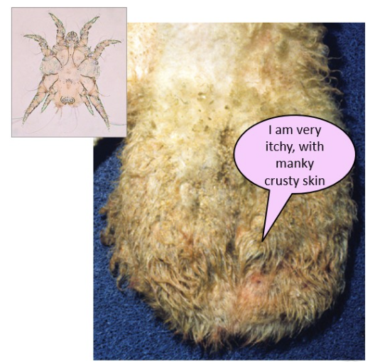
scrotal dermatitis - scrotal mange
key cause in rams in the non-burrowing mite Chorioptes bovis
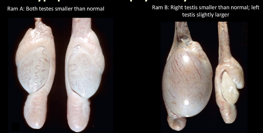
left = hypoplasia; right = atrophy
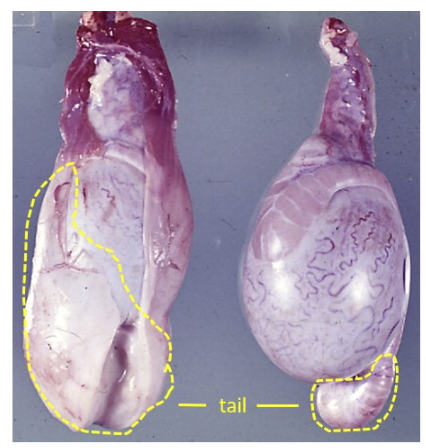
left = advanced epididymitis predominantly affecting the tail due to Brucella ovis
epididymitis caused by gram-negative pleomorphic bacteria
→ expansion of the body and tail of the epididymis due to large amounts of thick, yellow creamy exudate
→ there is also expansion of the tunic by edema and inflammation
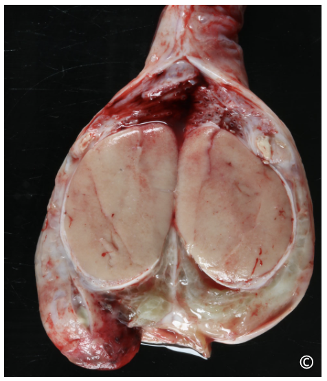
where is the lesion?
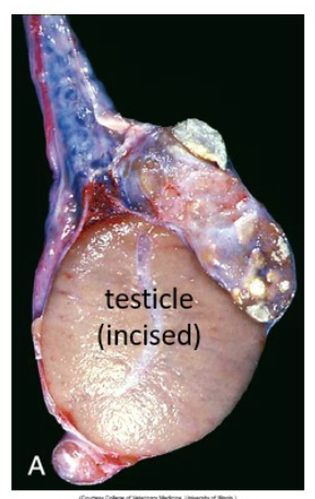
lesion is in the head of the epididymis (in a dog) → markedly enlarged due to multiple spermatic granulomas/abscesses (white-yellow masses)
body and tail of the epididymis are small, as the granulomas have obstructed the flow of sperm
where is the lesion?
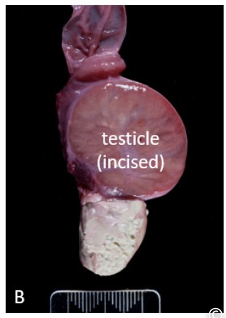
lesion is in the tail of the epididymis, which is effaced by a large granuloma
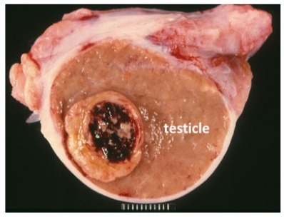
interstitial (leydig) cell tumor → well-demarcated, soft, yellow-orange masses that bulge on incision, often with areas of hemorrhage and necrosis
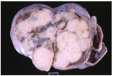
Sertoli cell tumor → firm, white, lobulated masses with prominent fibrosis; often cause marked testicular enlargement
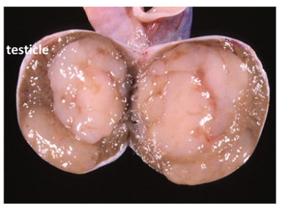
Seminoma → soft, homogenous white to beige masses with a gelatinous appearance, may resemble lymphoma
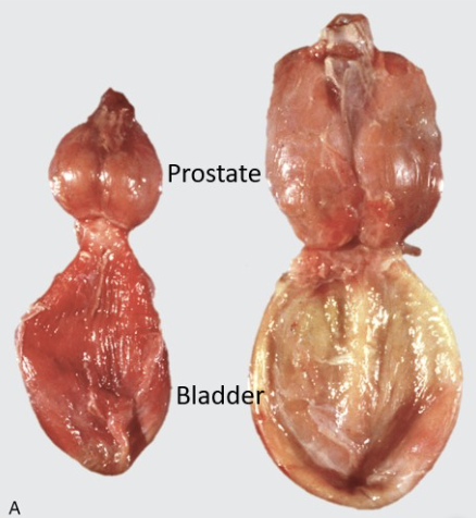
right - bilaterally and symmetrical enlarged prostate due to benign prostatic hyperplasia (BPH)
left - relatively normal prostate
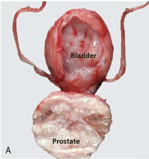
prostatic carcinoma in a dog (incised)
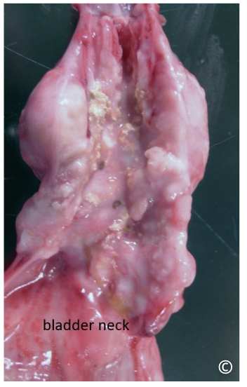
prostatitis - the prostate is enlarged and edematous with multiple foci of inflammation
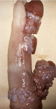
penile neoplasia - fibropapilloma: lesions are typically wart-like, multiple and found on the glans penis
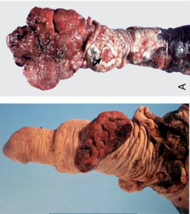
penile neoplasia - SCC: lesions arise on the glans penis and present as firm, irregular, nodular masses that frequently ulcerate and appear red, wet, and friable
penile neoplasia - transmissible venereal tumor (TVT: lesions appear as solitary or multiple masses on the penis or prepuce, often with ulceration and bleeding
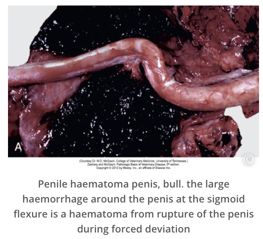
penile trauma - common in bulls due to forced deviation of the penis during mating; can result in rupture of the tunica albuginea with associated hemorrhage and hematoma formation
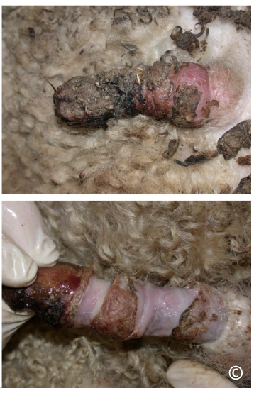
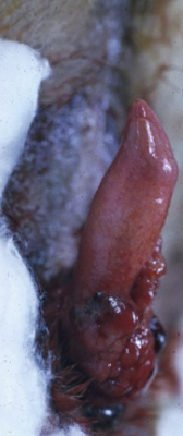
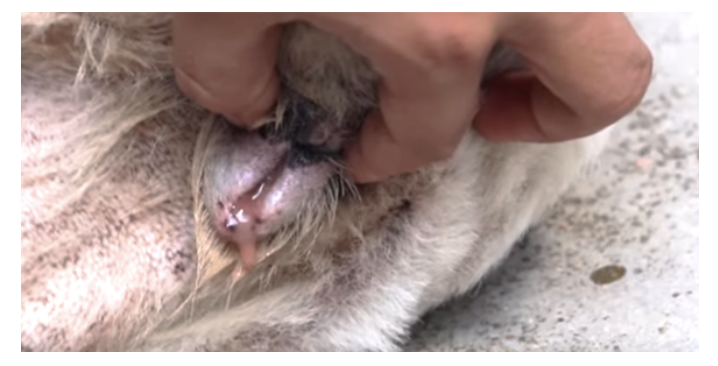
ovine posthitis (pizzle rot) - ovine balanoposthitis with inflammation, crusting and ulceration of the glans penis and prepuce
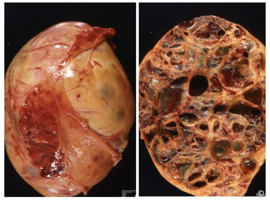
Ovarian granulosa cell tumor in a mare.
Large (>25cm!) lobulated granulosa cell tumor effacing normal ovarian structure.
Right = incised tumor with cystic + solid areas
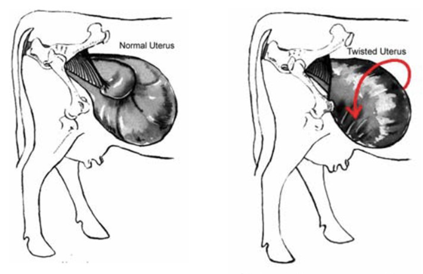
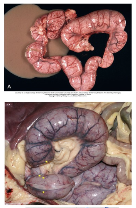
uterine torsion
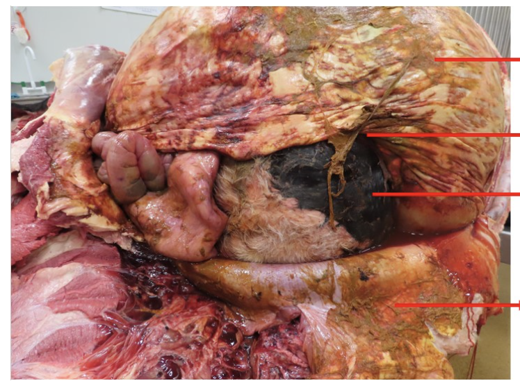
uterine rupture
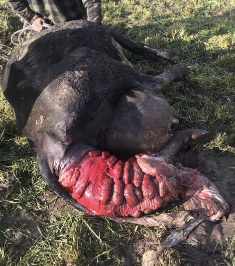
extreme case of uterine prolapse in a cow
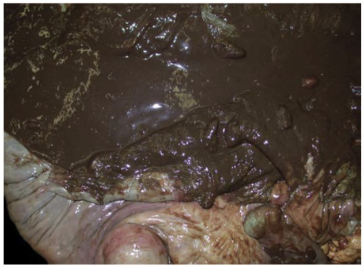
postpartum metritis in a cow → distended uterus filled with foul-smelling dark brown fluid; red-black, dull endometrium indicates bacterial infection
suppurative endometritis and pyometra in a cow
open pyometra - pus is able to drain through the cervix, often purulent or bloody
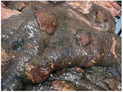
pyometra in canines → horns and body of both uteruses are grossly distended and tubular, with a dark red to purple serosal surface
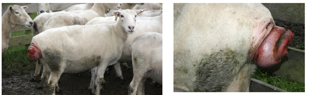
ewes with relatively recent vaginal prolapse (or ‘bearings’); tissue is congested but not yet dry, cracking and necrotic
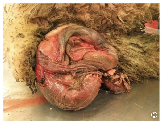
older vaginal prolapse and signs of necrosis, rupture and inflammation are present
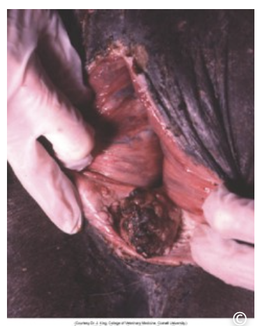
SCC in the vulva of a mare