Human Eye Plaque Model
1/63
There's no tags or description
Looks like no tags are added yet.
Name | Mastery | Learn | Test | Matching | Spaced | Call with Kai |
|---|
No analytics yet
Send a link to your students to track their progress
64 Terms
#10) Lateral Rectus Muscle: moves eye laterally
A

#18) Ciliary Body: contains the ciliary muscle & ciliary process
B

Anterior Segment: contains anterior & posterior chambers
C
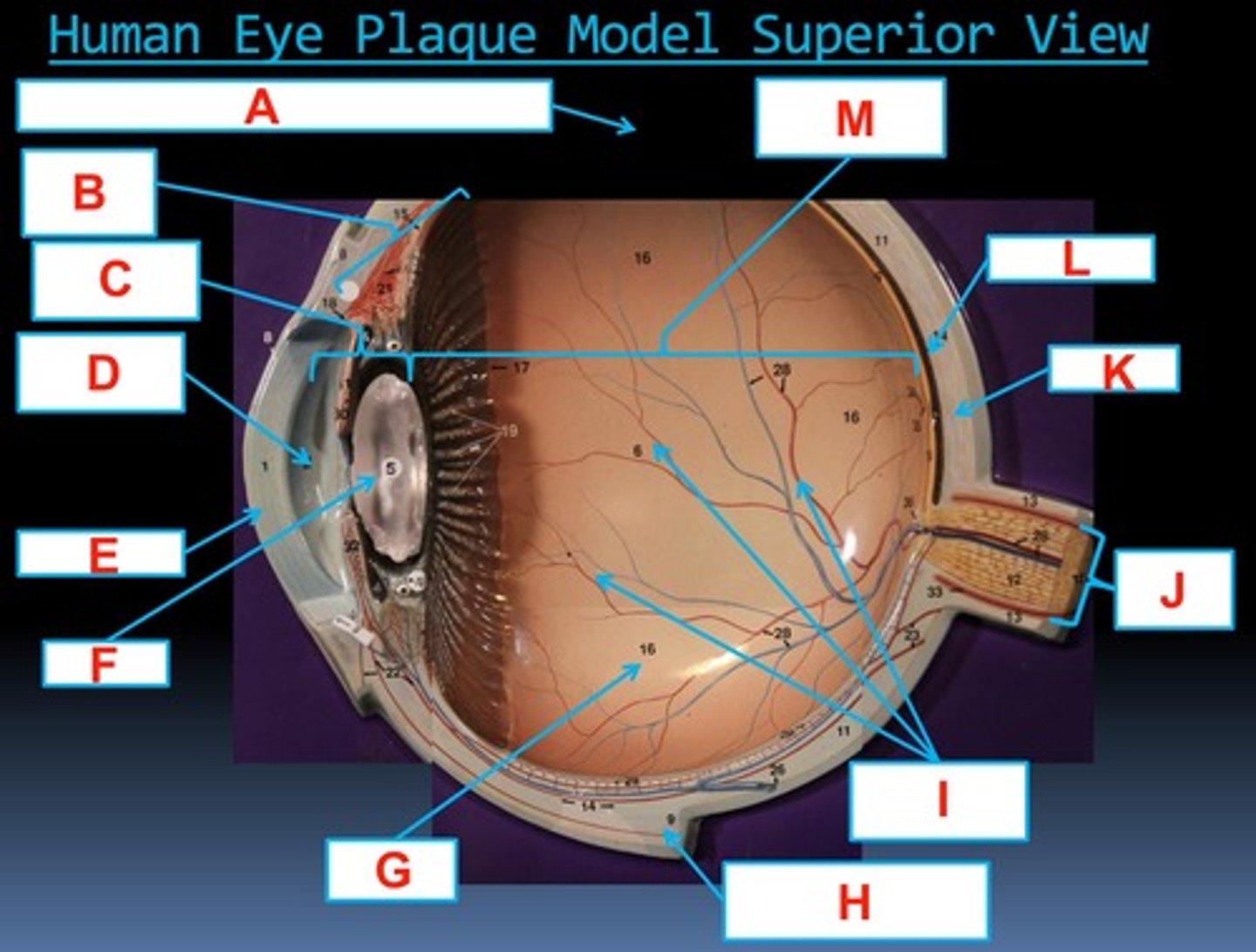
Aqueous Humor: nourishes internal eye
D

#1) Cornea: refracts light
E

#5) Lens: refracts light
F

#16) Retina: vision
G

#9) Medial Rectus Muscle: moves eye medially
H

#6) Vitreous Humor: helps keep retina in place (against wall of eyeball)
I

#12) Optic Nerve: CN#2, transmits visual signals from optic disc
J

#11) Sclera: helps maintain eyeball shape
K

#14) Choroid: vascularizes eye
L

Posterior Segment: contains vitreous humor
M

#18) Ciliary Body: contains the ciliary muscle & ciliary process
A

#4) Posterior Chamber: contains aqueous humor
B

#8) Conjunctiva: secretes mucus to lubricate eye
C

#31) Sphincter and Dilator Pupillae Muscles
D

Pupil: lets light enter eye
E

#2) Anterior Chamber: contains aqueous humor
F

#1) Cornea: refracts light
G
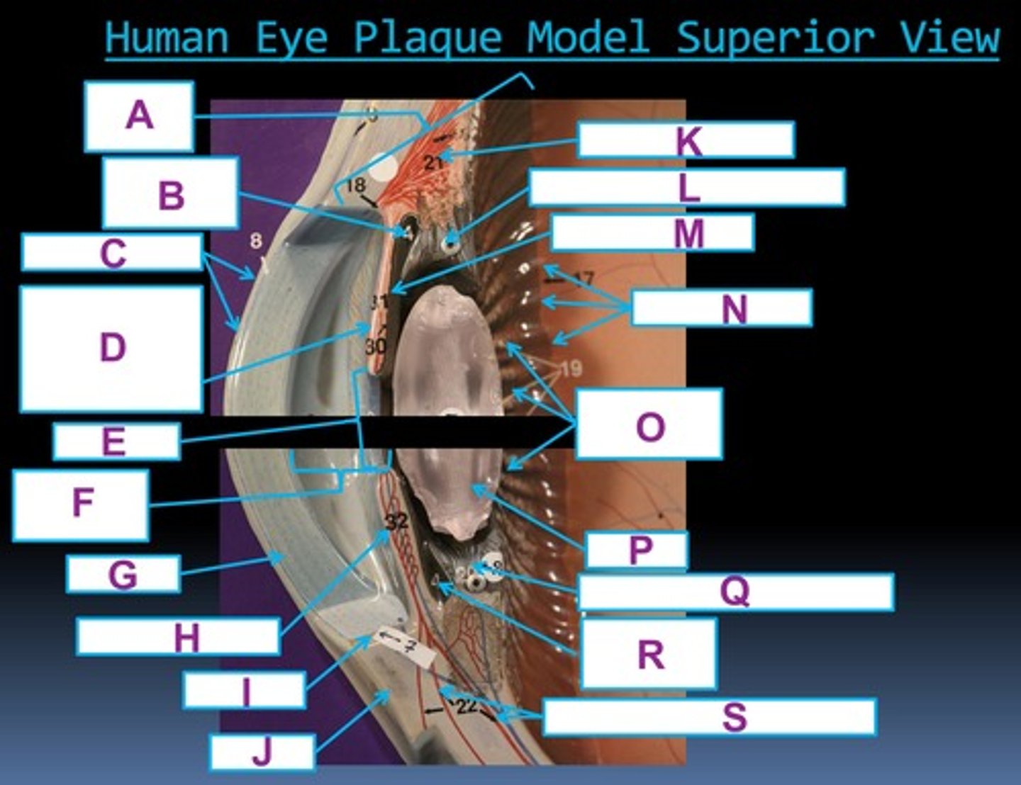
#32) Iridial Arteries
H

#7) Limbus
I

#11) Sclera: helps maintain eyeball shape
J

#21) Ciliary Muscle: helps change lens shape
K

#20) Suspensory Ligaments: attaches the lens & ciliary processes
L

#30) Iridial Retina
M

#17) Ora Serrata
N

#19) Ciliary Processes: secretes aqueous humor
O

#5) Lens: refracts light
P

#20) Suspensory Ligaments: attaches the lens & ciliary processes
Q

#4) Posterior Chamber: contains aqueous humor
R

#22) Anterior Ciliary Arteries
S

#28) Superior Central Vein of the Retina: drains into superior ophthalmic vein
A

#6) Vitreous Humor: helps keep retina in place (against wall of eyeball)
B

Retina: vision
C

#28) Inferior Central Artery of the Retina: supplies inner layers of the retina
D
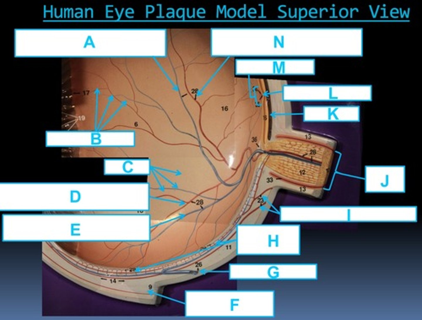
#28) Inferior Central Vein of the Retina: drains into superior ophthalmic vein
E

#9) Medial Rectus Muscle: moves eye medially
F

#26) Vorticose Vein
G

#29) Choroid Capillaries
H

#23) Posterior Ciliary Arteries
I

#12) Optic Nerve: CN#2, transmits visual signals from optic disc
J

Choroid: vascularizes eye
K

#35) Fovea Centralis: provides sharp central color vision
L

#34) Macula Lutea: area of high cone density in retina
M

#28) Superior Central Artery of the Retina: supplies inner layers of the retina
N

Choroid: vascularizes eye
A

#13) Dural Sheath
B
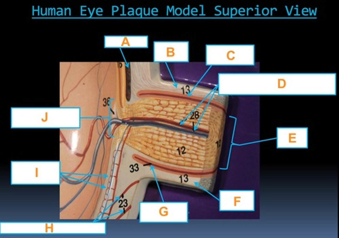
Arachnoid Mater
C

#28) Central Artery and Vein of the Retina: supplies inner layers of the retina
D

#12) Optic Nerve: CN#2, transmits visual signals from optic disc
E

#13) Dural Sheath
F

#33) Arachnoid Mater
G

#23) Posterior Ciliary Arteries
H

#29) Choroid Capillaries
I

#36) Blind Spot: total insensitivity to light
J

#44) Macular Zone: the center of direct vision
A

Optic Nerve: CN#2, transmits visual signals from optic disc
B

#38) Ganglion Cell Layer: receives depolarizing potentials from bipolar cells
C
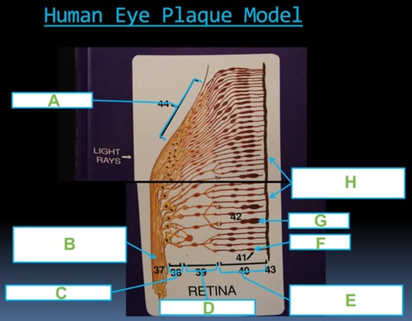
#39) Bipolar Cell Layer: sends depolarizing signals to RGCs in light
D

#40) Rods and Cones Cells Layer of the Retina: photoreceptor layer
E

#41) Rod Cell: no color, dim light & peripheral vision
F

#42) Cone Cell: color, bright light, & sharp central vision
G

Pigmented Layer of the Retina: absorbs excess light
H
