Micro. - Exam 1 Study Guide
1/44
There's no tags or description
Looks like no tags are added yet.
Name | Mastery | Learn | Test | Matching | Spaced |
|---|
No study sessions yet.
45 Terms
Microbes are ubiquitous - What does that mean? (Ch.1)
Ubiquitous = Found everywhere
An object that is free of all life forms = Sterile
The three domains of all life: Bacteria, Archaea, and Eukarya. Where did these cell types originate from? and how long ago?
The Bacteria & Archaea = Prokaryotes
Then Fungi, Helminths, Protozoans, Animals, Plants, Eukarya = Eukaryotes
Three domains originate from the last common ancestor (LCA)
How long ago? 3.8 billion
What makes the Prokaryotes different from the Eukaryotes? (Ch.1)
Prokaryotes = No nucleus, No organelles, cell wall - Peptidoglycan
Eukaryotes = Nucleus, organelles, ~ (sometimes) cell wall - Fungus
What is classification in phylogeny? What about nomenclature? (Ch. 1)
Phylogeny = “Tree of life”
Classification in phylogeny: The orderly arrangement of organisms into a hierarchy, placing them on the “branching tree of life” (Phylogeny)
Nomenclature: The assignment of scientific names for Genus and species, giving a proper name in 2 parts
Ex: “Tree of Life” - Phylogeny
3 Domains (Most general category): Bacteria, Archaea, Eukarya
Kingdom
Phylum
Class
Order
Family
Genus
species (Most specific)
(For Genus & Species: Need to know nomenclature)
What rules do we have to follow in nomenclature? (Ch.1)
Rules:
2 parts: Genus & species
Underlined (Both parts)
Genus is capitalized, species is lowercase
Ex: (Proper name in 2 parts)
“Genus species” - Handwritten
“Genus species” - Typed (Has to be italicized if typed)
What did the following scientists do or how did they contribute to the field of Microbiology? (Ch.1)
Joseph Lister
Ignaz Semmelweis
Robert Koch
Antonie van Leeuwenhoek
Louis Pasteur (he is mentioned in multiple chapters)
Joseph Lister:
Introduced hand-washing with disinfectants (Aseptic techniques)
The surgeon who advocated using disinfectants on hands and in the air prior to surgery (HW)
Ignaz Semmelweis: Handwashing (rinse hands in between patients)
Robert Koch:
“Koch’s Postulates” = a series of steps to identify a microbe in a disease (Postulates = Steps)
Famous for discovering the microbe that was causing Anthrax, he figured that out with his steps
Scientist that showed that anthrax was caused by the bacterium, Bacillus anthracis (HW)
Antonie van Leeuwenhoek:
Invented the microscope
The Dutch merchant who made and used quality magnifying lenses to see and record microorganisms (HW)
Louis Pasteur:
First to provide Germ Theory
Disproved “Spontaneous generation” with the swan-neck flask experiment
He came up with the Anthrax vaccine
Discovered viruses (Gave us the name virus)
The concept of abiogenesis was finally disproven by: Pasteur’s use of swan-neck flasks (HW)
What is genetic engineering? (Ch.1)
Manipulating DNA of an organism to create something in the industrial setting
“When humans manipulate the genes of microorganisms, the process is called?: genetic engineering” (HW)
4 main types of macromolecules that make up life?
Carbohydrates
Lipids
Proteins
Nucleic acids
Peptidoglycan, ATP, agar, enzymes, antibodies, DNA/ RNA
[Have a general idea of where these are / what their purpose is] (Ch.1)
Peptidoglycan: Component of bacterial cell wall (The Structure that’s inside bacterial cell walls) specific to just bacteria in the cell wall
ATP: Energy for the cell
Agar: An Important component of culture media; a substance that solidifies our media
Enzymes: Catalysts for all chemical reactions in cells. Speeds up reactions. (Proper description: “Biological catalyst”; speeds up reaction)
Antibodies: Products of our immune cells; glycoproteins with specific regions of attachment for bacteria, viruses, and other microorganisms
DNA: Cell instructions
RNA: Copy of cell instructions
Prokaryotes
Bacterial structures – know where they are found and their purpose: (Ch.3)
Which ones listed are found in ALL bacteria?
Flagella
Pili
Fimbriae
Glycocalyx (slime layer, capsule)
Outer membrane & LPS layer (gram -)
Cell wall
Cytoplasmic membrane
Cytoplasm
Ribosomes
Cytoskeleton
Chromosome
Plasmid
Endospores
Flagella = Structure for movement
Pili = Conjugation (swapping DNA between bacteria)
Fimbriae = Attachment
Glycocalyx (slime layer or capsule) = Outer layer for protection
It can look like a slime layer or capsule
Capsule protects bacteria from phagocytosis (white blood cells eating bacteria)
Outer membrane & LPS layer (gram -): Extra layer in the cell wall for gram -’s
Only found in gram - bacteria
Cell wall = Structure & Support
Cytoplasmic membrane = Metabolism (Found in ALL bacteria)
Cytoplasm = Water (Found in ALL bacteria)
Ribosomes = Protein synthesis; making proteins (Found in ALL bacteria)
Cytoskeleton = Extra Support (Found in ALL bacteria)
Chromosome = DNA (Found in ALL bacteria)
Plasmid = Extra DNA for extra traits
Endospores = for survival
To protect the genetic material (Protect DNA); during harsh or unfavorable conditions
Know the shapes and arrangements: (Ch.3)
Coccus, bacillus, vibrio, spirillum & spirochete
Diplococcus
Staphylococcus
Streptococcus
Streptobacillus
Palisades
Coccus: Round-shaped
Bacillus: Rod-shaped
Vibrio: Curved rod-shaped
spirillum & spirochete: Spiral-shaped
Diplococcus: Pair of round cells
Staphylococcus: Irregular cluster (Clump)
Streptococcus Chain of round cells
Streptobacillus: Chain of rod-shaped cells
Palisades: Random arrangement of bacilli
What does pleomorphic mean?
Variations in cell wall shape
Know the names for different arrangements of flagella
Polar: flagella at one or both ends
Monotrichous (One): Single flagellum
Lophotrichous: small bunches of flagella emerging from the same site
Amphitrichous: Flagella at both ends of the cell
Peritrichous: Flagella are dispersed randomly all over the cell
Cell wall differences: Gram + vs. Gram - (Ch.3)
Why do they stain different? What’s different in their cell walls?
What are the Basic steps of gram stain?
Why do they stain different?
Because of their cell wall differences
What’s different in their cell walls?
Gram + : Thick peptidoglycan layer; Purple (“Positively purple”)
Gram - : Thin peptidoglycan layer, outer membrane (Pink / Red)
Decolorize w/ alcohol (Decolorize on 3rd step)
Basic steps of gram stain:
1) Primary stain = Crystal violet
2) Mordant = Iodine
3) Decolorizer = Alcohol
4) Counter stain = Safranin
What makes the ‘special’ groups different [Mycobacteria & Mycoplasma]? (Ch.3)
Mycobacteria: Thick Cell layer wall of Mycolic acid *Very Resistant* (Has a cell wall)
Mycoplasma: They have No cell wall
How do flagella move?
What is chemotaxis? (Ch.3)
How do flagella move? - They move by Chemotaxis
Chemotaxis: movement of bacteria in response to chemical signals
Positive chemotaxis: movement toward favorable chemical stimulus
Negative chemotaxis: movement away from a repellent
What makes the bacterial ribosomes different than those in eukaryotic cells? (Ch.3)
Size difference & units
Prokaryotes: (Bacterial Ribosomes) “Odd #’s” or “Bacteria are odd little organisms”
50S (Large subunit)
30S (Small subunit)
70S (Large and small subunits together = One ribosome)
Eukaryotes: (Eukaryotic cells) “Even #’s Eukaryotes”
60S
40S
80S (Eukaryotic ribosome)
What is an endospore? (Ch.3)
Why do some bacteria make one? What is the purpose?
What is an Endospore? = Survival little shell for DNA
Why do some bacteria make one? = So that they survive during really bad conditions, so one cell survives, single cell surviving
Dinstiction of endospores:
Fungalspores: Fungal spores are meant for reproduction (1 spore can make multiple fungal spores)
Endospores for bacteria: Not multiplying (no reproduction), just a single cell surviving
What is the purpose?
Eukaryotes: (Ch.4)
Name structures Eukaryotes have that Prokaryotes don’t
What is the function of each organelle listed above?
Mitochondria: Energy or metabolism for the cell
The mitochondria is not just metabolism (Metabolism is just the summary of the process)
(TQ!) The proper term is aerobic respiration = process of extracting energy (happening in mitochondria) from chemical compounds
Golgi apparatus: Modifying proteins
Nucleus: Holds DNA
Endoplasmic Reticulum (Rough): Protein synthesis (Where proteins are made)
Differences in DNA / Chromosomes: Chromatin & histone proteins (Ch.4)
DNA:
Eukaryotes: Have Chromatin & Histone proteins
Prokaryotes: Do not have Chromatin & Histone proteins
Chromosomes:
Eukaryotes: DNA has histone proteins, helps it squish down and condense down into chromatin
Prokarytes: Don’t have that
Which groups of Eukaryotes have cell walls? Which groups have cilia? (Ch.4)
Which groups of Eukaryotes have cell walls?
Fungal group (Fungus): cell walls made of chitin
Which group have Cilla?
Protozoans
Fungus: (Ch.4)
Know the diffrence between molds vs. yeast, or dimorphic
Molds: have Hyphae
Yeast: round-shaped cells, yeast are yeast
Dimorphic: can be both (Yeast & Hyphae), depending on the temperature
Fungus: (Ch.4)
Know the structures: hyphae (septate vs aseptate), mycelium, budding
Hyphae:
If hyphae has a line going down the middle = Septate
if hyphae doesn’t have line down the middle = Aseptate
Mycelium: buddle of hypahe, a mask of hyphae
Budding: process of the yeast reproducting, forming a new yeast cell
Fungus: (Ch.4)
Know the terms: heterotrophic, saprobic, and parasitic
Heterotrophic: Get nutrients from a wide variety of organic substrates
Saprobic: get nutrients from dead plants and animals -Decomposing fungi
Parasitic: grow on the bodies of living animals or plants
Fungus: (Ch.4)
What makes up fungal cell walls that is different than bacterial cell walls?
Chitin
Helminths: (Ch.4)
Know the name for the 3 main groups, both the proper and common name
The Helminths: 3 main groups, both proper & common names
Tapeworms (Common name) - Cestodes (proper name)
Flukes (Common name) - Trematodes (Proper name)
Roundworms (Common name) - Nematodes (Proper name)
Parasitic = means that these organisms require what? (Ch.4)
Require a living host to survive
Basic life cycle: (Ch.4)
What is the basic life cycle of helminths?
Intermediate host vs definitive host, what is the difference between the two?
Basic life cycle of helminths:
Fertilized egg → Larvae → adult worm
Intermediate vs. Definitive:
Intermediate host: is where larvae develop
Definitive host: is where the adult worm develops
Porozoans:
Kingdom Protista with algae
2 basic life stages:
1) Cyst - asleep, dormant
2) Trophozoite - active, moving & feeding
Viruses = Obligate intracellular parasites, what does that mean? (Ch.5)
Have to be inside of a living host to multiply
History of viruses - who first proposed term virus and then what is meant by filterable virus? (Ch.5)
Who first proposed the term “virus”? - Louis Pasteur
What is meant by a filterable virus? - The virus passes through the filter, virus is smaller than bacteria, it passes through the filter
What are the viral structures of these?: (Ch.5)
Nucleic acids
Capsid
Envelope
Glycoprotein spikes
Naked vs Enveloped virus
Nucleic acids:
DNA or RNA, double stranded or single stranded
Capsid:
Protein shell surrounding nucleic acid
different capsid types:
Nucleocapsid: the capsid + nucleic acid
Naked viruses: nucleocapsid only
Envelope:
External covering of a capsid, usually a peice of the hosts cell membrane
Glycoprotein spikes:
Can be found on naked or enveloped viruses
Naked vs Enveloped virus:
Naked virus: nucleocapsid only
Enveloped virus: Take some of the cell membrane when released from a host cell
How do they get an envelope? - From stealing it from the host’s cell membrane
Know the order of the steps of viral replication (Ch. 5)
Adsorption
What is ‘host range’ and ‘tropism’ (Adsorption)
Host range = the limited range of cells that a virus can infect
Tropism = specificities of viruses for certain tissues
Penetration
What are the two ways viruses can get in? (Penetration)
A) Endocytosis - Cell eats virus
B) Fusion - Virus envelope fuses with host
Uncoating
Synthesis
Assembly
Release
What are the two ways viruses can get out? (Release)
A) Cell Lysis/ Rupture
B) Budding
What are Bacteriophages? (Ch.5)
Viruses that infect bacteria (the weird looking ones)
Cytopathic effects (CPE) - what is this? what do they look like & why do we look for them? (Ch.5)
Cytopathic effects (CPE) what is this?
Damage to a cell from a virus that we can see under a microscope
CPE’s all have to be viewed under a microscope
Types of CPEs:
Syncytia: cells fused together as a clump or mass
Plaque: This is clear spot where the cells are destroyed
Inclusion body: Debris from dead cells
Why do we look for them?
They tell us if a virus was present, evidence of a viral infection
Extras:
Latent viruses
Oncogenic viruses
Satellite viruses
Viroid?
What is a prion? What do prions results in [general description of disease]
Antiviral treatments:
• Have to target step in replication cycle
• Ideally would harm virus and not host
• Easier to prevent with vaccine than treat after infected
Latent viruses = dormant/asleep
Oncogenic virus = Tumor-producing virus
Satellite viruses = Dependent on other viruses for replication
Viroid = incomplete virus or plant
What is a prion? = it is an infectious protein
What do prions result in? = is progessive fatal neurogeical disease
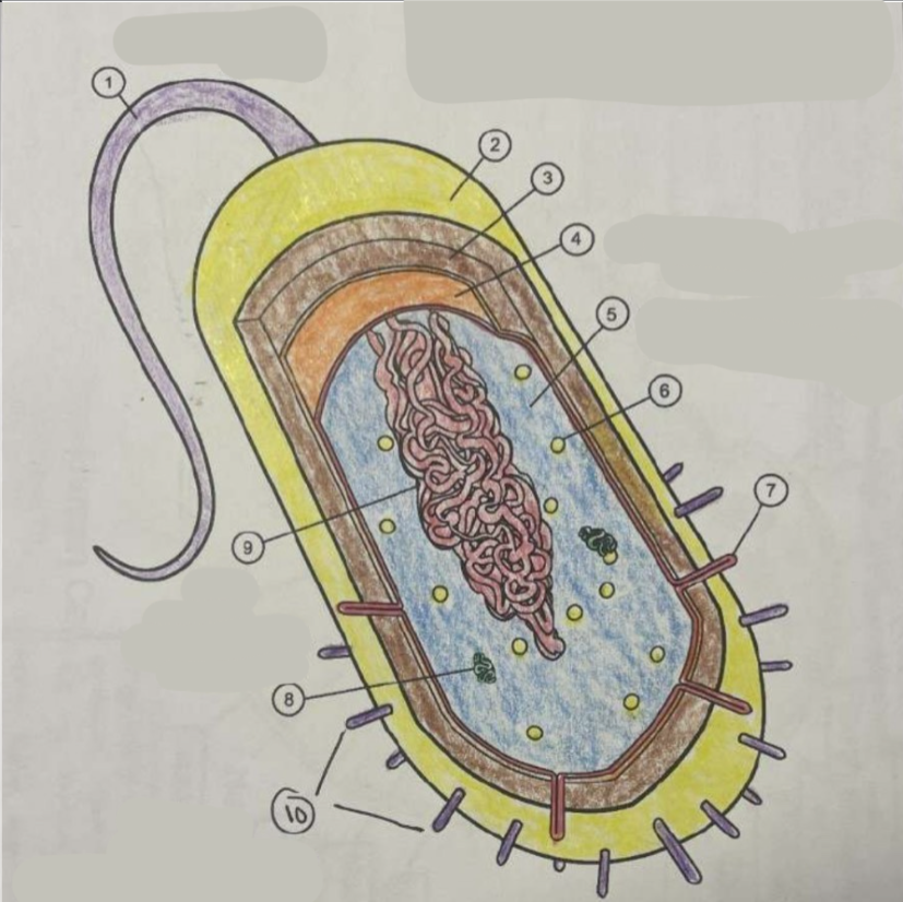
What is 1? (Name & Function)
Name: Flagellum
Function: Movement

What is 2? (Name & Function)
Name: Capsule
Function: Protection

What is 3? (Name & Function)
Name: Cell wall
Function: Structure & Support

What is 4? (Name & Function)
Name: Cytoplasmic membrane
Function: Metabolism

What is 5? (Name & Function)
Name: Cytoplasm
Function: Water

What is 6? (Name & Function)
Name: Ribosome
Function: Make proteins

What is 7? (Name & Function)
Name: Fimbriae
Function: Attachment

What is 8? (Name & Function)
Name: Plasmid
Function: Extra traits

What is 9? (Name & Function)
Name: DNA
Function: Cells instructions