TAMU BIOL 319 LAB Preface and Lab 1
1/140
There's no tags or description
Looks like no tags are added yet.
Name | Mastery | Learn | Test | Matching | Spaced | Call with Kai |
|---|
No study sessions yet.
141 Terms
Sagittal plane/section
divide the body or organ into right and left portions
Midsagittal plane/section
divides the body or organ into equal right and left sides
Frontal (coronal) plane/section
divides the body or organ into anterior and posterior portions
Transverse (cross section)
divides the body or organ into superior and inferior sides
Oblique
passes through the body or organ at an angle
Anatomical position
To stand erect with arms at the sides and palms of the hands turned forward
Superior (cranial)
toward the head end or upper part of a structure or the body; above
Inferior (caudal)
away from the head end or toward the lower part of a structure or the body; below
Anterior (ventral)
Nearer to or at the front of the body
Posterior (dorsal)
Nearer to or at the back of the body
Medial
Nearer to the midline of the body
Lateral
Further from the midline of the body
Intermediate
Between two structures
Ipsilateral
On the same side of the body as another structure
Contralateral
On the opposite side of the body as a structure
Proximal
Closer to the trunk or point of attachment
Distal
Farther from the trunk or point of attachment
Superficial
Toward or on surface of the body
Deep
Away from the surface of the body
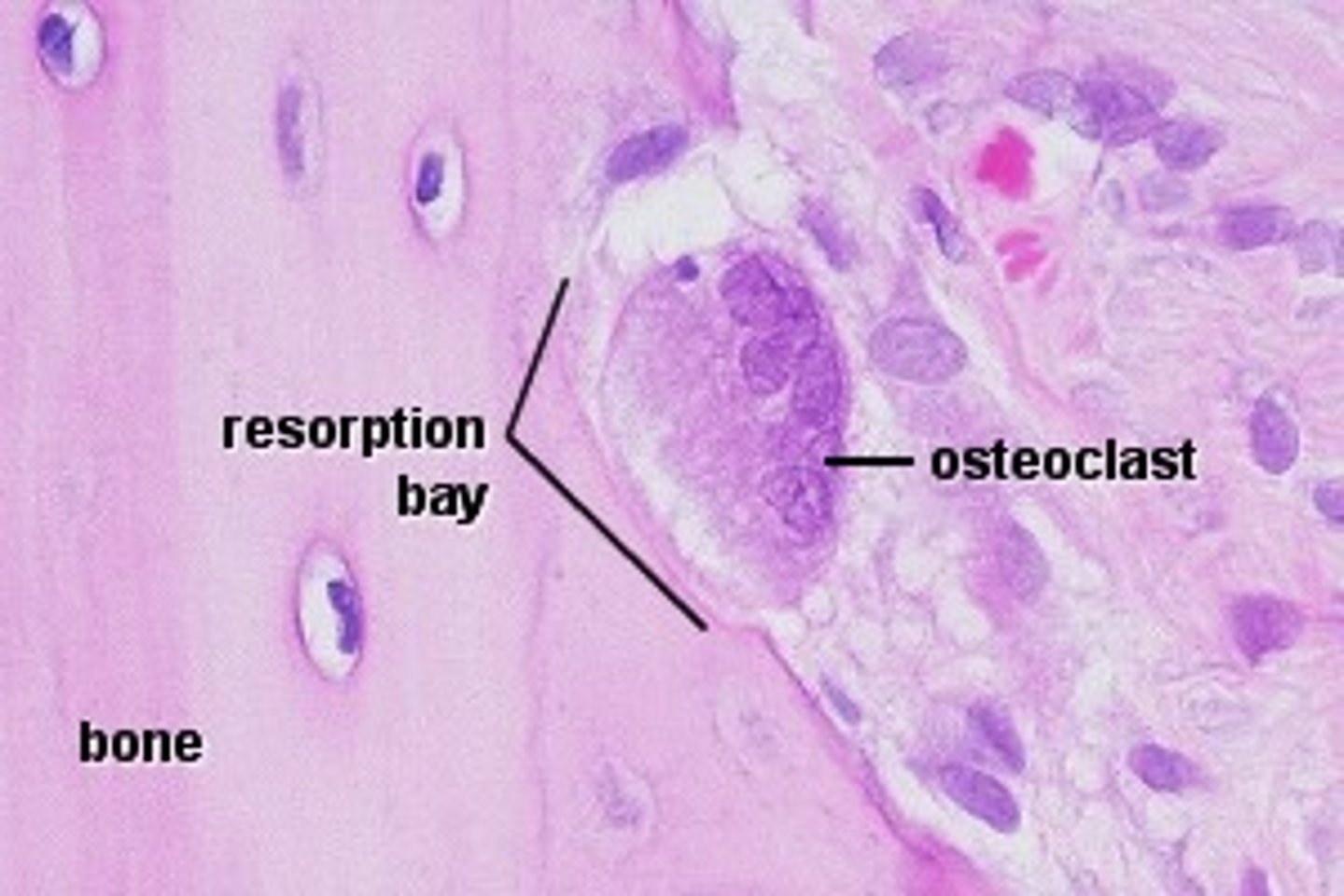
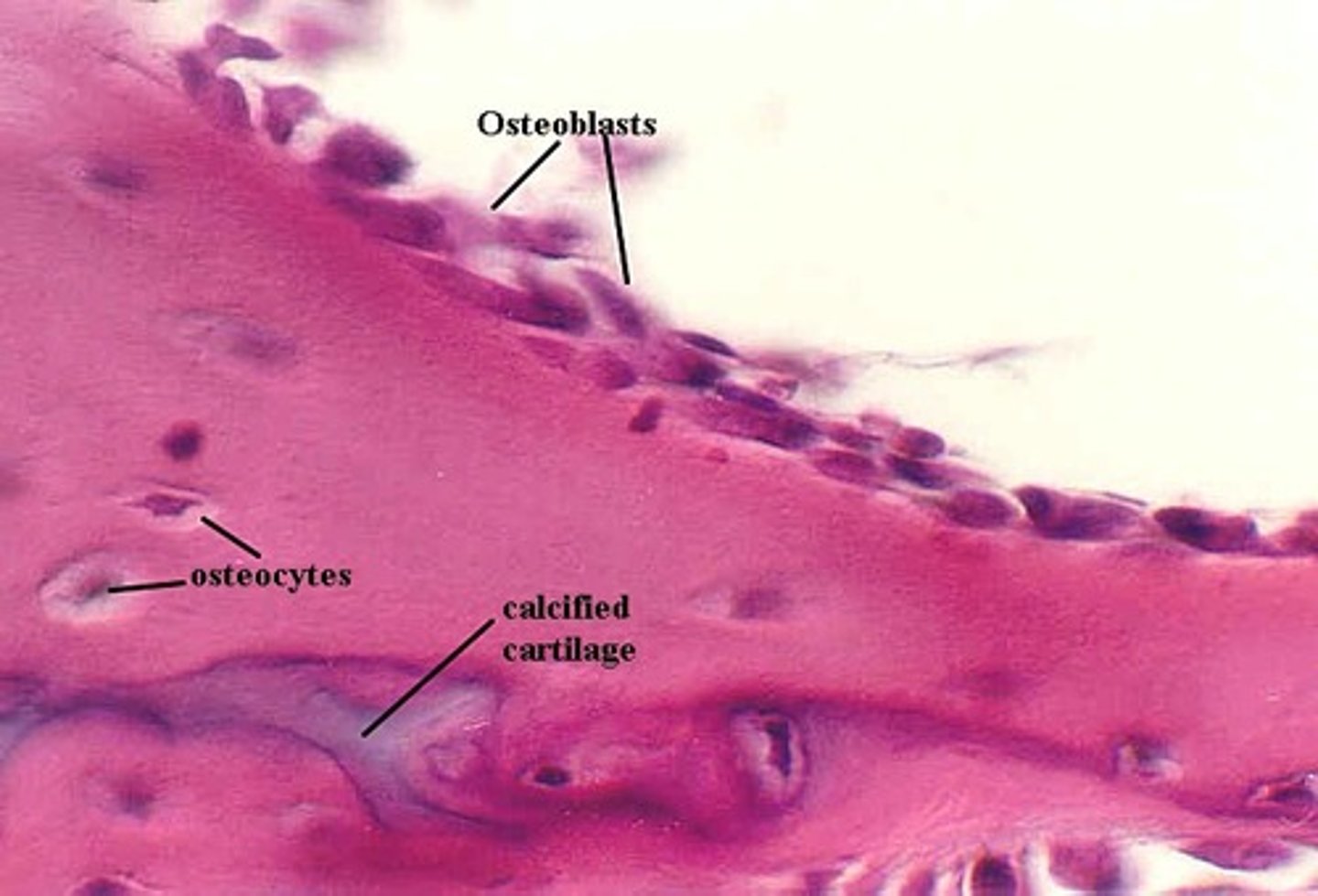
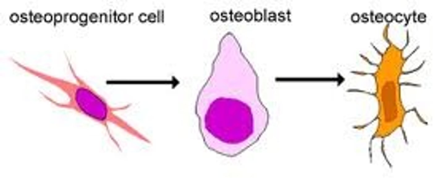
Osteocyte
derived from osteoblasts- involved in bone maintenance
Osteoclast
Multinucleate cell that secretes acids and enzymes to dissolve bone matrix
Osteoblast
Derived from osteoprogenitor cells - secretes organic components of matrix
Osteoprogenitor cells
stem cells that differentiate into osteoblasts
Histology
study of tissues
Tissues
group of cells that are similarly structured and work together to accomplish specific function
epithelial tissue
lines and covers organs, their internal passageways, and forms glands
Surfaces in epithelial tissues
Basal (basement) and apical (top)
epithelial tissues structure
sheet of cells tightly joined together by tight junctions
Simple Cuboidal Epithelium function
secretion and absorption
Stratified Squamous Epithelium function
protect underlying tissues in areas subjected to abrasion
Tight Junction
connections b/t cells that prevent fluid from going between cells and making them go through it
desmosomes
connections made up of protein b/t cells that holds them together
Epithelial Functions
filter, absorb, protect, secrete, excrete, sensory reception
Apical (free) surface
Surface of the cells that are exposed to the external environment or to an internal passage way or cavity
Basal Lamina
how epithelium attaches to body, functions as filter at base of epithelium
Simple epithelium
one single layer of cells
Goblet Cells
secrete mucus that coats cells to protect epithelia at free surface
Stratified epithelium
Composed of more than one layer of cells
Most common type of stratified epithelium
stratified squamous
Classifying stratified epithelium with 1+ epithelial cells
type at free surface determines classification of tissue
Regeneration of tissues
How easily the tissues can be regenerated; based on the rate of mitosis and the amount of blood supply
These tissues have GOOD or EXCELLENT regeneration
Epithelial tissue, bone, areolar tissue, dense irregular connective tissue, and blood forming tissue
These tissues have MODERATE regeneration
Smooth muscle and dense regular connective tissue
These tissues have WEAK regeneration
Skeletal muscle and cartilage
These tissues have NONE or ALMOST NO regeneration
Cardiac muscle tissue and (central nervous system) nervous tissue
Reasons why regeneration of tissue can be high
high rate of mitosis and adequate/abundant blood supply
reticular lamina
made up of fine collagen fibers
Basement Membrane
helps epithelia resist tearing/stretching, resists structural integrity and creates boundary
Avascular
no blood vessels or supply
Innervated
supplied by nerve fibers for regulation
Function: simple squamous epithelium
allow passage of materials by diffusion and filtration in sites where protection isn’t important; secrete lubricating substances in lining

Description: simple squamous epithelium
single layer of flattened cells with disc-shaped central nuclei and sparse cytoplasm; the simplest form of epithelia
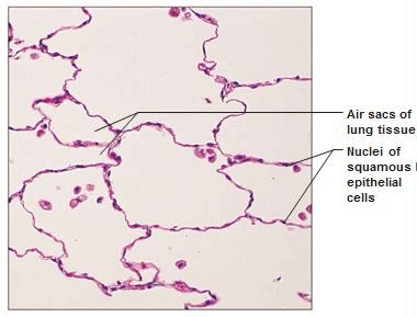
Location: simple squamous epithelium
Kidney glomeruli, air sacs of lungs, lining of heart, blood vessels, lymphatic vessels, lining of ventral body cavity
Avelous
Air sack present in between linings of the simple squamous in the lung

Endothelium
provides a slick, friction-reducing lining in hollow organs that transport fluids
Mesothelium
the epithelium found in serous membranes lining the ventral body cavity and covering its organs
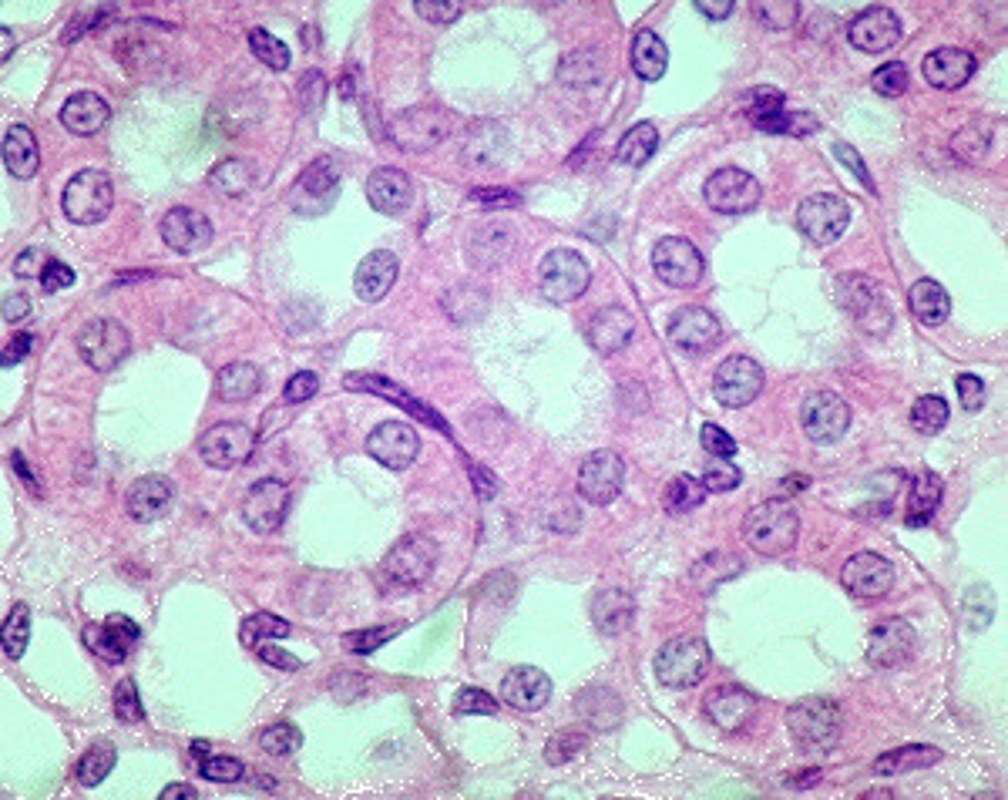
Description: simple cuboidal epithelium
single layer of cube-like cells with large, spherical central nuclei

Function: simple cuboidal epithelium
Secretion and absorption

Location: simple cuboidal epithelium
Kidney tubules; ducts and secretory portions of small glands; ovary surface
Description: simple columnar epithelium
single layer of tall cells with round/oval nuclei; some cells bear cilia; layer may contain mucus-secreting unicellular glands (goblet cells)
Function: simple columnar epithelium
absorption; secretion of mucus, enzymes, and other substances; ciliated type propels mucus/reproductive cells by ciliary action
Location: simple columnar epithelium
Nonciliated type lines most of the digestive tract (stomach to rectum), gallbladder, and excretory ducts of some glands; ciliated variety lines small bronchi, uterine tubes, and some regions of the uterus.
Description: stratified squamous epithelium
thick membrane composed of several cell layers; basal cells are cuboidal or columnar and metabolically active; surface cells are flattened (squamous); in the keratinized type, the surface cells are full of keratin and dead; basal cells are active in mitosis and produce the cells of the more superficial layers
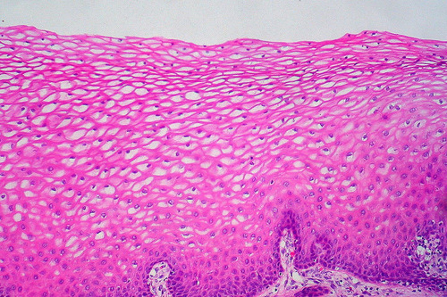
Function: stratified squamous epithelium
protects underlying tissues in areas subjected to abrasion

Location: stratified squamous epithelium
nonkeratinized type forms the moist linings of the esophagus, mouth, and vagina; keratinized variety forms the epidermis of the skin, a dry membrane

Basement membrane
Cells at the base of an epithelial layer are attached to this
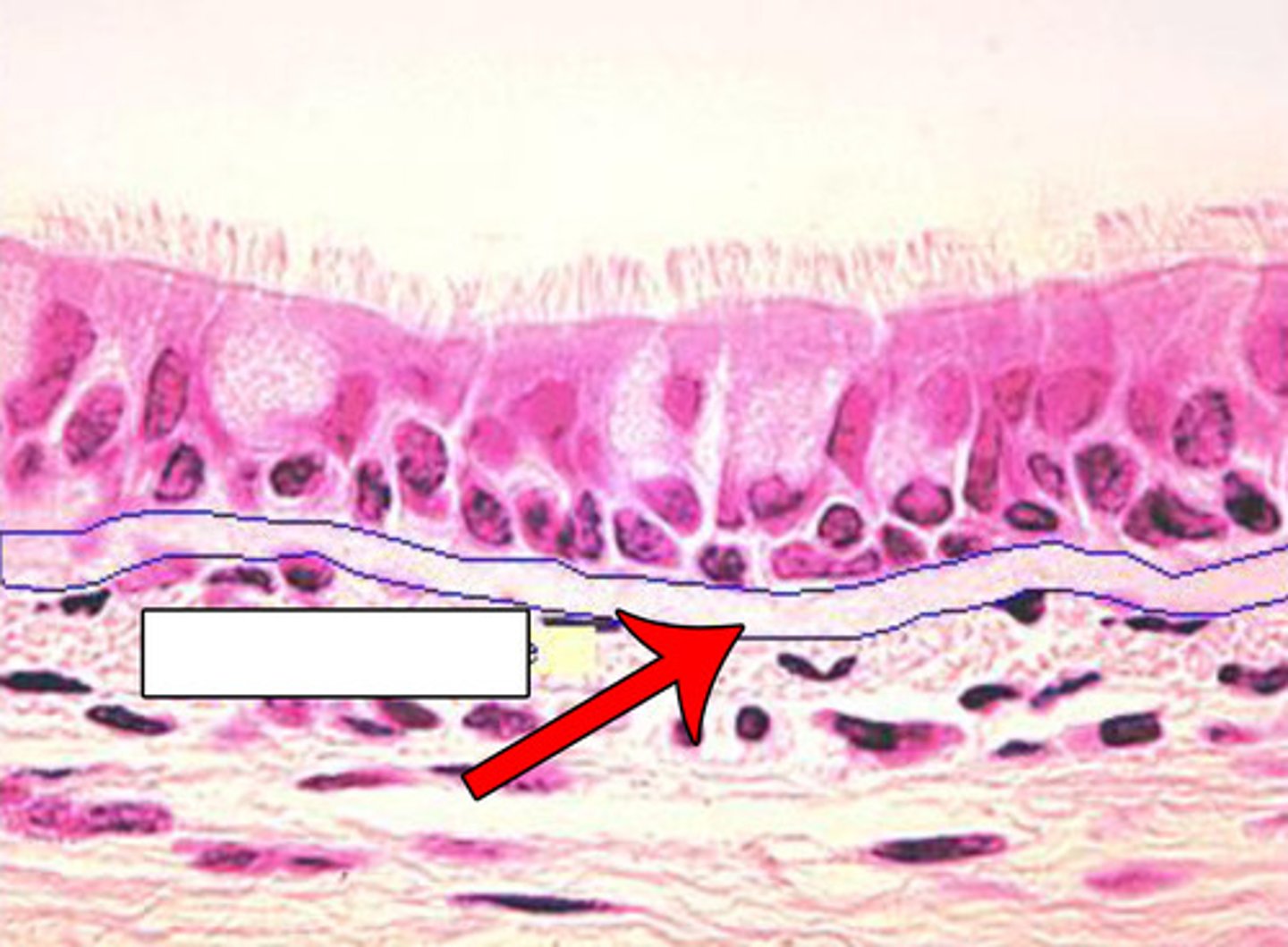
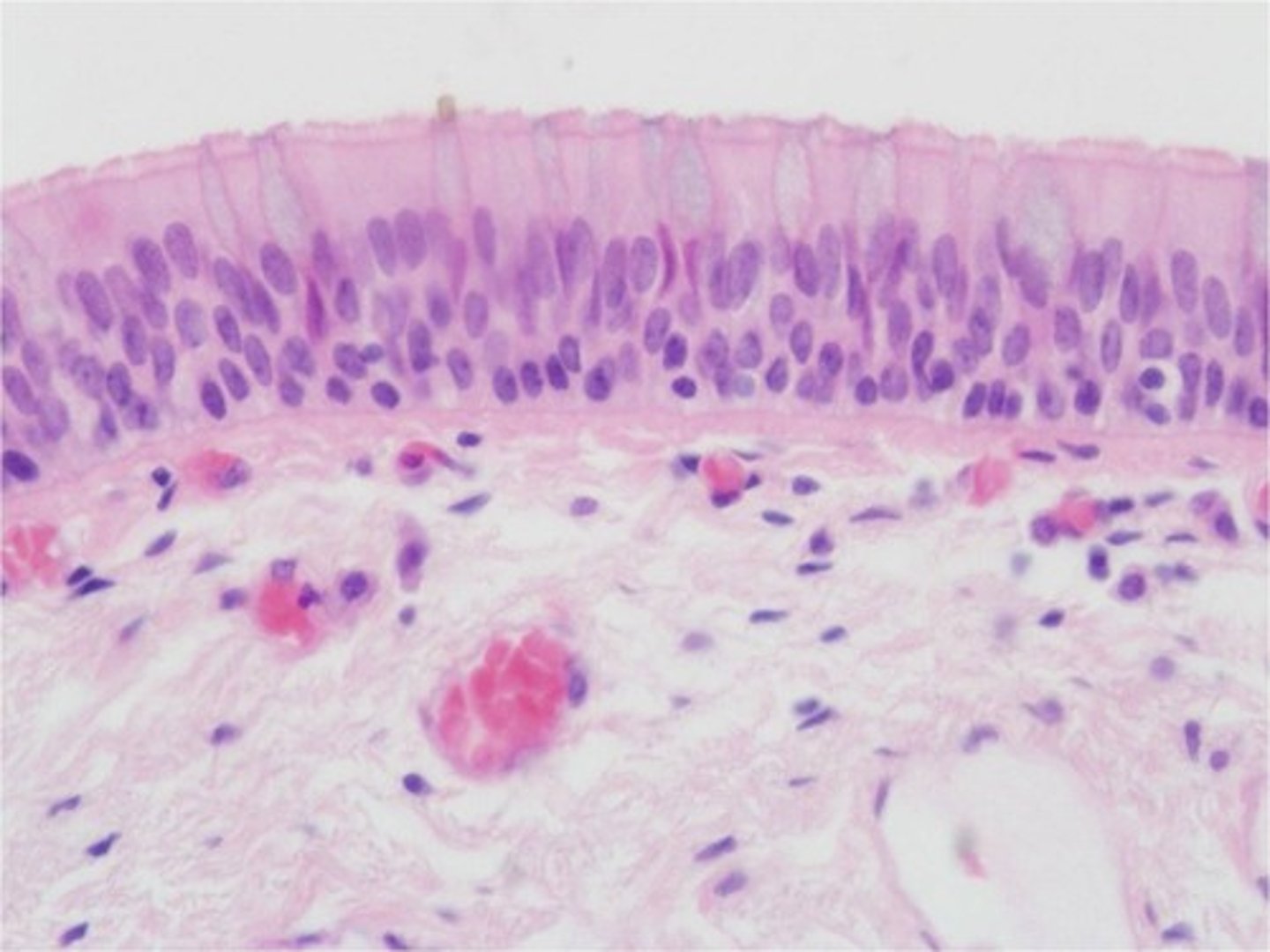
Description: pseudostratified ciliated columnar epithelium
Single layer of cells of differing heights, some not reaching the free surface; nuclei seen at different levels; may contain goblet cells and bear cilia

Function: pseudostratified ciliated columnar epithelium
Secretion of mucus; propulsion of mucus by ciliary action
Location: pseudostratified ciliated columnar epithelium
Nonciliated type in male's sperm-carrying ducts and ducts of large glands; ciliated variety lines the trachea, most of the upper respiratory tract

Description: transitional epithelium
resembles both stratified squamous and stratified cuboidal; basal cells cuboidal or columnar; surface cells dome shaped or squamous-like, depending on degree of organ stretch

Function: transitional epithelium
stretches readily and permits distension of urinary organ by contained urine
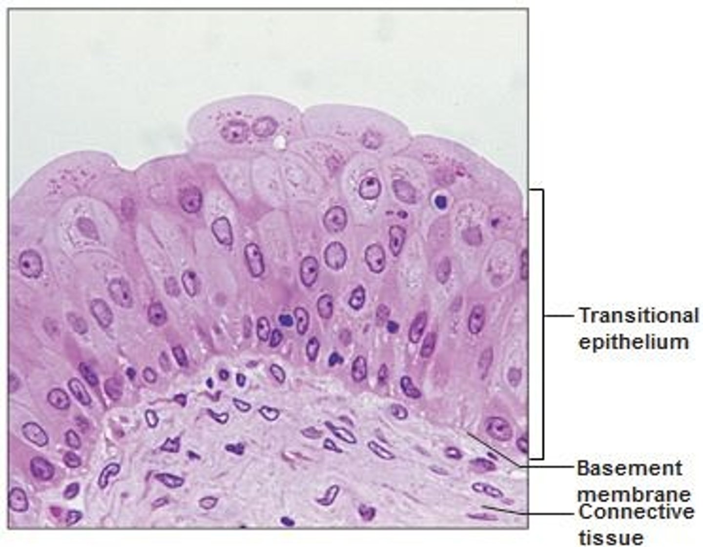
Location: transitional epithelium
lines the ureters, bladder, and part of the urethra

Connective tissue
provides structural support and connects all of its parts
Fibroblasts
cells that secrete the proteins that join other molecules in the matrix to from fibers
Mast cells
Detect foreign microorganisms and initiate immune responses against them
Adipocytes
fat cells and contain vacuoles for storage of lipids
Loose connective tissue
has an open network of protein fibers in a thick, syrupy ground substance and is divided into three groups
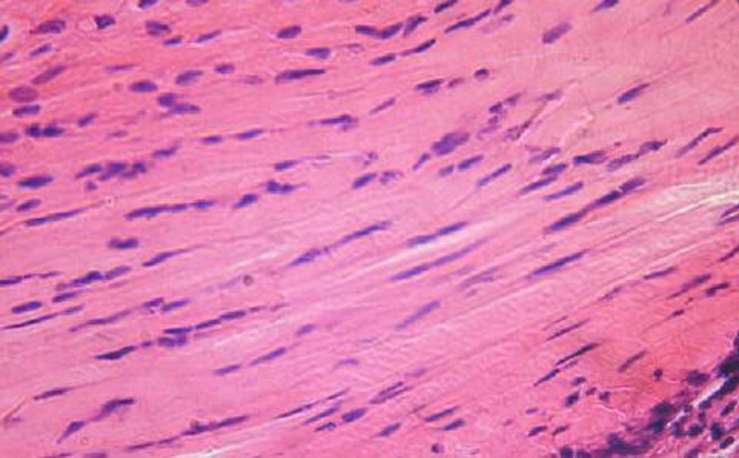
Dense connective tissue
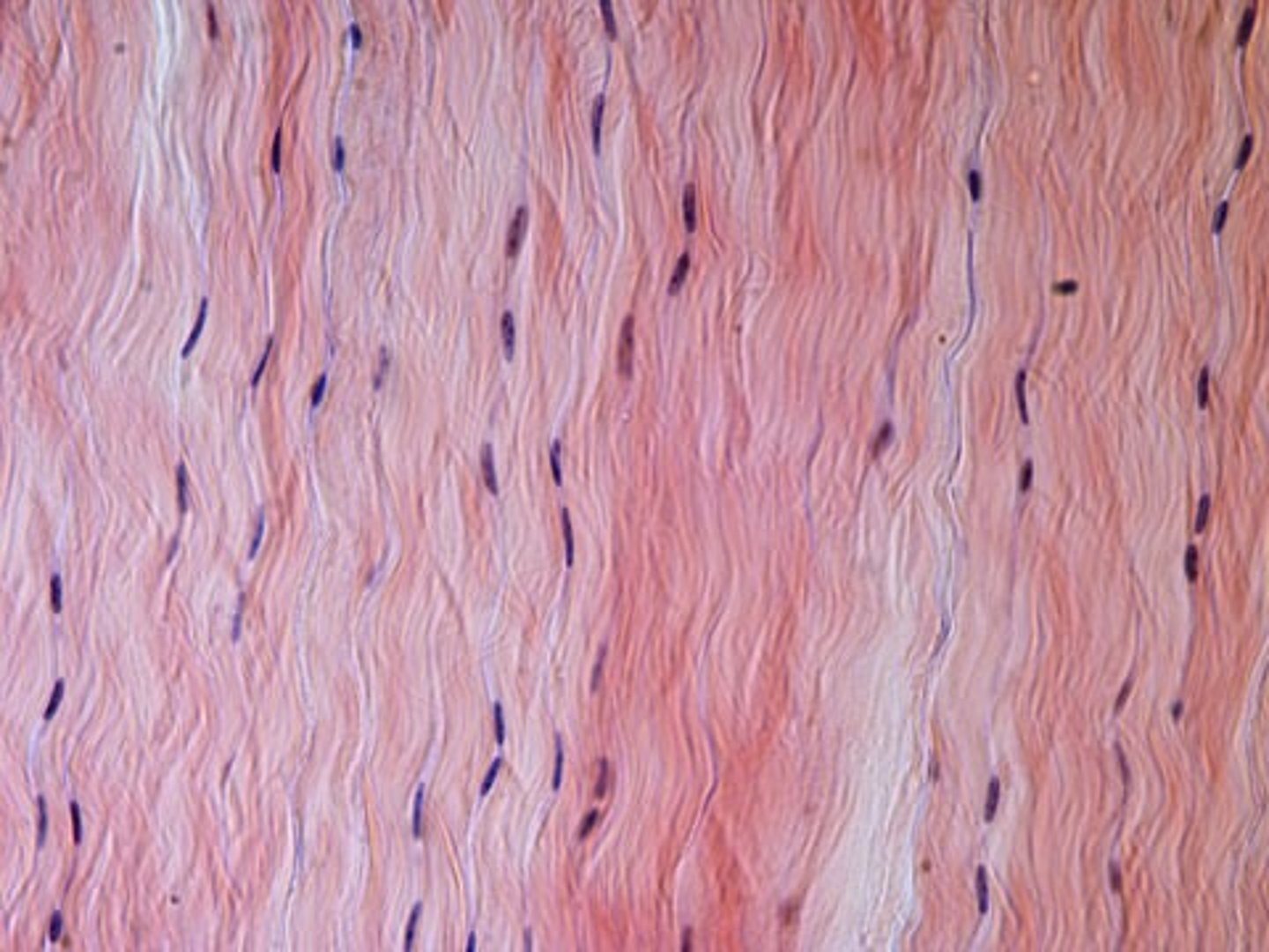
Made up of two types of fibers: protein fibers assembled into thick bundles of collagen and elastic fibers with widely scattered cells
Dense regular
Protein fibers in the matrix are arranged in parallel bands (same direction)
Dense irregular
fibers are interwoven run in many directions
What are the two types of fluid connective tissue?
blood and lymph
What are the two types of supporting connective tissue?
bone and cartilage
Perichondrium
surrounds all supporting connective tissue in cartilage and produces chondroplasts
What are the three types of cartilage?
hyaline, elastic, fibrocartilage
Which tissue has all 3 fibers present?
Areolar tissue
Description: Areolar Connective Tissue
gel-like matrix with all three fiber types; cells: fibroblasts, macrophages, mast cells, and some white blood cells; loose arrangement of fibers

Function: Areolar Connective Tissue
wraps and cushions organs; its macrophages phagocytize bacteria; plays important role in inflammation; holds and conveys tissue fluid

Location: Areolar Connective Tissue
Widely distributed under skin
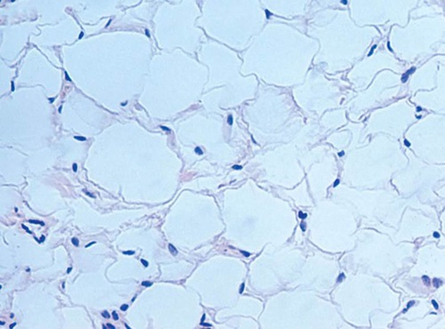
Description: Adipose tissue
matrix as in areolar, but very sparse; closely packed adipocytes/fat cells have nucleus pushed to the side by large fat droplet

Function: Adipose tissue
provides reserve food fuel; insulates against heat loss; supports and protects organs

Location: Adipose tissue
under skin, in butt, breasts, and abdomen
Description: Reticular tissue
network of reticular fibers in a typical loose ground substance; reticular cells lie on the network

Function: Reticular tissue
forms a soft internal skeleton (stroma) that supports other cell types, including white blood cells, mast cells, and macrophages
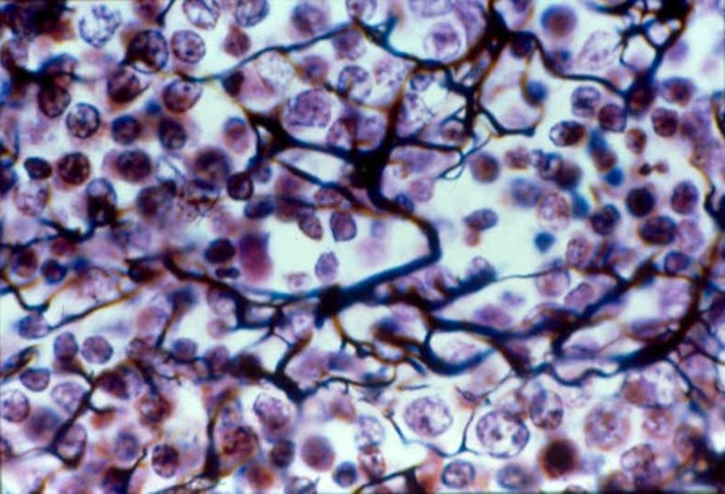
Location: Reticular tissue
lymphoid organs (lymph nodes, liver, bone marrow, and spleen)

Description: Dense regular connective tissue
Primarily parallel collagen fibers; a few elastic fibers; major cell type is the fibroblast
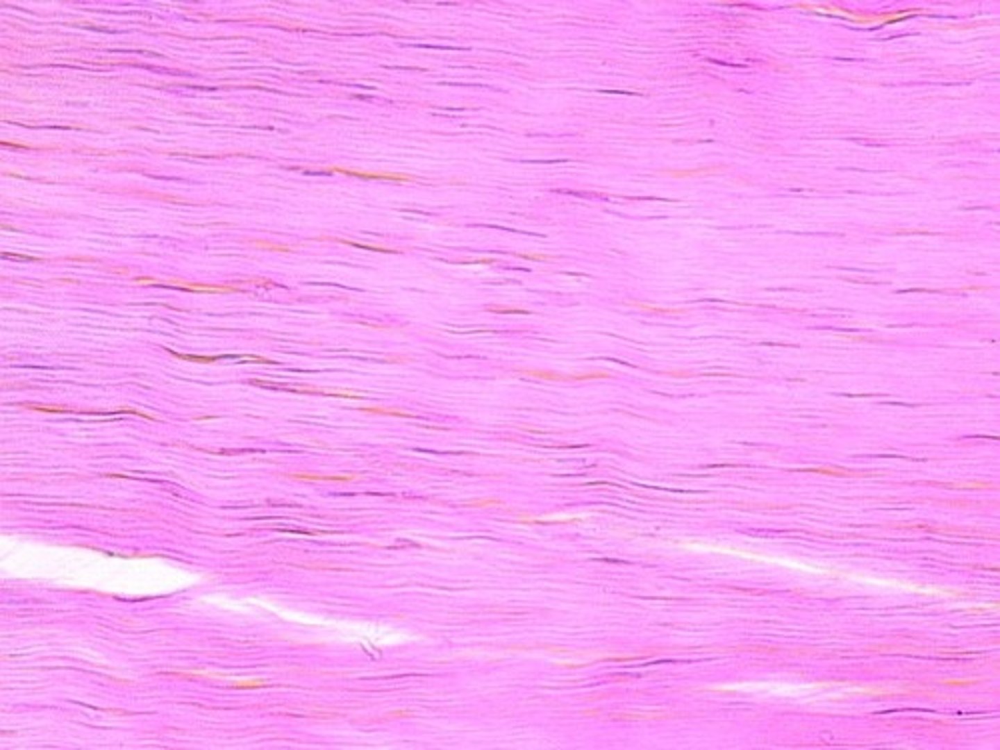
tendons
muscle to bone
ligaments
bone to bone
Function: Dense regular connective tissue
attaches muscles to bones/muscles; attaches bones to bones; withstands great tensile stress when pulling force is applied in one direction

Location: Dense regular connective tissue
Tendons (connect muscle to bone); most ligaments (connect bone to bone); aponeuroses
Description: Dense regular elastic tissue
Dense regular elastic tissue containing a high proportion of elastic fibers