Psychology - biopsychology
1/89
There's no tags or description
Looks like no tags are added yet.
Name | Mastery | Learn | Test | Matching | Spaced | Call with Kai |
|---|
No analytics yet
Send a link to your students to track their progress
90 Terms
What is the nervous system divided into?
The central nervous system
The peripheral nervous system
What is the central nervous system divided into?
the brain and the spinal chord
What is the periphiral nervous system divided into?
The Automatic nervous system
The somatic nervous system
What is the peripheral nevous system and what is it divided into?
The system is all the nerves that branch out the spinal chord
The symapthetic nervous system, when activated it prepares the body for fight or flight which is almost in miliseconds (automatic response)
The parasympathetic nervous system, the dominant nerve which is a resting state, once a threat disappears the parasympathetic nervous system overides the sympathetic nervous system
What is the somatic nervous system?
Is originated from the brain and the spinal chord
Controls voluntary movement, it takes information via the senses and movement happens in response to sensory info e.g taste, smell, touch
the brain interprets information and makes response to the enviromental stimuli
What are neurons?
Neurons are specialised cells and they transmitt info from one place to another
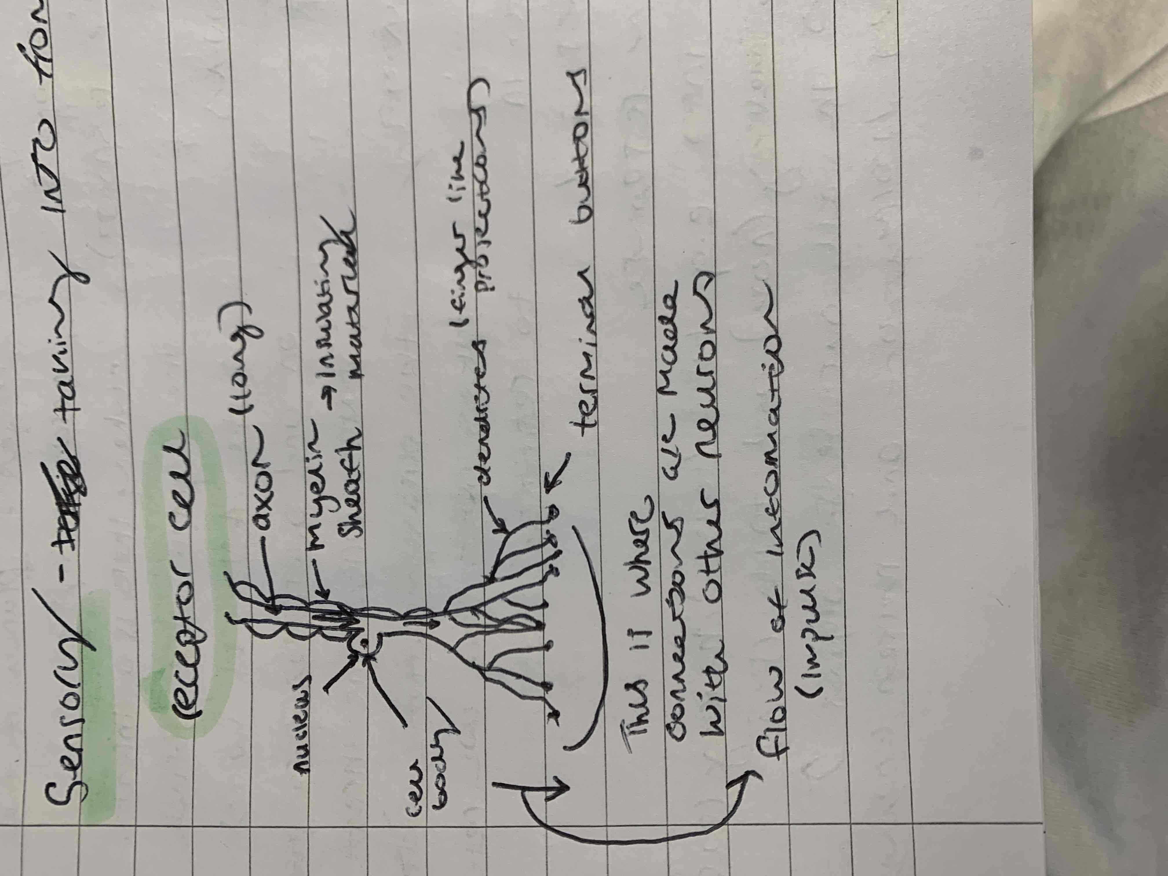
What is the sensory neuron? descibe its function and characteristics
It takes sensory information to the C.N.S
The axon which alows the flow of information to other neurons can be very long and is protected by a myelin sheath that acts as an insulating material
The nucleus is found along the axon in the cell body
The axon splits off into dendrites and end with terminal buttons where connections are made with other neurons
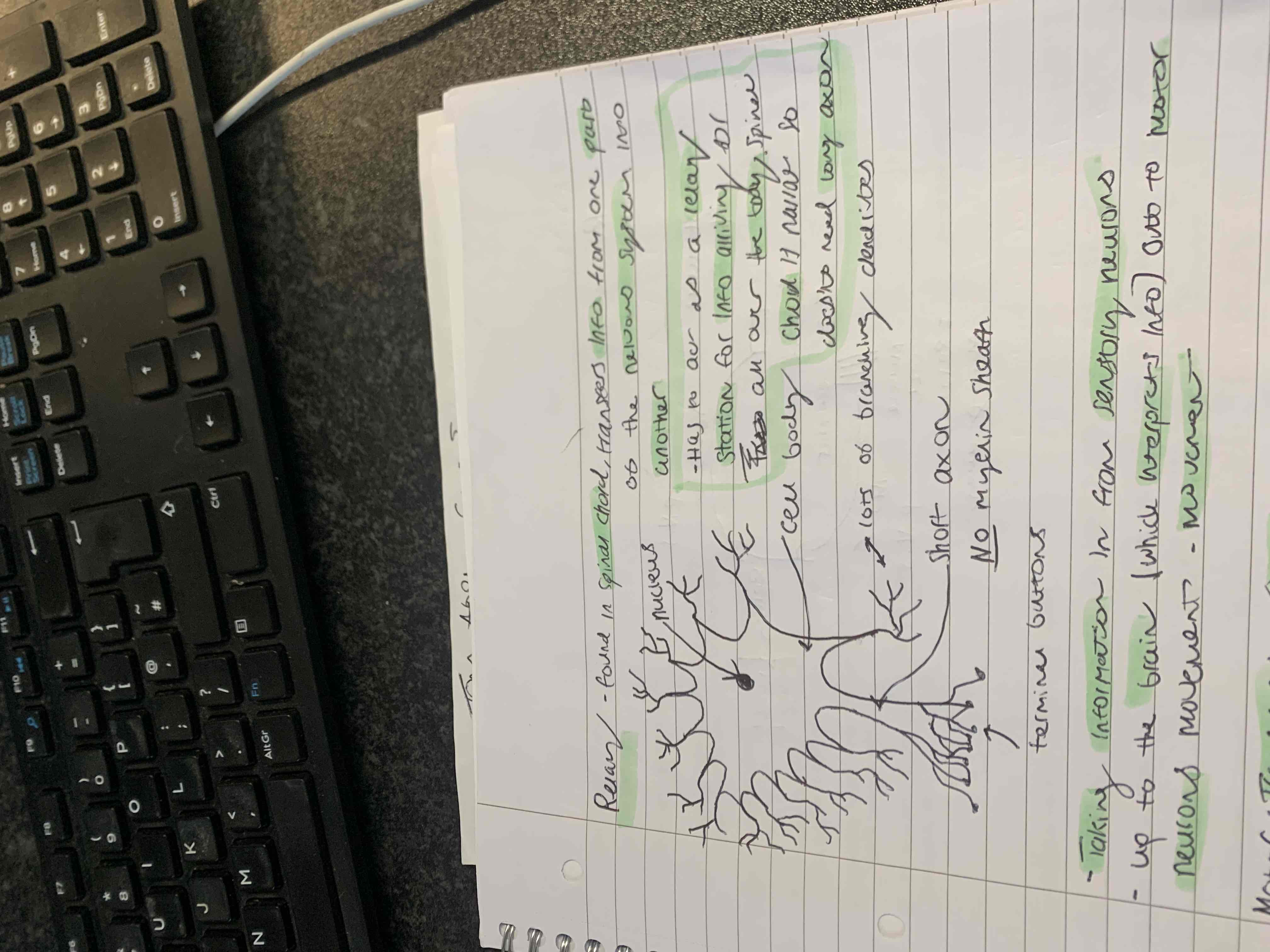
What is a Relay neuron? Describe its function and characteristics
Found in the spinal chord and transfers information from one part of the nervous system to another
Takes information in from sensory neurons and up to the spinal chord which interprets information to moter neurons to create a response
Has to act as a ‘relay station’ for information arriving all over the body, the spinal chord is narrow so the neuron does not need a long axon
Dendrites are split off from all over the neuron and there is a high proportion of them
The nucleus is found in the middle of the cell body
They have a short axon with no myelin sheath leading to dentrites and terminal buttons
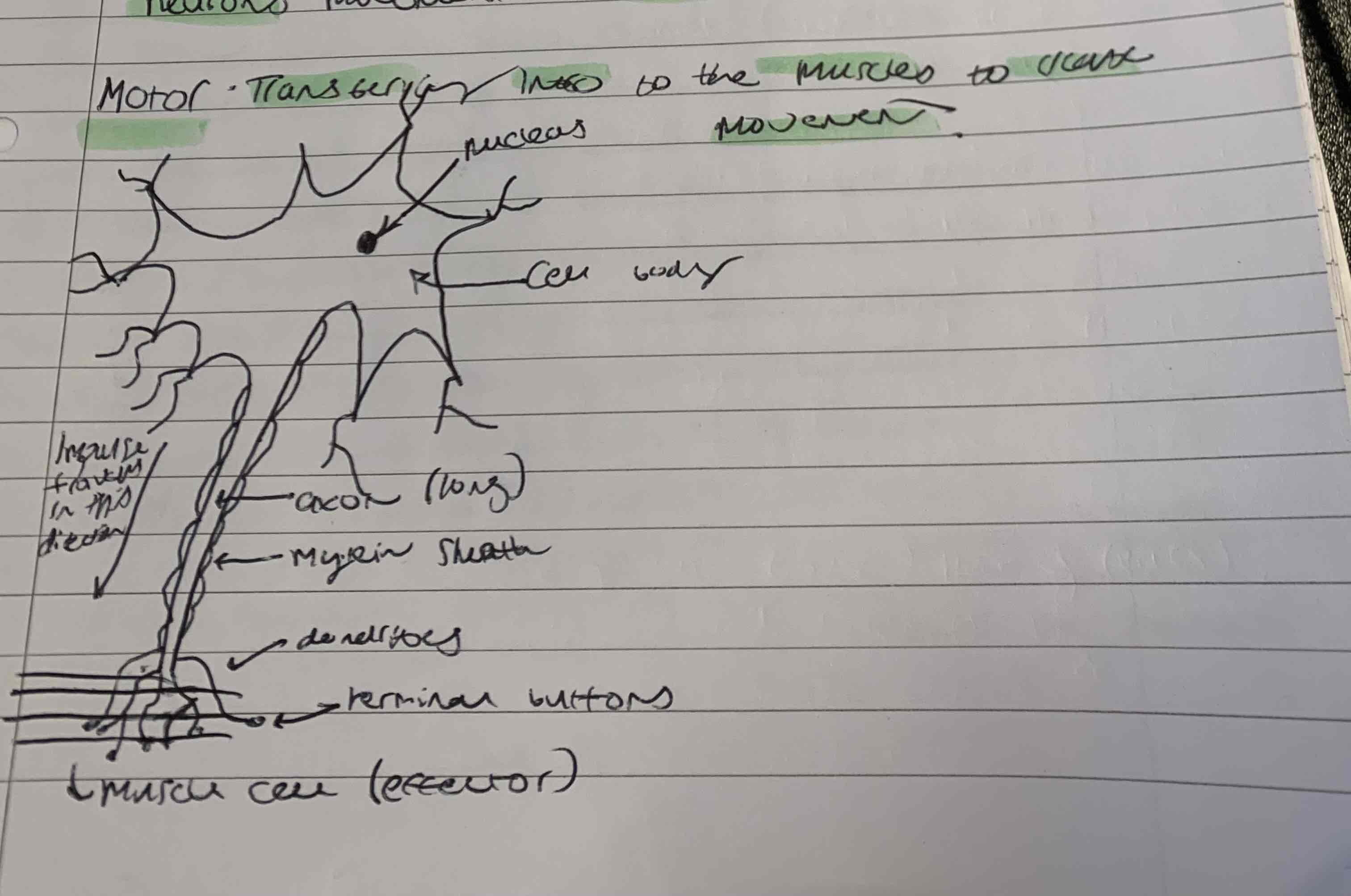
What is a motor neuron? Describe its function and characteristics
Transfers information to the muscles to create movement
Similar to the relay neuron but has a longer axon with a myelin sheath
The nucleus is found in the centre of the cell body
There are less dendrites that branch off than the relay neuron
The terminal buttons end in the muscle which is an effector that creates movement
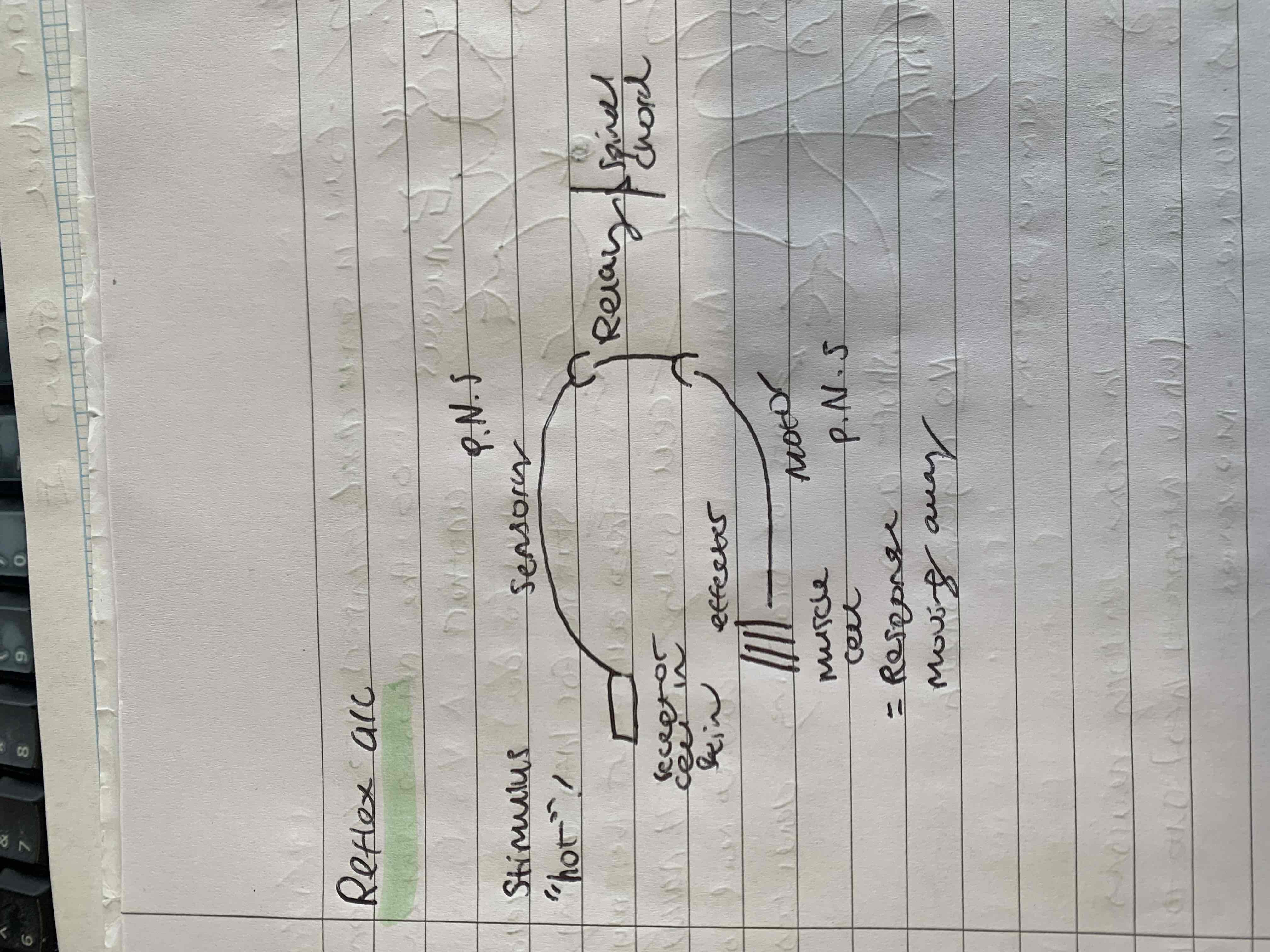
How does the reflex arc work? Describe how all the neurons work together
The stimulus detects things like heat and the receptor which in this case is the skin cells transmit nerves through the sensory neuron which lies in the periphiral nervous system
Then they reach the relay neuron which interacts with the spinal chord allowing it to react and pass a message to the motor neuron
Then through the motor neuron in the periphiral nervous system the nerves move to the effector which is the muscle cell allowing movement
What are neurotransmitters?
Chemical messengers which have two possible actions, Inhibitory and exicitatory
What are inhibitory and excitatory types of neurotransmitters?
Inhibitory - reduces the liklihood that post-synaptic transmission will fire (stop message)
Excitatory - have the opposite action, they increase the liklihood that post-synaptic neurons will fire (Go message)
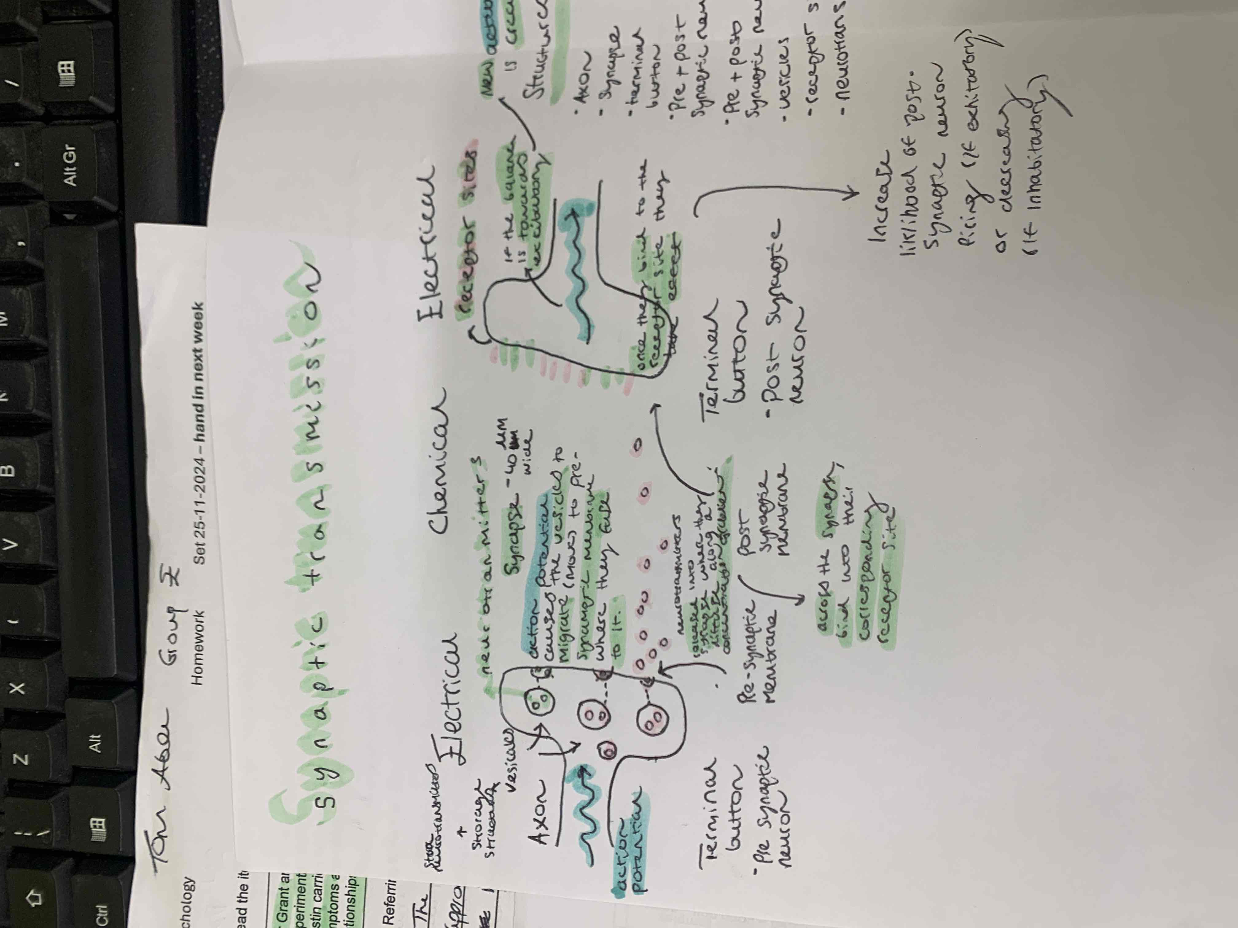
Synaptic transmission, How does the process of sending neurotransmitters across the gap work? what does it look like?
The terminal buttons act as an electrical impulse to prepare neurotransmitters to cross the synaptic cleft which is around 40um wide (nano-metres)
The vesicles store the neurotransmitters, action potential causes the vesicles to migrate to pre-synaptic membrane which the neurotransmitters fuse to.
This means neurotransmitters are released into the synapse along a concentration gradient across the synapse bind into the receptor site
Once they bind to a receptor they take effect which increases the liklihood of post-synaptic firing if exhitatory or decreases if inhibitory
If the balance is towards exhitatory new action potential is created
What is summation?
if we have more exhitatory to inhibitatory messages the neuron fires and vice verse
What is the fight or flight response? What two parts rof the body respond?
the fight or flight response prepares the body to respond to threat and expend energy
It is controlled/activated by the sympathetic nervous system and hormones
How does the sympathetic nervous system prepare the body for fight or flight?
The hypothalamus (A small of cells in the brain) percives a threat that is either physical or psychological and activates the sympathetic nervous system to prepare the body for fight or flight. This provides symptoms (on another flashcard)
How do hormones prepare the body for fight or flight?
The hypothalumus sends a message to the piturity gland releasing the hormone ACTH (adreno-cortico-trophic hormone)
The ACTH circulates in the blood until it reaches the adrenal glands and causes the adrenal medella to release adreneline.
What effects does the fight or flight response have on us?
the heart starts beating harder and faster to circulate more blood around the body
breathing increases, deepens and goes faster. more o2 in the body and more co2 out
Pupils dialate to allow better vision
Blood flow redirects to muscles for strength and energy.
sweating from excess heat from preparing body for fight or flight
How does the body transition back to a resting state (through sympathetic nervous system and hormones)?
when the threat has passed the sympathetic nervous system is overidden by the parasympathetic nervous system (rest and digest) this is an almost automatic response
The piturity gland stops producing ACTH and thus in turn adrenaline stops being produced, this can last longer
What are the main features of the brain?
4 lobes, frontal, parietal, occipital and temporal
2 strips/bands, motor cortex and sensory cortex (somatosensory strip)
2 areas, Brocha’s area and wernickes area
2 hemispheres, right and left
bundle of fibres joining the two hemispheres - corpus callosum
cerbral cortex, folded coat of brain tissue 5mm thick
What is the frontal lobe?
Located at the front of the brain and bilateral
Planning voluntary movement and exeutive movement and functions like making judgements, organising behaviour, logic, creativitity, personality
what is the parietal lobe?
at the top towards the back of the frontal lobes, intergrates sensory information from the body to the face
What are the occipital lobes?
at the back, processing visual information e.g the skill of reading, understanding language
what are the temporal lobes?
auditary infromation and speech hippocampus, new memories
What is the motor cortex?
Back of the frontal lobes, co-ordinated voluntary movement
What is the sensory cortex?
somatosensory strip, front of the parietal lobe or behind the motor cortex. processes sensory information from the body and face
The cortex has a map of the body, the more sensitive the area, the greater the area of the cortex is dedicated to it.
What is brocas’s area?
in the frontal lobe in the left at the junction between the motor cortex and temporal lobe for right handed people. responsible for production of speech, moving tounge or mouth
What is wernickes area?
back of the temporal lobes, interpretation of spoken language, comprehension of speech, lateralised on the left (right handed)
what is the corpus calcosum?
bundle of fibres at the bottom of the hemisphere that connects them together, allows for communication between hemispheres
operation that cuts the corpus results in two independantly
what is the cerebral cortex?
folded brain tissue that is about 5mm thick
has higher cognitive functions e.g
language
problem solving
descion making
imagination
creativity
maths
what does the left and right hemisphere do?
largely semetrical but not the same, logic and reasoning is lateralised to the left, the right hemisphere is more holistic e.g face processing
What is the laterlisation of function?
The left and right hemispheres are not the same it comes to stucture and function, some structues and functions are only found in one side of the brain and this is called the laerlisation of function.
for example, language is laterilised on the left for most people who are right handed
Broca’s and wernickes area is found in the left
research suggests the right and left hemispheres have a different form of processsing which has led to the idea of having a right or left brain having differant types of brains would affect your problem solving abilities and certain other unique abilities
having a left or right brain, what is each brain better at?
left hemisphere - where logical, rational, analytical and thoughts rise
right hemisphere - where creative, imaginitive, holistic abilities rise
What was sperry’s research into lateralisation on the left side of the brain?
His research is called split-brain research because he used participants who had their corpus callosum cut in order to reduce the impact of epilepsy
What was Sperry’s aim and method throughout his experiment?
Aim
to illustrate that language is lateralised on the left.
Method
There were 11 male, right handed participants. They all had their corpus callosum cut surgically to reduce the impact of epilepsy
What was the procedure Sperry used in his research?
Highly controlled lab experiment
Participants will sit in front of a screen with a cross in the middle, two words would be shown left and right of the cross.
The participants were asked to fixate on the cross.
The words would flash briefly and the participants were asked what they saw.
they were given a pen in their left hand and asked to draw what they see when a word has flashed to the left of the screen
what is contralateralisation?
the left hemisphere controls the right side of the body/ vice versa
information from the right side of the body goes to the left hemisphere and vice versa
the corpus callosum being cut means no communication can be made
what did sperry find during his research?
He found that when reading the word ‘toad’ on the left side of the screen, the participants could not say what it is, whereas when reading on the right side they could read the work ‘stool’ out. However, on the left side they could draw a toad instead of reading it out.
How can we conclude sperrys research?
These findings show that language is laterlalised on the left (at least for right handed people)as info arriving from the right hemisphere can’t be expressed verbally as there is no language centre. also the corpus callosum has been cut so it means that communicaton with language centre on the left is not possible. However the right centre still recieves the infromation as it an be expressed verbally e.g through drawing
Studying the brain
What is post mortem/autopsy examination?
What are three examples of post mortem/autopsy examinations?
researchers will study the brain of a person who displayed paticular behaviour patterns whilst they were alive which suggested possible brain damage
Allow for detailed examination of anatomical and neurochemical aspects of the brain. Allows researchers to examine deeper regions like the hypothalamus and the hippocampus
Broca, examined the brain of a man with speech problems whilst alive. It was discovered he had a lesion in an area of the frontal lobe important for speech production. Later known as Broca’s area
Wernicke, discovered a region in the left temporal lobe important for language comprehension and processing
Iverson, examined the brain of deacesed schizophrenic patients and found that they all had a higher concentration of dopamine in the limbic system
Studying the brain
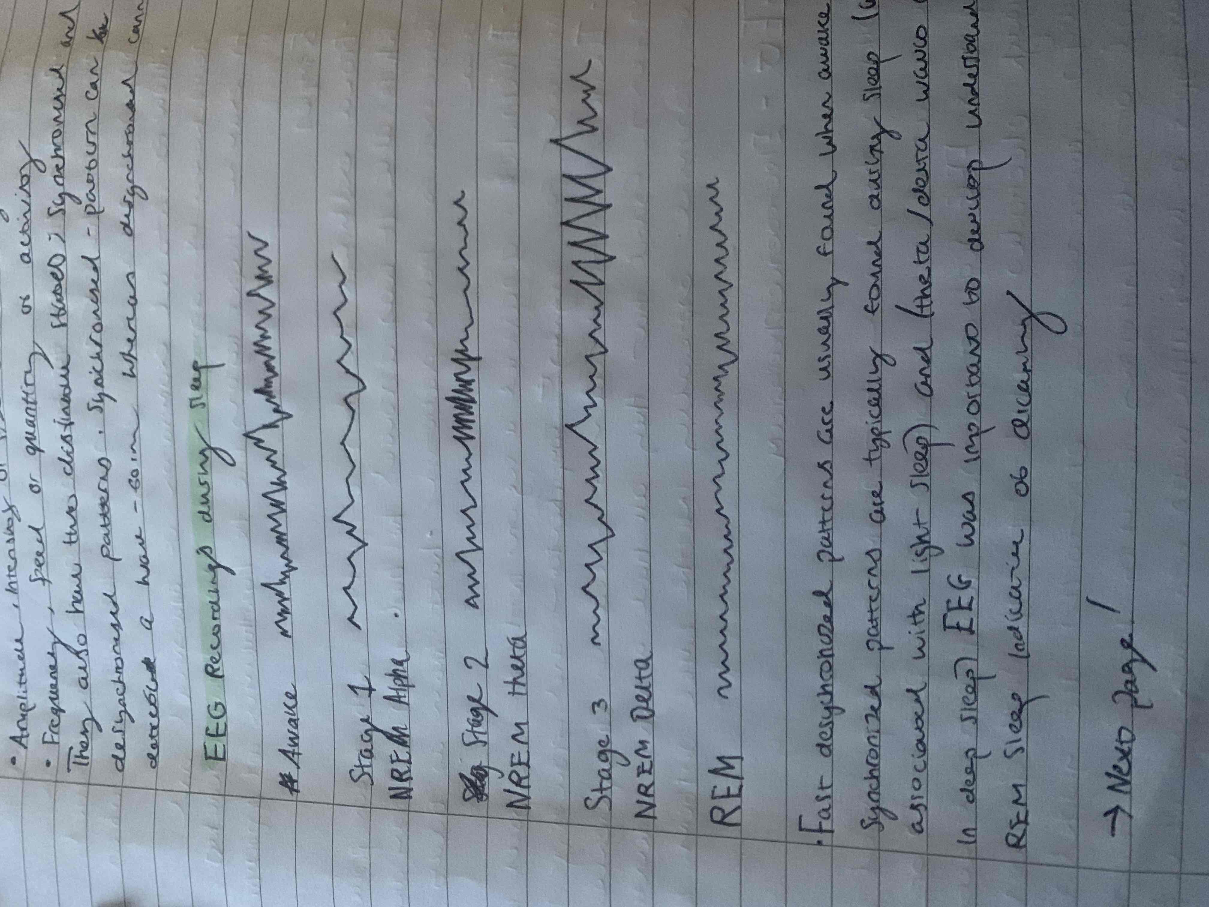
What is an EEG?
Electorencephalogram
Works on the premise that information is processed as electrical activity in a form of action potential or nerve impulses. EEG scanners measure this electrical activity through electrodes attatched to the scalp connected to EEG machine. Small electrical charges were detected by electrode that are graphed over a period of time as real time wave patterns, indicating levels of activity in the brain
There are four types of waves; alpha waves, beta, theta and delta. Each of these properties have two basic properties psychologists can examine, amplitude and frequency
They also have two states, synchronised and desynchronised patterns, synchronised can occur in wave form whereas desynchronised cannot
EEG readings during sleep, what are the stages, how does this work?
Fast desynchronised patterns are usually found when awake and synchronised patterns are typically found during sleep, alpha is associated with light sleep and theta and delta is associated with deep sleep. EEG’s are important to develop understanding of REM sleep that is indicative of dreaming
Studying the brain
What is an ERP
event related potentials
Use similar equipment to EEGs, electrodes attached to the scalp, computor equipment drowns out distractions
Key difference is that a stimulus presented to participant like picture or sound and the researchers look at activity related to stimulus. The stimulus can be presented hundreds of times and an average response is graphed. This is called ‘averaging’
Latency is the time period between stimulus and response, it is very short and divided into two broad catagories
Waves that occur within 100 miliseconds following a stimulus are sensory ERP’s.
ERP’s that occur after 100 milliseconds are called cognitive ERP’s as they demonstrate some information processing
Studying the brain
what are FMRI scans?
Functional magnetic resonance imaging
Scanning measuring the haemodynamic response (blood flow in the brain when an indivdual preforming a task, the scan works on the idea that neurons in the brain that are most active use the most energy
Energy requires glucose and oxygen, oxygen is carried in the blood stream attached to the haemoglobin and is responsible for release by active neurons, at this point the haemoglobin becomes deoxygenated
An FMRI can detect that a deoxygenated haemoglobin has a differant magnetic quality from oxygenated haemoglobin. a FMRI can be used to create a 3D map of the brain highlighting differant neural activity
FMRI images show activity 1-4 seconds after it occurs with an accuracy of 1-2 mm
An increase in blood is to the need for more oxygen in the area of a brain when there is more neural activity
Red blood flow has the most activity and blue has the least
Modern version of autopsy
What is the method the carry out a post-mortem/autopsy?
Remove the brain from the skull
preserve it in tormaldehyde
slice into sections, look under electron microscope, do visual inspection to look for obvious damage
They correlate psychological difficulties in life with structured damage observed at autopsy
What was Broca’s study into ‘tan’? How is it an example of a autopsy?
A french patient ‘loborge’ who is nicknamed ‘tan’ aided Paul Broca’s research
Tan could only understand spoken language but couldn’t produce any coherant language. He could only say ‘tan’
After tan’s death Broca conducted an autopsy on tans brain. He found tan had a lesion in his left frontal lobe
allowed broca to conclude that the area was responsible for language production. People with damage in this area have Broca’s aphesia resulting in slow, innaccurate speech
Demant and Kleitman (1957) what was the aim of their research? What did they use to study? What is REM and NREM?
Aim
The research aimed to find obective methods and demonstate the correlation between dream content and physiological indicators of dreaming like eye movements
Does dream recall differ between REM sleep and NREM stages of sleep?
Is there a positive correlation between dream duration and REM period length?
Are eye movements related to dream content
What did they use?
EEG’s tracing cyclical changes that occur in brain activity during sleep. Electrodes are placed around the skull to anaylse brain waves
EOG’s traces eye movement during sleep, electrodes are placed around the eye region
REM - rapid eye movements
NREM - non REM
What was the sample and procedure of Demant and Kleitman’s study?
7 males and 2 females, joined through oppurtunity sampling. 5 spent 6-17 nights in the lab and 4 spent 1-2 nights in the lab
participants would report to the lab before their own bedtime and asked to avoid caffine and alchohol.They slept in a dark, quiet room and a doorbell would be used at random from REM and NREM
procedure 1, the participants were woken at various times to test dream recall. Dream narritives would be recorded on tape and only counted if recall was clear
procedure 2, the participants were woken 5 to 15 minutes into REM sleep, the participants guessed how long they dreamt for
procedure 3, the participants eye direction movements was detected from the EOG, ppts were woken up and reported their dream
What conclusions could be made from Dermant and Kleitmans experiment?
Dreams occur in REM sleep only. Dreams for NREM sleep are from previous REM episodes
Dream duration and REM sleep is similar, showing they are not instantaneous, they are experianced in real time
Eye movements correspond to where dreamers are looking at and not random events
What are the strengths of an autopsy?
allows for detailed examination of the brain down to a single neuron (using an electron microscope)
Conditions like schizophrenia can be understood
they can help future generations
What are the limitations of autopsy?
when understanding the impact of brain damage on psychological processes, autopsy works well if there is a single psychological deficit and a single area of damage. If a person has multiple psychological problems and multiple sites of damage we don’t know what corresponds to the problem
The technique doesn’t have any value to the person during their life, as it’s carried out once they have died
consent is required to carry out an autopsy and this limits the number of brains available to research
the most interesting brains for scientists are the most damaged but this raises an ethical concern that people with brain damage may not fully know what they are consenting too
What are the strengths of FMRI’s?
you can investigate localisation of function without harming the indivdual as it is a non-invasive method compared to PET scans which involves injecting the ppts with a radioactive substance
allows us to understand precisly where certain functions are located
allows us to investigate unconscious mental activity e.g day dreaming
created a detailed map of the brain
What are the limitations of FMRI’s?
patients have to lie very still up for an hour or so, more difficult to use with children or people woth learning difficulties and people who are claustrophobic
very small sample sizes as they are very expensive makes findings less reliable
poor temporal resolution, 5 second delay between the brain activity and it being displayed on screen for us to look at
What are the strengths of EEG’s?
Non-invasive
possible to fall asleep with them on, so can be used to diagnose sleep disorders
far broader application than FMRI as you don’t have to lie still in small space
used to diagnose epilepsy due to characteristic abnormal wave patterns
excellent temporal resolution recording activity as it occurs compared to FMRI with 5 second delay
What are the limitations of EEG’s?
Can’t be used to precisely locate functions, it has no spacial resolution compared to FMRI
electrodes may not be placed accuartly or move reducing the reliability of the recording
What are the strengths of ERP’s?
As ERP software cancels out background noise, we can use it to identify precisely when a cognitive function occurs, and general location
A more advanced version of an EEG technique
What are the limitations of ERP’s?
diffarent software programmes produce differant results so it makes it tricky to compare results from studies
it may not be possible to get rid of all background activity making identifying ERP’s unreliable
What are the positives of sperry’s split brain research?
Highly controlled experiment so sperry could be confidant that words flashed to the left or right if the screen were only arriving in the right or left hemisphere. We can be confident that the results demonstrate the effect of cutting the corpus callosum plus the lateralisation of language
Sperry’s results are reliable as it is a highly controlled lab study with a set procedure, this means they can be replicated and have similar results
Clearly demonstrated the lateralisation of function (at least for right handed males)
What were the weaknesses for sperry’s split brain research?
small unrepresentative sample, only 11 male right handed participants, researchers need to be cautious when understanding when generalising findings as left handed people tend to be bilateral
Sperry didn’t have a control group of people without epilepsy cut who gave their corpus callosum cut. The epilepsy itself may change the organisation of the brain in relation to language. He couldn’t separate the effect of epilepsy as all his participants had the condition
What are the strengths of the theory of lateralisation of function?
sperry’s research with split brain patients has been replicated (e.g Gazzaniga) and researchers are mostly in agreement that language at least for right handed people is lateralised on the left.
Modern FMRI studies support the conclusion but for some people language is bilateral - particularly left handed people
What are the limitations of the theory of lateralisation of function?
Whilst there is evidence that the left hemisphere tends to process information in an analytical way and the right in a more holistic way. The idea that a person is either left brained or right brained is not too accurate. People do seem to have a preference for processing information in a particular way but it’s not either/or as the two hemispheres are in constant communication with each other
What is aphesia, what are the two types?
the difficulty in communication and usually caused by stroke
Brocas aphasia, damage to the left hemisphere
Wernickes aphasia, damage to the left temporal lobe
What are the issues with the two aphasias?
brocas, difficulty producing speech, speech is hesitant and disjointed (missing words and phrases) person knows what they want to say, but have difficulty forming words
Wenickes, difficulty with speech comprehension, speech is fluent but meaningless - a jumble of words - they can’t understand what is said to them or produce a meaningful answer
What is plasticity? what do we mean by structure?
The ability for the brain to change it’s structure in response to enviromental demands.
When talking about structure we are meaning;
Connections between neurons
Locations between functions
Amount of brain tissue dedicated to a certain function
What does plasticity involve?
Neural growth, more connections between existing neurons, no neurons are forming but the connections between them are becoming more dense
Neural pruning, connections being deleted between neurons as they are no longer used. Dormant neural connections are re-established and re-used
How does Neural growth occur overtime?
Neural growth occurs quickly in the first five years of life when we are exposed to so much so quickly
After five years, growth will slow down and continue throughout life
At puberty and into early adolesence nearul pruning occurs in large swathes
What is functional recovery?
when a person is able to regain functions they had lost due to brain damage, it is possible due to brain plasticity
How can functional recovery occur?
axonal sprouting, when the cell body of a damaged neurons grows a new axon meaning it can form new connections
Reformation of blood vessels to supply oxygen and glucose and allow cell’s to repair themselves
Homologous areas (an area that is in the same place but on the opposite side of the brain) They take over functions of the damaged area e.g Brocas aphasia due to a stroke, corresponding area on the right will take over the function
Dormant pathways, they can be re-activated to take over functions
Where is there evidance for the brains plasticity?
The London taxi driver study by Maguire
Brain scans, MRI scans (static black and white detailed imagery of the brain)
Controlled the experiment by using all male, right-handed participants who all had their brain scanned in the past for medical reasons
They found that there was a greater volume of grey matter in the posterior hippocampus in those who had attempted to learn the knowledge
They found the longer the taxi drivers had spent learning the greater volume of grey matter
Why?
London taxi drivers have to pass the knowledge test (learning all the streets of London), this involved using spatial and navigational skills with learning the knowledge increasing the size of this area
Maguire et al concluded that the study shows the plasticity of the brain to adapt and change according to the demands it faces
How can we evaluate functional recovery? what is an example of someone who has experianced functional recovery?
case study of Jody, she had severe epilepsy as a young child they found it originated in her right cortex, when she was three she had a surgery called a hemishereoctamy which removed her right hemisphere
She made a relative recovery as her brain re-located functions to the left hemisphere, there were issues in her arm and hip
What is CIMT? What is it an example of?
The brain will spontaneously start to preform functional recovery but eventually it slows down, for maximum recovery many indivduals need neurorehabilitation like CIMT
CIMT (contraint-induced-movement-therapy)invloves preventing the participant to stop using their functioning limb and require them to use the non-functioning limb
The functioning limb is placed into a cast so they are made to use their non-functioning limb
An example is where children with hemeplygia go through intensive therepy of 3 weeks, 5 days of each week for 3 hours a day
What are the strengths of the idea of localisation of function?
evidence e.g Broca’s area, wernickes area, aphasia is caused by damage to these areas showing a particular function
Language located on the left, sperry’s split brain research
Methods of studying the brain
What are the limitations to the idea of localisation of function?
the brain is able to re-organise it’s structure in response to demands made upon it e.g maguire and his study on london taxi drivers
What is a biological rhythm?
A change in bodily processes or behaviour that repeated regularly
What is a circadian rhythm?
Is 24 hour cycle
An example is the sleep/wake cycle, on average we spend 8 hours asleep and 16 hours awake and sleep is synchronised with night and being awake is synchronised with daylight
What is a infradian rhythm?
they take longer than 24 hours to be completed
For example, the menstrual cycle takes 28 days and seasonal affective disorder takes a year
What is a ultradian rhythm?
A cycle that takes less than 24 hours to complete and can occur at multiple times
an example of a ultradian rhythm is the stages of sleep which can happen around 4-5 times per night
How do the stages of sleep work?
We ‘descend down the sleep elevator’ through stages 1-4.
Stage 1 is known as drowsy sleep
Stage 2 is known as hypoglobic sleep
Stage 3 is known as deep sleep
Stage 4 is known as very deep sleep
Then we ascend the sleep elevator back to stage 3 and 2 until we reach REM sleep which is associated with dreaming
The cycle takes around 90 minutes to complete and can be repreated
What is an endogenous pacemaker?
An internal body clock, regulating the biological rhythms
What is a exogenous zeitgeber?
External cues that influence and reset our internal body clocks
What is the SCN?
What does the SCN do?
Suprachiasmatic nucleus
is a group of nerve cells that are located in the hypothalamus, it lies above the optic chiasm where optic nerves cross
The SCN is believed to be the ‘master clock’
The pineal gland is instructed by the SCN to produce melatonin which makes us drowsy, this happens when light levels fall
When light levels rise, the production of melatonin is halted, we wake up and hopefully feel alert
How can exogenous zeitgebers influence the SCN?
Light levels act as an external cue which in turn tells the SCN when to stop or start instructing the pineal gland to produce melatonin
The idea of light levels resetting our body clock everyday is called Entrainment
What was Decoursey’s study into circadian rhythms?
A field experiment that was designed in 2000
The experimental condition was 30 chipmunks who had their SCN destroyed
The control condition was 20 chipmunks who had their SCN intact
They placed a tag on the chipmunk to track them after they released the chipmunks back into the wild and observed them for 80 days
The researchers found the chipmunks in the experimental condition were killed in a higher proportion as their body clocks were confused so they were out when they were at risk
How can we evaluate Decoursey et al’s study?
strengths
as it was a field experiment, we can see the chipmunks behave in their natural settings
Limitations
we need to be cautious when generalising our findings from the chipmunk’s sleep/wake cycle to our sleep/wake cycle, it may be slightly different or more complex
However, we also know that this type of study cannot take place on humans as it would be unethical
What was Michel siffre’s case study of circadian rhythms?
1962
He spent two months underground in caves within the alps, he had no sunlight or clocks
He monitered a number of biological processes including the sleep/wake cycle
He found that despite the absence of natural daylight he maintained a regular sleep/wake cycle of around 25 hours
Conclusions were made that, we have an internal biological clock that controls our circadian rhythm, it appears our sleep cycle is closer to 25 hours than 24
It shows our need for exogenous zeitgebers such as light levels to regulate our 24 hour sleep/wake cycle
What are the limitations of Michel siffre’s study?
previously, it was thought that the SCN wasn’t influenced by artificial light. However, other research demonstrates that without artificial light the free body runing clock is closer is closer to 24 than 25
What is chronotherepy?
practical application, chronothereputics
it is the study of how timing effects drug treatments and other interventions
e.g cancer treatment has an optimal time for delivery maximising the effect of drug and minimising the side effects
What is the endocrine system?
this is the system that works with the nervous system to control functions in the body, it is controlled by the hypothalamus in the brain. The system works more slowly than the nervous system but sill has powerful and important effects
What do glands in the endocrine system produce?
glands will produce hormones, these are chemicals secreted into the blood stream affecting the cells in the body that contains a receptor for that hormone
Hormones effect cells in the heart increasing heart rate and they effect cells throughout the body increasing metabolic rates - in turn this affects growth rates
What are three examples of glands?
adrenal gland, responsible for hormones regulating the bodys response to stree
Thyroid gland, regulates metabolism, influences heart rate and energy levels
Pituitary gland, known as the master gland as it controls the release of hormones for all the glands in our body