Anatomy and Physiology II - Section 1, Lesson 3
1/139
There's no tags or description
Looks like no tags are added yet.
Name | Mastery | Learn | Test | Matching | Spaced | Call with Kai |
|---|
No analytics yet
Send a link to your students to track their progress
140 Terms
what is systemic circulation
vasculature responsible for providing the functional blood supply to all body tissue
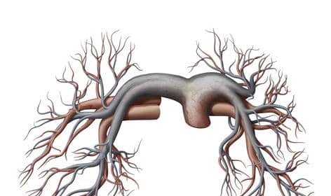
What is pulmonary circulation
these are connected to the lungs
What is portal circulation
vasculature responsible for returning deoxygenated blood to the lungs for reoxygenation
What is the most common route of circulation?
systemic circulation, where blood flows from the heart via arteries, through a capillary bed, and back to the heart via veins
Blood relatively high in oxygen concentration is returned to the left atrium via the four pulmonary veins. What circuit does it use?
pulmonary circuit
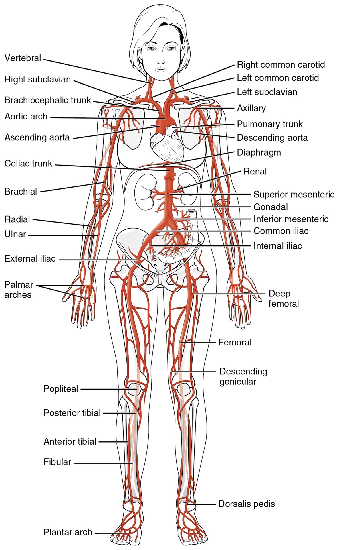
What artery is the source of all systemic arteries?
The aorta, which is the largest

After the aorta arises from the left ventricle of the heart, arches backwards over the heart, and descends through the thorax and abdomen, what does it divide into?
the common iliac arteries.
The heart has its own blood supply. What is this circulation called?
the coronary circulation
What does coronary circulation do?
supplies oxygen and fuel-rich blood to the heart muscle itself. In fact, the coronary arteries are the very first branches of the main supply artery of the body, the aorta. Any restriction to this blood supply will compromise the ability of the heart to pump blood effectively.
what are 3 distinct regions that the aorta has?
the ascending aorta, aortic arch, and the descending aorta, which includes the thoracic and abdominal regions.

what are the first vessels that branch from the ascending aorta
the paired coronary arteries
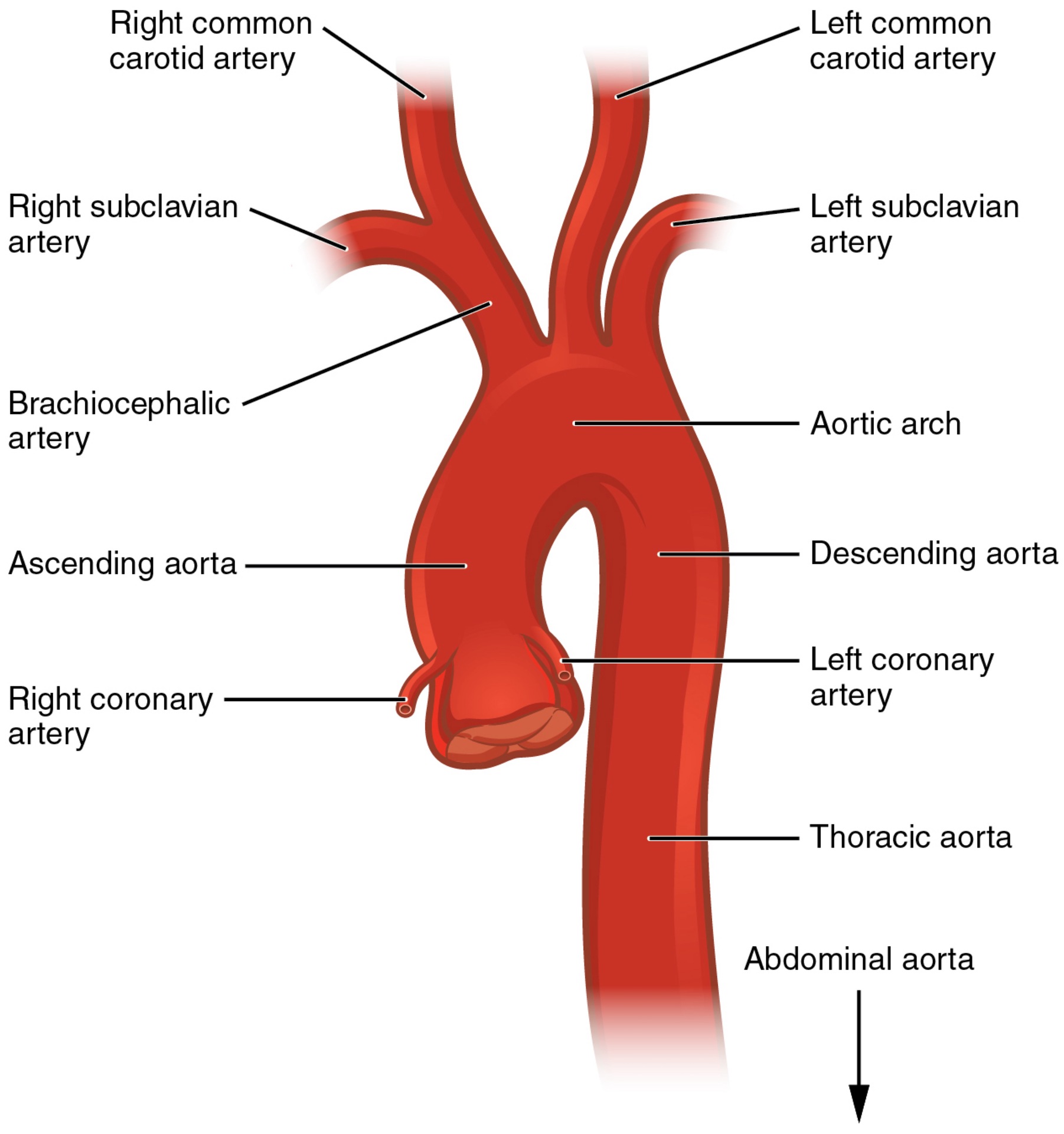
Where do the paired coronary arteries arise from?
two of the three sinuses in the ascending aorta just superior to the aortic semilunar valve. These sinuses contain the aortic baroreceptors and chemoreceptors critical to maintain cardiac function

Where specifically does the left coronary artery arise from
the left posterior aortic sinus

Where specifically does the right coronary artery arise from
the anterior aortic sinus. Normally, the right posterior aortic sinus does not give rise to a vessel.

What vessels encircle the heart, forming a ring-like structure that divides into the next level of branches that supplies blood to the heart tissues?
The coronary arteries
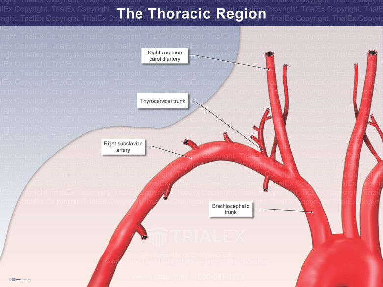
what are the three major branches of the aortic arch
the brachiocephalic artery, the left common carotid artery, and the left subclavian (literally “under the clavicle”) artery
what type of arteries are the three major branches of the aortic arch
based on proximity to the heart, each of these vessels is classified as an elastic artery.
The brachiocephalic artery is located only on which side of the body
the right; there is no corresponding artery on the left

The brachiocephalic artery branches into
the right subclavian artery and the right common carotid artery.
The left subclavian and left common carotid arteries arise independently from the aortic arch but otherwise follow a similar pattern and distribution to the corresponding arteries on the right side.


what do the subclavian arteries supply blood to?
the arms, chest, shoulders, back, and central nervous system
aorta→subclavian arteries what do the subclavian arteries branch into?
gives rise to three major branches: the internal thoracic artery, the vertebral artery, and the thyrocervical artery

aorta→subclavian artery→internal thoracic artery where does the internal thoracic artery, or mammary artery, supplies blood to
the thymus, the pericardium of the heart, and the anterior chest wall.
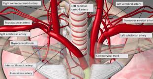
aorta→subclavian artery→vertebral Where is the vertebral artery located?
passes through the vertebral foramen in the cervical vertebrae and then through the foramen magnum into the cranial cavity

aorta→subclavian artery→vertebral Where does the vertebral artery supply blood to?
the brain and spinal cord.The paired vertebral arteries join together to form the large basilar artery at the base of the medulla oblongata.
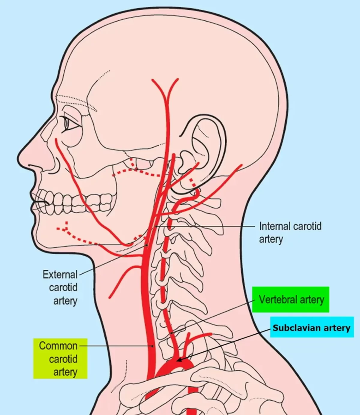
The paired vertebral arteries join together to form the large basilar artery at the base of the medulla oblongata. what is this an example of?
an anastomosis
aorta→(left) subclavian artery/brachiocephalic to right subclavian→thyrocervical artery What does the thyrocervical artery provide blood to
the thyroid, the cervical region of the neck, and the upper back and shoulder.
aorta → (left) common carotid artery/brachiocephalic to right common→?
internal and external carotid arteries. The right common carotid artery arises from the brachiocephalic artery, and the left common carotid artery arises directly from the aortic arch.

aorta → (left) common carotid artery/brachiocephalic to right common→internal and external carotid arteries. What do external carotid arteries supply blood to?
numerous structures within the face, lower jaw, neck, esophagus, and larynx. These branches include the lingual, facial, occipital, maxillary, and superficial temporal arteries

aorta → (left) common carotid artery/right from brachiocephalic to right common carotid→internal and external carotid arteries. What do internal carotid arteries supply blood to?
the brain and eyes
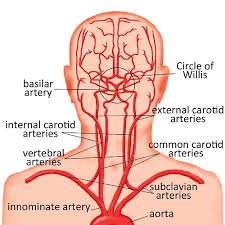
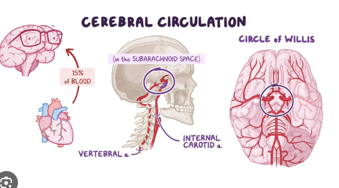
What are the two primary suppliers of blood to the human brain?
The internal carotid arteries, along with the vertebral arteries,
Why is it critical that blood supply to the brain remain uninterrupted
Given the central role and vital importance of the brain to life
What percent of blood flow is directed to the brain at any given time?
approximately 20%. remarkably constant

What will happen if there is a loss of blood flow for longer periods, typically between 3 and 4 minutes?
It will likely produce irreversible brain damage or a stroke, also called a cerebrovascular accident (CVA)
What happens when blood flow is interrupted, even for just a few seconds,
a transient ischemic attack (TIA), or mini-stroke, may occur, resulting in loss of consciousness or temporary loss of neurological function. In some cases, the damage may be permanent.
why are the locations of blood to the brain beneficial?
The locations of the arteries in the brain not only provide blood flow to the brain tissue but also prevent interruption in the flow of blood.
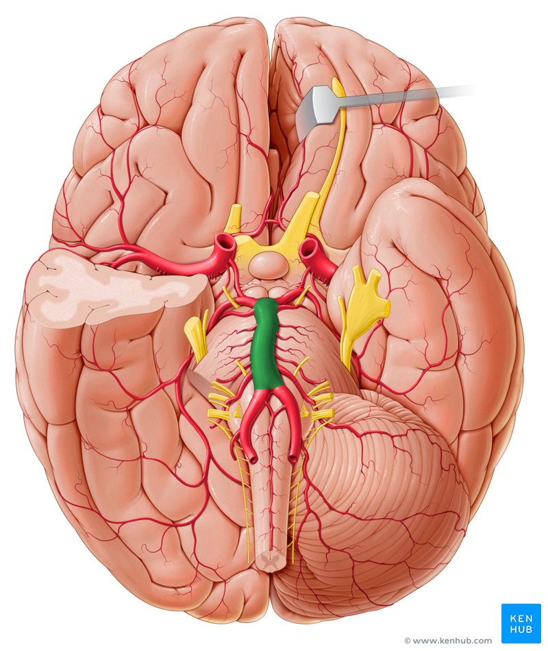
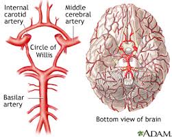
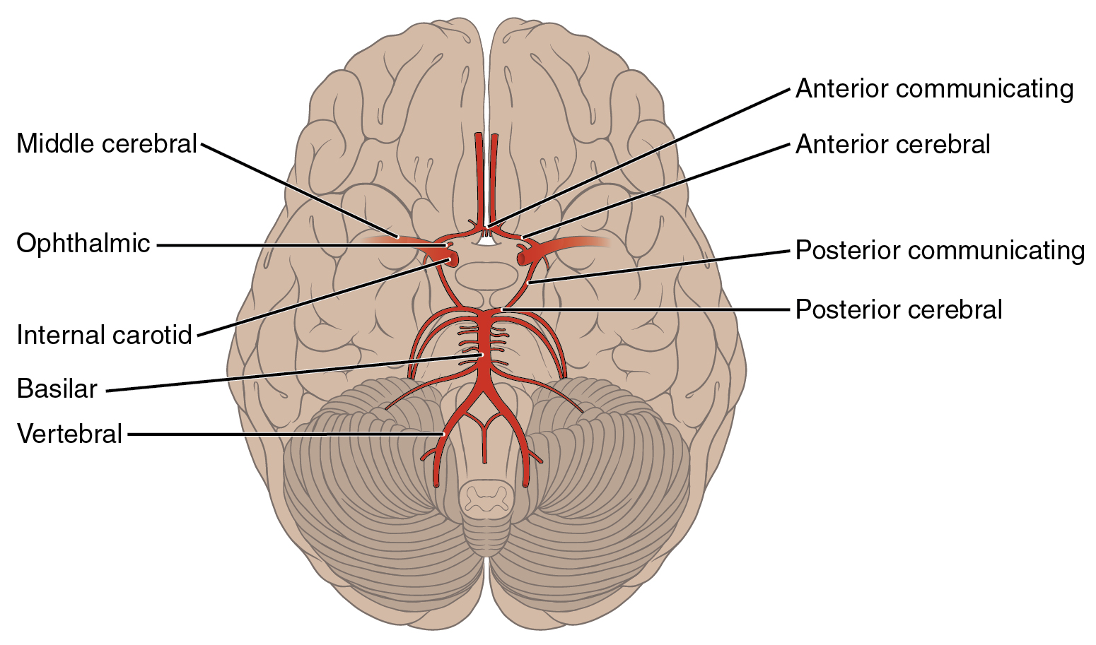
Both the carotid and vertebral arteries branch once they enter the cranial cavity, and some of these branches form a structure known as the arterial circle. What is this called?
circle of Willis, an anastomosis that is remarkably like a traffic circle that sends off branches (in this case, arterial branches to the brain). As a rule, branches to the anterior portion of the cerebrum are normally fed by the internal carotid arteries; the remainder of the brain receives blood flow from branches associated with the vertebral arteries.

How does the internal carotid artery connect to the base of the brain?
continues through the carotid canal of the temporal bone and enters the base of the brain through the carotid foramen, where it gives rise to several branches.
aorta → (left) common carotid artery/brachiocephalic to right common→internal carotid artery →anterior cerebral artery what does this supply blood to?
the frontal lobe of the cerebrum
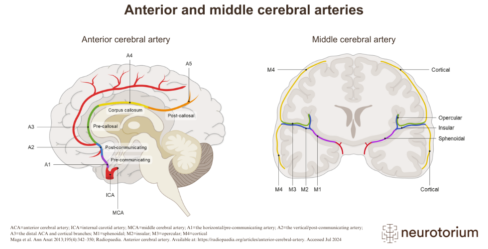
aorta → (left) common carotid artery/brachiocephalic to right common→internal carotid artery →middle cerebral artery what does this supply blood to?
the temporal and parietal lobes

What is the most common site of a CVA
the temporal and parietal lobes

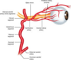
aorta → (left) common carotid artery/brachiocephalic to right common→internal carotid artery →ophthalmic artery what does this supply blood to?
the eyes.
aorta → (left) common carotid artery/brachiocephalic to right common→internal carotid artery →right and left anterior cerebral arteries join together to form
an anastomosis called the anterior communicating artery
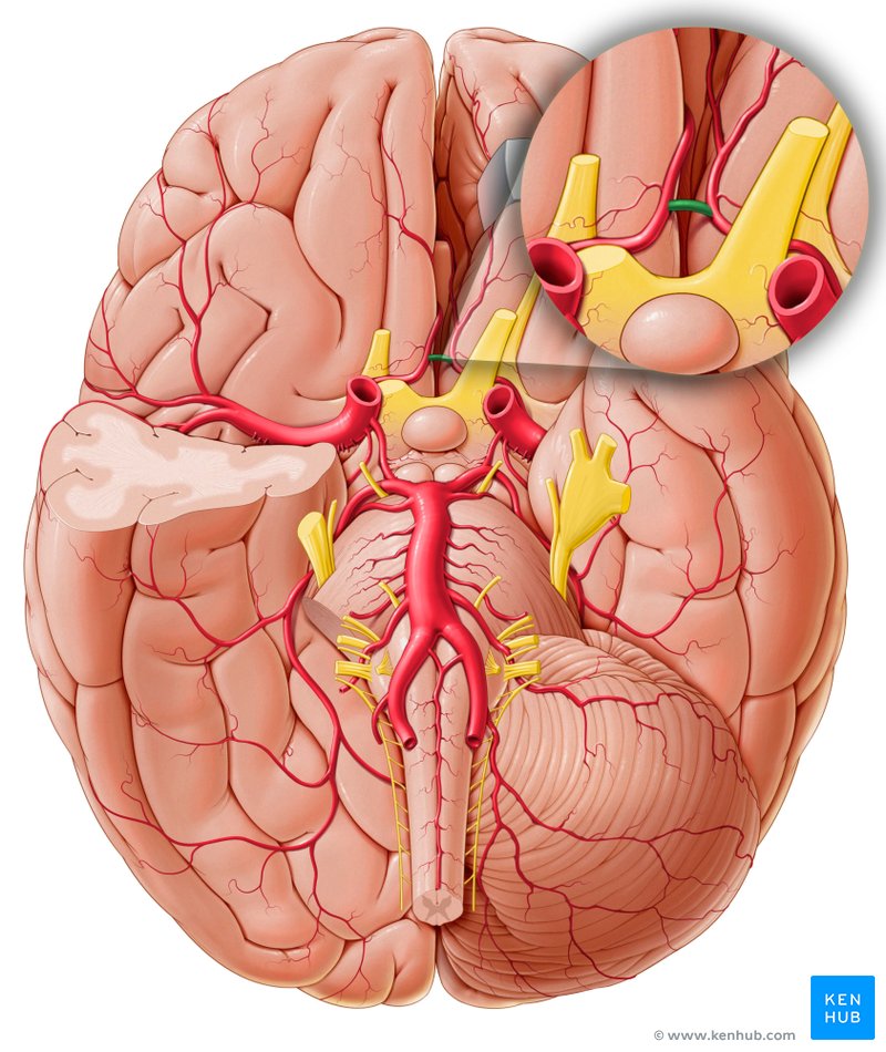
aorta → (left) common carotid artery/brachiocephalic to right common→internal carotid artery →right and left anterior cerebral arteries/initial segment anterior communicating artery→
the anterior portion of the arterial circle/circle of willis
The posterior portion of the arterial circle is formed by
The posterior portion of the arterial circle is formed by a left and a right posterior communicating artery ← posterior cerebral artery← basilar artery

basilar artery →posterior cerebral artery→posterior communicating artery→ posterior portion of the arterial circle supplies blood to what
The posterior portion of the cerebrum and brain stem

What is the basilar artery
an anastomosis that begins at the junction of the two vertebral arteries and sends branches to the cerebellum and brain stem.
what do the left and right coronary arteries and their main branches follow
the atrioventricular and interventricular grooves on the surface of the heart and are often embedded in pericardial fat.

how do the left and right coronary arteries and their main branches communicate
by joining (anastomosing) together around the heart to form continuous loops.
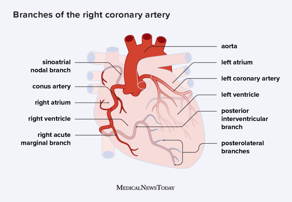
the left and right coronary arteries and their main branches communicate by joining (anastomosing) together around the heart to form continuous loops. why is this important
if one of the vessels becomes blocked, as it provides an alternative route for blood to get to the myocardium to prevent ischemia or lack of oxygen and blood to the tissue.

From which part of the aorta does the coronary arteries branch off?
Ascending aorta

Venous blood is blood is relatively low in oxygen and relatively high in
carbon dioxide. since much of the oxygen has been extracted for use by the tissues and the waste gas carbon dioxide was picked up to be transported to the lungs for elimination.
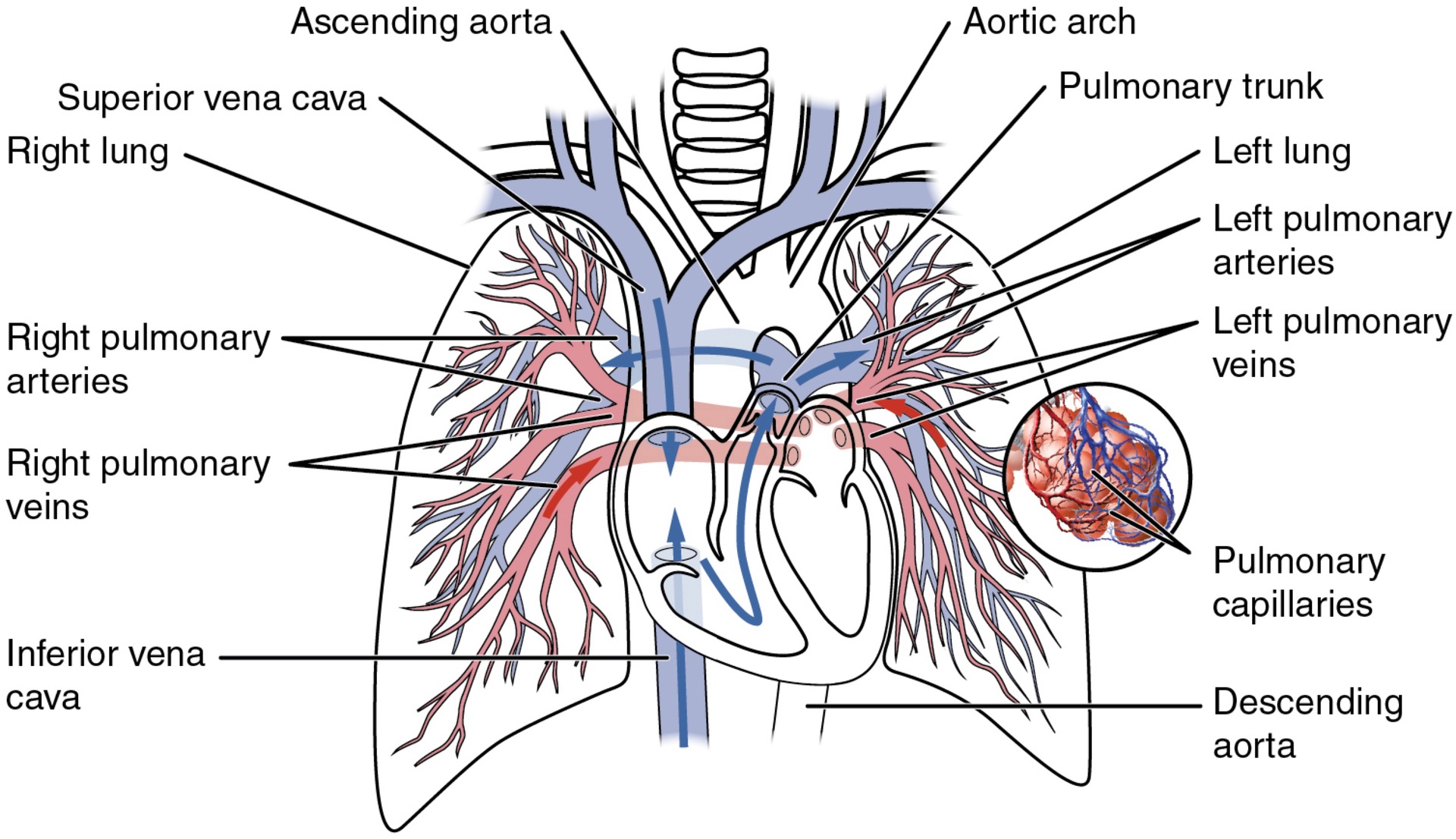
The single vessel exiting the right ventricle is
the pulmonary trunk
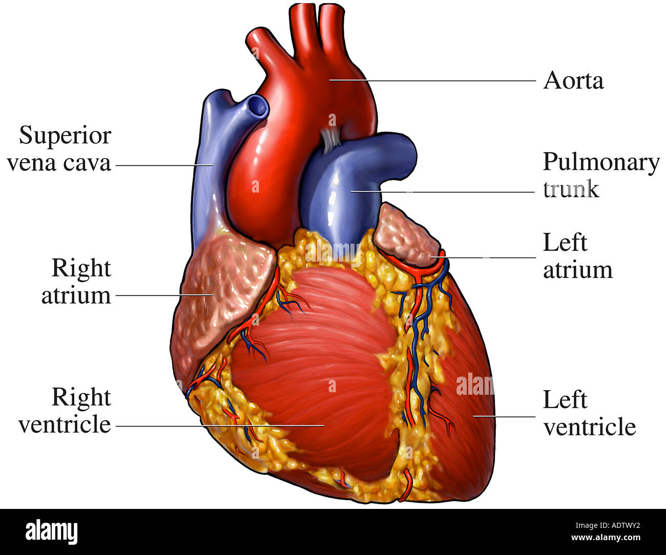
What is at the base of the pulmonary trunk
the pulmonary semilunar valve, which prevents backflow of blood into the right ventricle during ventricular diastole.
Where do the left and a right pulmonary arteries come from?
a bifurcation from the pulmonary trunk
how is blood oxygenated once it is in the lungs?
The pulmonary arteries in turn branch many times within the lung, forming a series of smaller arteries and arterioles that eventually lead to the pulmonary capillaries. The pulmonary capillaries surround lung structures known as alveoli that are the sites of oxygen and carbon dioxide exchange.

How many pulmonary veins (returning oxygenated blood from the veins to the heart via route to the left atrium) are there?
Four pulmonary veins, two on the left and two on the right, return blood to the left atrium. These vessels are the only veins in the body that carry oxygenated blood. At this point, the pulmonary circuit is complete.
The liver is a complex biochemical processing plant. How is this?
It packages nutrients absorbed by the digestive system; produces plasma proteins, clotting factors, and bile; filters blood, removing toxins and bacteria; and disposes of worn-out cell components and waste products
Instead of entering the circulation directly, what happens to absorbed nutrients and certain wastes
absorbed nutrients and certain wastes (for example, materials produced by the spleen) travel to the liver for processing, via the hepatic portal system
How do portal systems begin and end?
in capillaries
Where do the initial capillaries from the stomach, small intestine, large intestine, and spleen lead to
the hepatic portal vein and end in specialized capillaries within the liver, the hepatic sinusoids.
What is the only other portal system
the hypothalamic-hypophyseal portal vessel (endocrine system)
What does the hepatic portal system consist of?
the hepatic portal vein and the veins that drain into it

The hepatic portal vein itself is relatively short. Where does it begin?
beginning at the level of L2 with the confluence of the superior mesenteric and splenic veins
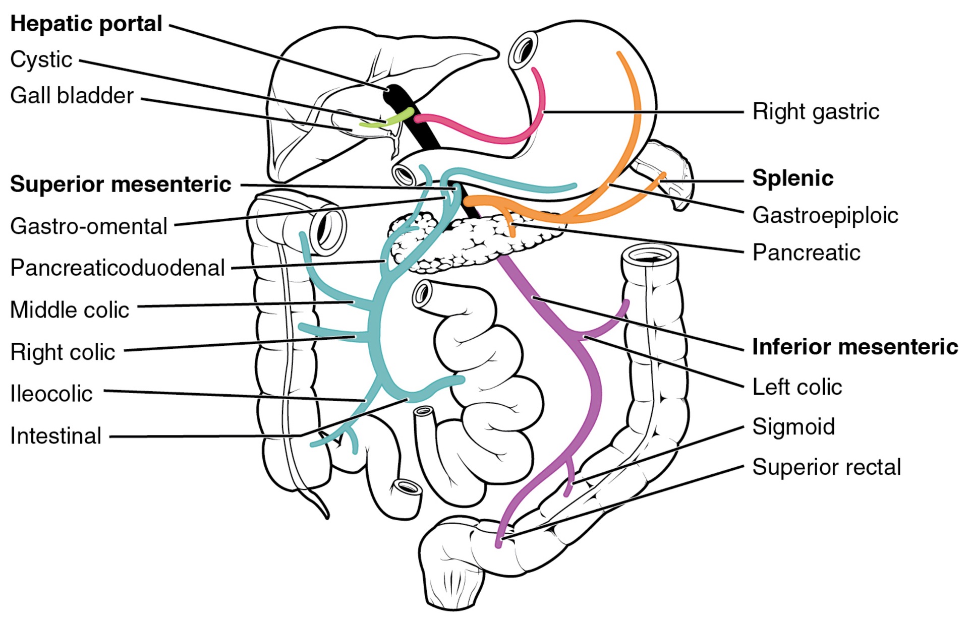
The hepatic portal vein itself is relatively short, beginning at the level of L2 with the confluence of the superior mesenteric and splenic veins. Where does it also receive branches from?
the inferior mesenteric vein plus the splenic veins and all their tributaries.

Where does the superior mesenteric vein receives blood from
the small intestine, two-thirds of the large intestine, and the stomach

What does the inferior mesenteric vein drain
the distal third of the large intestine, including the descending colon, the sigmoid colon, and the rectum.

What is the splenic vein formed from
branches from the spleen, pancreas, portions of the stomach, and the inferior mesenteric vein

branches from the spleen, pancreas, portions of the stomach, and the inferior mesenteric vein →
splenic vein
where does the liver receives its blood supply from
from normal systemic circulation via the hepatic artery and from the hepatic portal vein.

The liver processes the blood from the portal system to remove certain wastes and excess nutrients, which are stored for later use. How does this processed blood, as well as the systemic blood that comes from the hepatic artery, exit the liver
via the right, left, and middle hepatic veins and flows into the inferior vena cava.

Which main blood vessel brings blood that is filtered through the hepatic portal system back to the heart?
Inferior vena cava
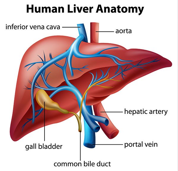
Blood from the digestive organs is transported to the liver by which vessel?
The hepatic portal vein
The liver has many functions. Identify 3 functions discussed here
Removes many toxins from the blood
Uses the blood from the digestive system for nutrient metabolism
Oxygenates the blood
Facilitates white blood cell production
Returns deoxygenated blood to the right atrium
Removes bacteria
Removes many toxins from the blood
Uses the blood from the digestive system for nutrient metabolism
Removes bacteria
hepatic portal system
specialized circulatory pathway that carries blood from digestive organs to the liver for processing before being sent to the systemic circulation
hepatic vein
drains systemic blood from the liver and flows into the inferior vena cava
hepatic artery proper
branch of the common hepatic artery; supplies systemic blood to the liver
What is the placenta?
a temporary organ about 20 cm in diameter and 3 cm thick that forms inside the uterus, providing a functional link between mother and fetus.
What is the fetal surface of the placenta like?
smooth and continuous with the umbilical cord.
What is the uterine surface of the placenta like
consists of placental villi carrying fetal blood, surrounded by capillaries carrying maternal blood
While the two bloods (maternal and fetal) do not mix directly, the exchange of substances between maternal and fetal blood occurs. How?
across capillary walls via diffusion. Diffusion across the placenta increases with fetal development.
In a developing embryo, when has the heart developed enough to begin beating
by day 21 post-fertilization
By what week of pregnancy are circulation patterns clearly established
the fourth week of embryonic life
Why is it critical for the circulatory system to form early during human development?
Because it supplies growing tissues with nutrients and gases and removes waste products, which is essential for the survival of the developing human.
When and where does blood cell and vessel production begin during early human development?
Around 15–16 days after fertilization, blood cells and vessels begin forming in structures outside the embryo (yolk sac, chorion, and connecting stalk). Development within the embryo itself starts about 2 days later.
When and from where do blood vessels begin to form during early embryonic development?
Blood vessels start forming around 15–16 days after fertilization in extraembryonic structures. Within the embryo, vessel development begins about 2 days later, originating from the embryonic mesoderm.
What is the mesoderm in embryonic development?
one of the three primary germ layers in the early embryo.
What does the mesoderm form and what does it give rise to?
The mesoderm forms between the ectoderm and endoderm. It gives rise to muscles, bones, blood vessels, the heart, and connective tissue.
What are the ectoderm and endoderm, and where are they located in the embryo?
The ectoderm is the outermost germ layer and forms structures like the skin and nervous system. The endoderm is the innermost germ layer and forms the lining of the digestive and respiratory systems. Both are part of the three-layered structure of the early embryo, with the mesoderm in between.
What are hemangioblasts, and what role do they play in early blood vessel formation?
During the first few weeks of development, blood vessels begin to form from the embryonic mesoderm. The precursor cells responsible for this process are called hemangioblasts.
What do hemangioblasts differentiate into, and what do those cells become?
Hemangioblasts differentiate into angioblasts, which form blood vessels, and pluripotent stem cells, which develop into the formed elements of blood (like red and white blood cells and platelets).
What are blood islands, and how do they form?
Blood islands are clusters of developing cells formed by angioblasts and pluripotent stem cells. They appear as scattered masses throughout the embryonic disc during early blood and vessel formation.
What happens as spaces appear in the blood islands during blood vessel formation?
Spaces that appear in the blood islands develop into vessel lumens. The endothelial lining of these vessels arises from the angioblasts within the islands.
What do the surrounding mesenchymal cells and pluripotent stem cells give rise to during blood vessel and blood formation?
Surrounding mesenchymal cells give rise to the smooth muscle and connective tissue layers of blood vessels. Meanwhile, pluripotent stem cells begin to form the elements of blood as the vessels develop.
What are the vascular tubes that develop on the blood islands, and what do they connect to?
The vascular tubes are early blood vessels that form on the blood islands. They eventually connect to one another and to the developing tubular heart.
How does the initial pattern of blood vessel development occur in the embryo?
Blood vessel development begins in multiple regions at the same time, not from a single central vessel. These vessels later connect to form a network.
What is angiogenesis?
Angiogenesis is the process of creating new blood vessels from existing ones.
Does angiogenesis only occur during early development?
No, angiogenesis continues throughout life as the body grows and develops, and in response to needs like healing or increased tissue demand.
Why is early formation of the circulatory system critical in human development?
Because it supplies growing tissues with nutrients and gases and removes waste products, which is essential for the survival of the developing human.
When and where do blood cells and vessels first begin to form?
Around 15–16 days after fertilization in extraembryonic structures (yolk sac, chorion, connecting stalk). Development inside the embryo begins about 2 days later.
Why does the developing embryo need the placenta?
As the embryo grows, its demand for nutrients and gas exchange increases, requiring a more efficient support system.