week 9 - cell-cell communication I
1/39
There's no tags or description
Looks like no tags are added yet.
Name | Mastery | Learn | Test | Matching | Spaced | Call with Kai |
|---|
No analytics yet
Send a link to your students to track their progress
40 Terms
function of ECM
defines cell shape and either attaches it to another cell or acts as a first-line defense against the outside world
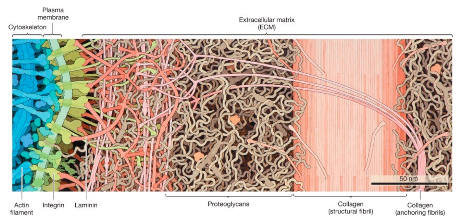
proteoglycans structure
consists of a core protein attached to many polysaccharides
where are ECM components synthesized?
in the rough ER, processed in the golgi apparatus, and secreted out of the cell through exocytosis
function of integrin
the intracellular portions of the integrins bind to proteins that are connected to the cytoskeleton, effectively linking the cytoskeleton and ECM
types of communication between eukaryotic cells
communication by direct contact between cells
indirect communication through signaling molecules
types of communication by direct contact between cells
tight junctions
desmosomes
gap junctions
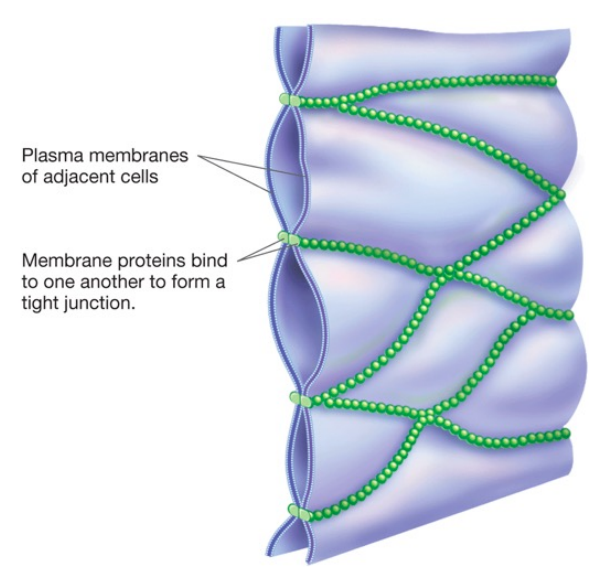
tight junctions structure
a tight junction is a cell–cell attachment composed of specialized proteins in the plasma membranes of adjacent animal cells. a long chain of these proteins forms on the surface of a cell that attaches to the same proteins on adjacent cells which pull the membranes of the two cells very close together this forms a watertight seal preventing solutions from flowing through the space between the two cells
where are tight junctions found?
between cells that form a barrier, such as the epithelial cells lining the stomach and intestines
tight junctions function
restrict passive movement of substances between the inside of the gut and the rest of the body
tight junctions are dynamic as they open and close in response to changes in environmental conditions, give an example of that
they loosen to permit more transport between epithelial cells lining the small intestine after a meal and then retighten later
define leaky gut syndrome
a syndrome of intestinal permeability due to disruption of tight junctions which allows for the entry of antigens into the bloodstream
desmosomes structure
consist of integral membrane attachment proteins forming bridges between anchoring proteins inside adjacent cells
intermediate filaments reinforce desmosomes by attaching to the anchoring proteins in the cytoplasm which forms a continuous structural support system between the cells in the tissue
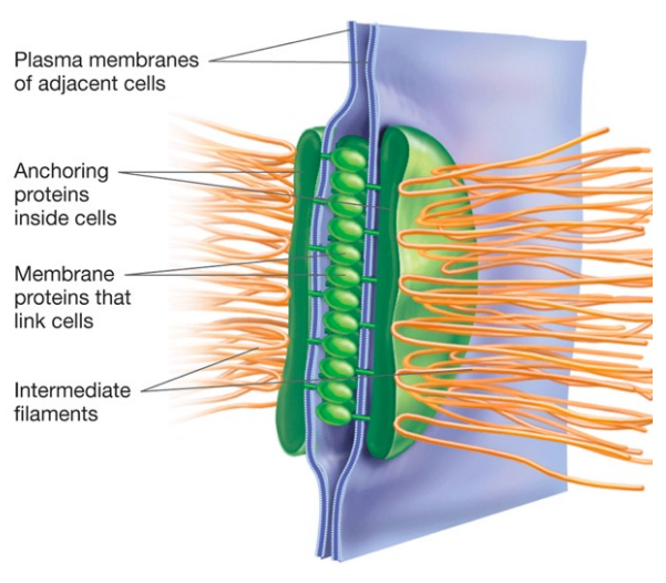
desmosomes function
stop separation during contraction by joining the cells together which is important for tissues that undergo strong mechanical stress
desmosomes found in
heart and intestinal tissue
gap junction structure
gap junctions connect cells through protein channels where specialized proteins assemble in the membranes of adjacent cells, creating interconnected channels that allow water, ions, and small molecules such as amino acids, sugars, and nucleotides to move between the cells
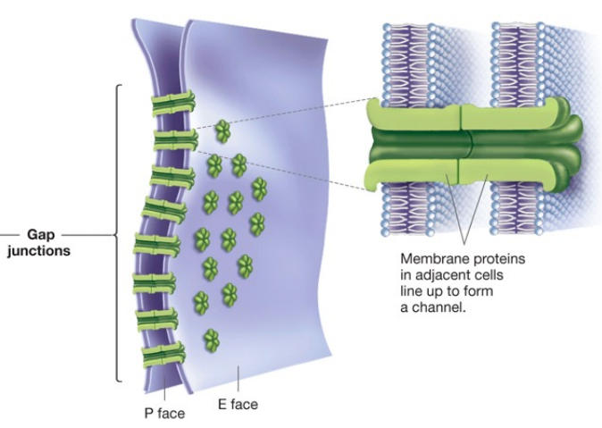
gap junction function
work as communication portals to help adjacent cells coordinate their activities by allowing the rapid passage of regulatory ions or small molecules
define pacemaker cells
a group of specialized cardiac cells that have automatic action potential generation capability
what is the role of gap junctions in cardiac action potential?
gap junctions allow action potentials to spread between cardiac cells by permitting the passage of ions between cells, producing depolarization of the heart muscle
therefore, all cardiac muscle cells are electrically linked together by gap junctions which allow the action potential to pass from one cell to the next
types of signals (no direct contact between cells)
endocrine - a ligand travels through the bloodstream to reach the receptor on the target cell
paracrine - signaling cell releases ligand to bind on the receptor on the target
neuronal - ligand travels across the neuron to the synapse to the receptor on the target cell
autocrine signaling - ligand released and bound on the same cell
steps of cell signaling
1- signal reception:
a chemical signal is detected when the signaling molecule binds to a receptor protein located at the cell surface (or inside the cell)
2- signal transduction:
the binding of the signaling molecule changes the receptor protein in some way which initiates the transduction
the transduction stage converts the signal to a form that can result in a specific cellular response
transduction sometimes occurs in a single step but more often requires a sequence of changes in a series of different molecules (a signal transduction pathway)
3- response:
The cell response may be almost any imaginable cellular activity such as:
catalysis by an enzyme
rearrangement of cytoskeleton
activation of specific genes in the nucleus
cells in a wide array of tissues may respond to the same signaling molecule, though, if they have the appropriate receptor, example:
if you are startled by a loud noise, cells in your adrenal glands secrete the hormone adrenaline (also called epinephrine), which carries the message “Get ready to fight or run”
In response, your heart and breathing rates increase and cells in your liver release glucose, which your muscles can use to power rapid movement which is the basis of an “adrenaline rush”
Cells in your heart, lung, and liver respond to adrenaline because they all have the receptor that binds to it
types of ligands and their receptors:
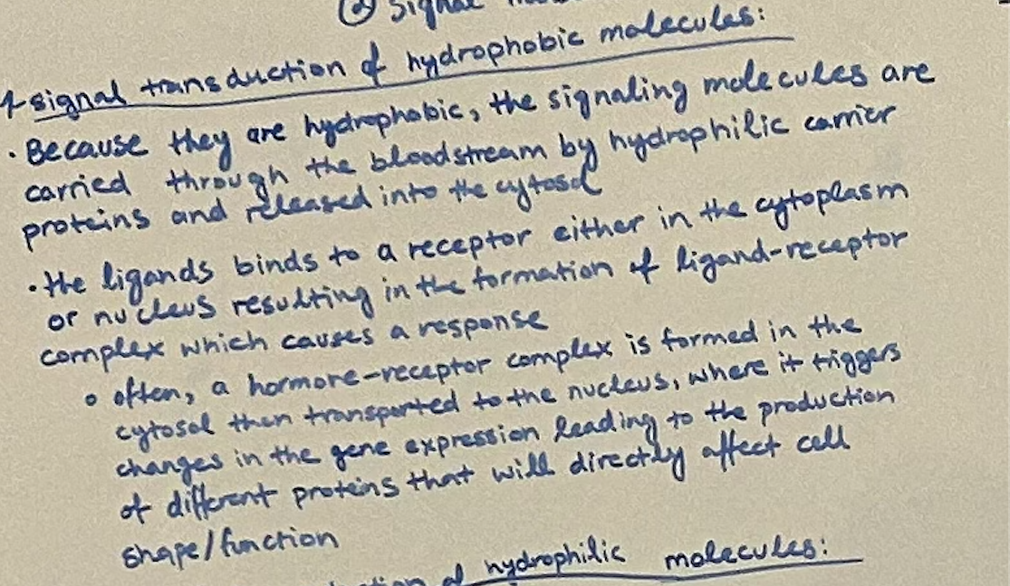
hydrophobic and small molecules - interact with the receptor inside or on the surface of the cell
intracellular receptors
ligand examples: steroid hormones including sex hormones, thyroid hormones, vitamin D, and NO (induce tissue relaxation)
hydrophilic molecules - interact with the receptor on the cell surface
ion channel receptors, receptor tyrosine kinase, G-protein-coupled receptors
ligand examples: neurotransmitters and non-steroidal hormones
define agonist
a substance that binds to a receptor and triggers the same response as the primary ligand
define antagonist
a susbstance that binds to a receptor to block or dampen the receptor’s normal response
explain the process of signal transduction of hydrophobic molecules
explain the process of signal transduction of hydrophilic molecules:
(a) ion channel receptors
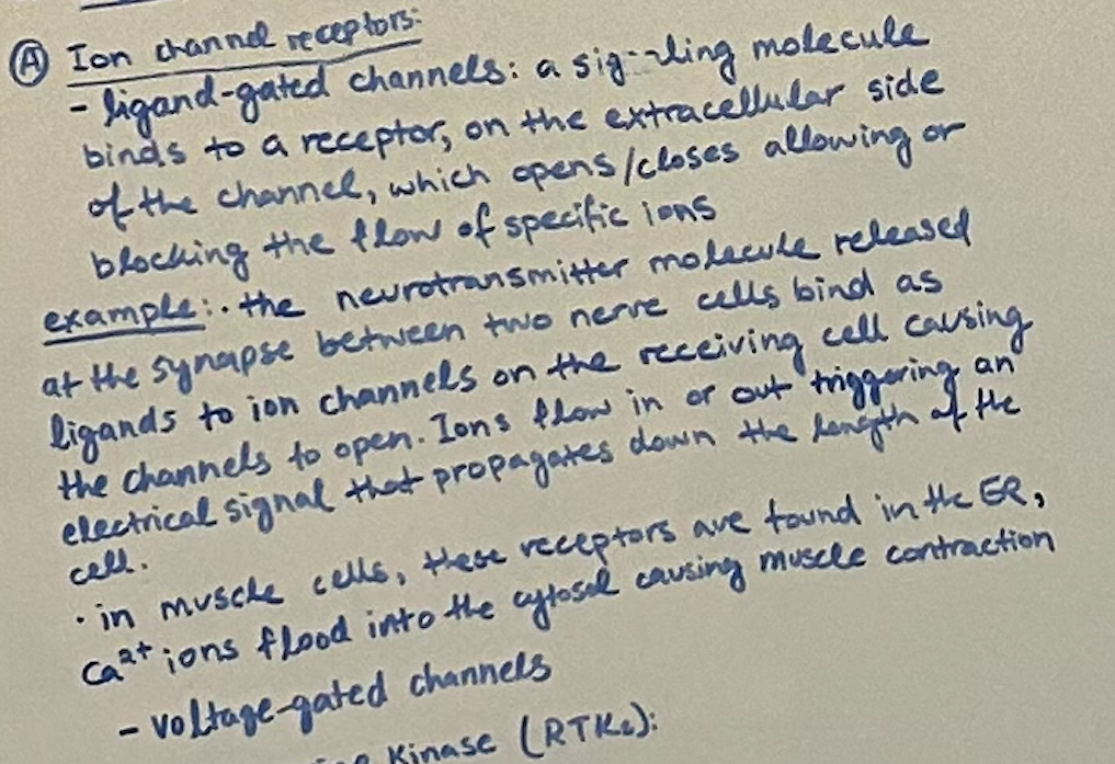
explain the process of signal transduction of hydrophilic molecules:
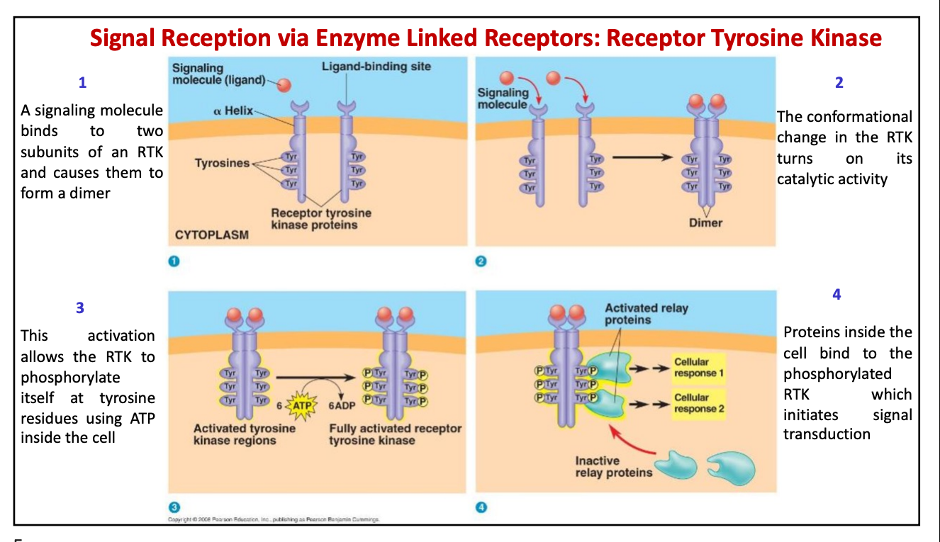
(b) receptor tyrosine kinase (RTKs)
general mechanism:
one ligand binds at each binding site of the two subunits of RTK causing them to form a dimer
conformational change in RTK turns on catalytic activity
this activation allows the RTK to phosphorylate itself at tyrosine residues using ATP in the cell
proteins inside the cell bind to the phosphorylated RTK which initiates signal transduction
define JAK
a kinase receptor bound on the receptor
define STAT
a transcription factor
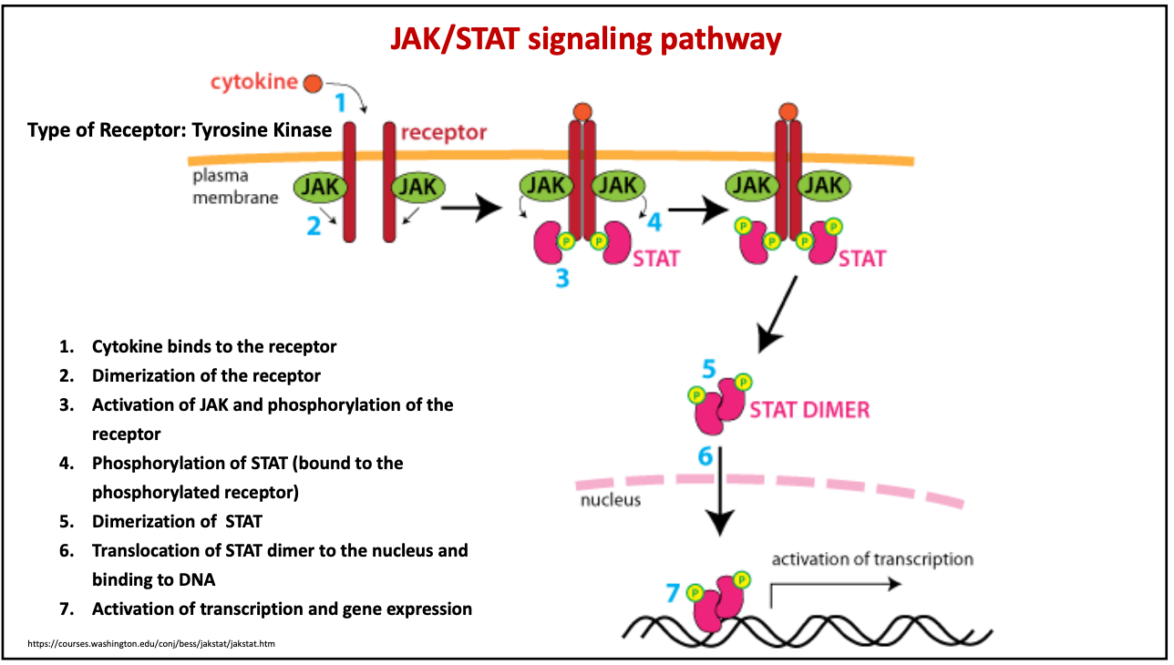
describe the JAK/STAT signaling pathway:
Cytokine binds to the receptor
Dimerization of the receptor
Activation of JAK and phosphorylation of the receptor
Phosphorylation of STAT (bound to the phosphorylated receptor)
Dimerization of STAT
Translocation of STAT dimer to the nucleus and binding to DNA
Activation of transcription and gene expression
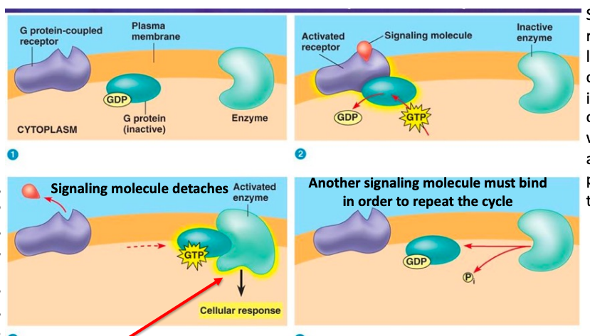
describe the g-protein-coupled receptor signaling pathway:
signaling molecule binds to the extracellular side of the receptor
G-protein on the intracellular side is bound with a GDP binds to the G-protein receptor which becomes active
when the G-protein binds, it becomes activated by replacing GDP with GTP
active G-protein dissociates from the receptor and binds to an enzyme
when it binds to an enzyme:
a. the enzyme becomes active and produces a secondary messenger which triggers a cell response
GTP is hydrolyzed to GDP and Pi which makes the G-protein inactive again
define secondary messengers:
intracellular signaling molecules released by the cell in response to exposure to extracellular signaling molecules (first messengers)
what is the function of cAMP?
activates certain protein kinases
what is the function of IP3?
opens Ca2+ channels, allowing stored Ca2+ to enter the cytosol
what is the function of Ca2+?
binds to receptor calmodulin (calcium modulating protein) which then activates kinases
stimulates muscle contraction
what makes cAMP?
made by adenyl cyclase using ATP
where is the phosphate group attached in cAMP?
both 5’ and 3’ carbons
how is cAMP inactivated?
by phosphodiesterase which converts it to AMP
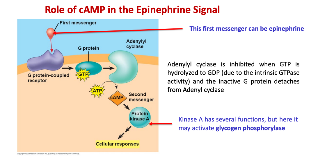
describe the role of cAMP in the epinephrine signal:
epinephrine (first messenger) binds to its alpha and beta receptors
inactive G-protein binds to the receptor and it becomes activated by replacing GDP with GTP
G-protein binds to adenyl cyclase which uses ATP to produce cAMP (secondary messenger)
cAMP binds to protein kinase A which eventually activates glycogen phosphorylase which would breakdown glycogen into glucose
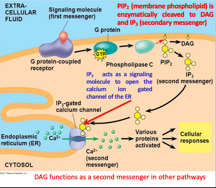
describe the role of Ca2+ and IP3 in cell signaling:
adrenaline binds to the receptor
G-protein binds to the receptor and is activated by the replacing GDP with GTP
activated G-protein activates phospholipase C which cleaves PIP2 to DAG and IP3
IP3 binds to a calcium-gated channel which allows Ca2+ stored in the RER into the cytosol
one of the functions of Ca2+ is to cause smooth muscle contraction