Microbiology 2: Prokaryotic Cells
1/58
There's no tags or description
Looks like no tags are added yet.
Name | Mastery | Learn | Test | Matching | Spaced | Call with Kai |
|---|
No analytics yet
Send a link to your students to track their progress
59 Terms
Discuss the essence of the prokaryotic cell
-They are invisible microorganisms that lack a nucleus and membrane-bound organelles.
- They are divided into 2 groups—bacteria & archaea
- They are much smaller than eukaryotic cells
- The adult body has about 10 trillion cells
Identify two prokaryotic microorganisms.
Escherichia coli (E. coli)
Methanogens (Archaea)
Escherichia coli (E. coli)
a bacterium commonly found in the intestinal tract
Methanogens (Archaea)
anaerobic microorganisms that produce methane as a byproduct of energy metabolism in low-oxygen environments like wetlands, sediments, and animal guts
Describe the shapes of Coccus
Spherical
Describe the shapes of Coccobacillus
short, rod-shaped
Describe the shapes of Vibrio
Curved, comma-shaped rods, often with a slight twist
Describe the shapes of Bacillus
cylindrical, rod-shaped, stick-like cells
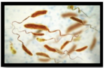
Describe the shapes of Spirillum
Rigid, corkscrew-shaped, capable of movement
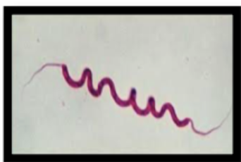
Describe the shapes of Spirochete
long, slender, flexible, spiral-shaped
Bacteria are identified by the arrangements they form. Describe the arrangements of Diplocci
Spherical bacteria (cocci) that remain in pairs after dividing in one plane.
Bacteria are identified by the arrangements they form. Describe the arrangements of tetracocci.
Groups of four spherical cells arranged in a square, formed by division in two perpendicular planes.
Bacteria are identified by the arrangements they form. Describe the arrangements of Sarcinae
Cuboidal packets of eight or more spherical cells, resulting from division in three regular planes.
Bacteria are identified by the arrangements they form. Describe the arrangements of Streptococci
Spherical cells arranged in chains, occurring when cells divide repeatedly in a single plane.
Bacteria are identified by the arrangements they form. Describe the arrangements of staphylococci.
Irregular, grape-like clusters of spherical cells formed by division in multiple, random planes.
Bacteria are identified by the arrangements they form. Describe the arrangements of Palisades
Rod-shaped bacteria that align side-by-side in a parallel, picket fence-like, or "V"-shaped pattern.
Bacteria are identified by the arrangements they form. Describe the arrangements of Streptobacilli
Rod-shaped cells that remain attached in chains end-to-end
Bacteria are identified by the arrangements they form. Describe the arrangements of Diplobacilli
Two rod-shaped bacteria (bacilli) that remain attached in pairs end-to-end after division.
What is the glycocalyx composed of?
It is a sticky gelatinous layer composed of a glycoprotein-polysaccharide complex that surrounds the cell walls of many bacteria
The glycocalyx can be a capsule or a slime layer. Explain.
It acts as either a capsule or a slime layer, based on its structure and adherence.
Capsules are organized, rigid, and tightly bound to the cell, protecting against phagocytosis.
Slime layers are disorganized, loose, and easily washed off, aiding in surface attachment and biofilm formation
treptococcus pneumoniae can cause pneumonia, ear infections, meningitis, and other diseases. It comes in two forms, encapsulated and non-encapsulated. When it is encapsulated, it is pathogenic and causes disease. When non-capsulated,, it loses its virulence ability. What does the capsule have to do with pathogenicity?
The capsule protects the bacterium from the host's immune system. The polysaccharide capsule prevents phagocytosis by inhibiting the binding of complement 𝐶3𝑏 to the cell surface, allowing the bacteria to survive, evade immune detection, and cause severe invasive diseases
Identify its three main subdivisions and functions of bacterial flageellum
The Basal Body— Anchors the flagellum to the cell wall/membrane and acts as a rotary motor, generating torque to spin the structure.
The Hook - Acts as a flexible universal joint, connecting the motor (basal body) to the filament and transmitting torque.
The Filament - A long, helical, propeller-like structure that generates propulsion by rotating, enabling bacterial swimming
What protein composes the bacterial flagellum?
Flagellin
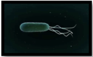
Describe each arrangement of flagella: Monotrichous:
a single flagellum located at one pole (end) of a bacterial cell
Describe each arrangement of flagella: Amphitrichous:
a single flagellum (or sometimes a tuft) at each of two opposite poles of the cell
Describe each arrangement of flagella: Lohotrichous:
a tuft (multiple) of flagella anchored at one (polar) or both ends of a bacterial cell
Describe each arrangement of flagella: Peritrichous
They rotate counter-clockwise to form a single, cohesive bundle for straight "runs" and clockwise to bundle-disrupt for "tumbles," allowing for efficient, directional movement
In Gram-negative bacteria.
Four protein rings (L P, MS, and C) in its basal body to anchor it into the cell envelope.
These include the L ring (embedded in the outer membrane), the P ring (located in the peptidoglycan layer), and the MS and C rings (located in the cytoplasmic membrane and the cytoplasm)
In Gram-positive bacteria
Two protein rings (MS and C)
Has a simpler basal body with only two rings. This is because gram-positive bacteria lack an outer membrane and periplasmic space. The 2 rings present are located in the cytoplasmic membrane and cytoplasm
Spirochetes have a type of modified flagellum called axial filaments.
Describe the structure and function of axial filaments.
Are specialized, internal flagella found in spirochetes, located in the periplasmic space between the inner membrane and outer sheath. Anchored at both cell poles, they wrap around the protoplasmic cylinder, enabling a corkscrew-like, twisting motion that allows movement through viscous environments.
Spirochetes have a type of modified flagellum called axial filaments. How does it generate locomotion?
Through thor axial filaments—also known as endoflagella—by rotating these internal structures between the cell wall and the outer membrane. It causes the entire helical-shaped, or corkscrew-shaped, bacterium to rotate, creating a characteristic twisting motility that allows them to move efficiently through viscous environments, such as connective tissue or mucus.
What is the function of fimbriae?
Used for adhering to surfaces, host tissues, and other cells to facilitate colonization and infection.
In human anatomy, fimbriae are finger-like projections at the end of the fallopian tubes that sweep ovulated eggs into the tube
Define each cell wall structure: Peptidoglycan
A polymer composed of sugar and amino acids, forming a mesh-like layer outside the cytoplasmic membrane that provides structural strength and shape.
Define each cell wall structure: N-acetylglucosamine (NAG) -
A glucose derivative and amino sugar that alternates with NAM to form the backbone of the peptidoglycan polysaccharide chain.
Define each cell wall structure: N-acetylmuramic acid (NAM) -
An amino sugar component of the bacterial cell wall backbone that acts as a sugar derivative of NAG; it is the attachment site for the peptide side chains.
Define each cell wall structure: Peptide cross-bridges:
Amino acid bridges (such as glycine) or direct peptide bonds that connect the peptide side chains of adjacent glycan strands, creating a 3D lattice structure.
Define each cell wall structure: Peptide side chains”
Short chains of amino acids (usually 3–5) are attached specifically to the N-acetylmuramic acid (NAM) residues within the peptidoglycan structure
Define each cell wall structure: wall teichoic acid
Polymers of polyol phosphate are found in Gram-positive bacteria, covalently linked to peptidoglycan; they provide flexibility, cell division support, and antigenicity.
Define each cell wall structure: Wall membrane (outer membrane)
A second lipid bilayer is found only in Gram-negative bacteria, located outside the thin peptidoglycan layer.
Define each cell wall structure: Lipopolysaccharides (LPS) -
Complex molecules anchored in the outer membrane of Gram-negative bacteria, functioning as a major antigen and endotoxin.
Define each cell wall structure: Porin Proteins -
Channel-forming proteins embedded in the outer membrane of Gram-negative bacteria that allow the passive diffusion of low-molecular-weight substances.

Define each cell wall structure: Periplasm (Periplasmic space)-
The concentrated gel-like matrix between the inner cytoplasmic membrane and the outer membrane in Gram-negative bacteria contains the peptidoglycan layer.
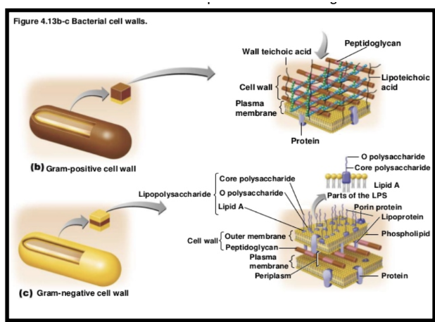
Gram-positive cell walls-
has a thick layer of peptidoglycan and contains teichoic and lipoteichoic acids
Gram-negative cell walls-
a thin peptidoglycan layer, an outer membrane containing lipopolysaccharides (LPS), and a periplasmic space between the 2 membranes

How does the selectively permeable membrane relate to the plasma membrane of bacteria?
it controls which substances can pass into and out of the cell. This regulation is crucial for maintaining the internal environment of the bacterium
How does the phospholipid bilayer relate to the plasma membrane of bacteria?
Forms a barrier between the inside and outside of the cell
How does the dual nature of phospholipid relate to the plasma membrane of bacteria?
A phospholipid is an amphipathic molecule, which has both a hydrophilic (water-loving) and a hydrophobic (water-fearing) tail. This dual nature allows the formation of the bilayer structure in a watery environment.
How does the hydrophobic interaction relate to the plasma membrane of bacteria?
Occurs between the nonpolar hydrocarbon tails of the phospholipids, which cluster together in the interior of the membrane, away from water
How does the integral proteins relate to the plasma membrane of bacteria?
Embedded within the membrane, often spanning the entire bilayer (transmembrane), and play roles in transport and signaling
How does the peripheral proteins relate to the plasma membrane of bacteria?
Attached to the surface pf the membrane, either on the inner or outer face, but do not extend into the hydrophobic core
How do the hopanoids relate to the plasma membrane of bacteria?
sterol-like molecules found in some bacterial membranes that help regulate membrane fluidity, similar to the function of cholesterol in eukaryotic cells
Where are you located in the cell?
In the cytoplasm, within an irregularly shaped region called the nucleoid
Explain what the nucleoid region is.
An irregular region within the prokaryotic cell cytoplasm. It's where the genetic material (DNA) is concentrated
Descibe the DNA of bacteria
In the form of a single, circular chromosome found in the nucleoid region.
What is a plasmid, and why are plasmids beneficial to bacteria?
A small, circular DNA molecule separate from the main chromosomes.
They are beneficial to bacteria because they carry genes that provide antibiotic resistance, which can be shared between bacteria.
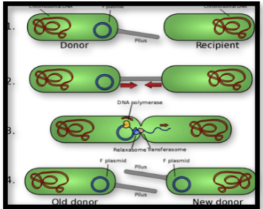
What is the significance of conjugation?
It allows for the transfer of genetic material between bacteria, a process known as horizontal gene transfer.
What role does the sex pilus have during conjugation?
Acts as a critical, hair-like appendage produced by donor bacteria to initiate conjugation by attaching to a recipient cell, drawing it closer, and facilitating the transfer of genetic material (usually plasmids.) It serves as a physical tender and, in some cases, a conduit for DNA, ensuring stable cell-to-cell contact.
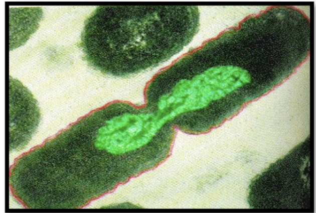
Describe binary fission in bacteria.
Subunits: The 70S ribosome consists of a 30S (small) and 50S (large) subunit.
Ribosomes are approximately 65% RNA and 35% protein, with ribosomal RNA (rRNA) acting as the catalytic ribozyme for protein synthesis.
Discuss ribosomes' function.
They are the site of protein synthesis, where they read messenger RNA (mRNA) and translate it into a specific amino acid chain.