KN 232 Week 5 Part 4: Anatomical Terms and Definitions for the Lower Limbs
1/42
There's no tags or description
Looks like no tags are added yet.
Name | Mastery | Learn | Test | Matching | Spaced |
|---|
No study sessions yet.
43 Terms
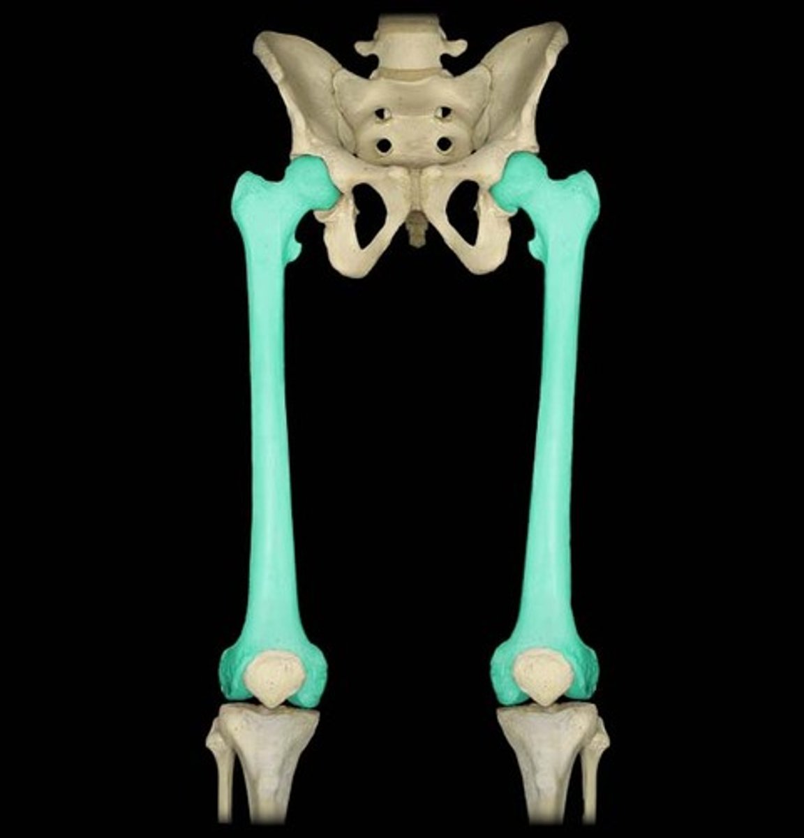
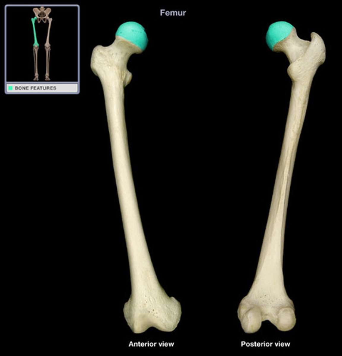
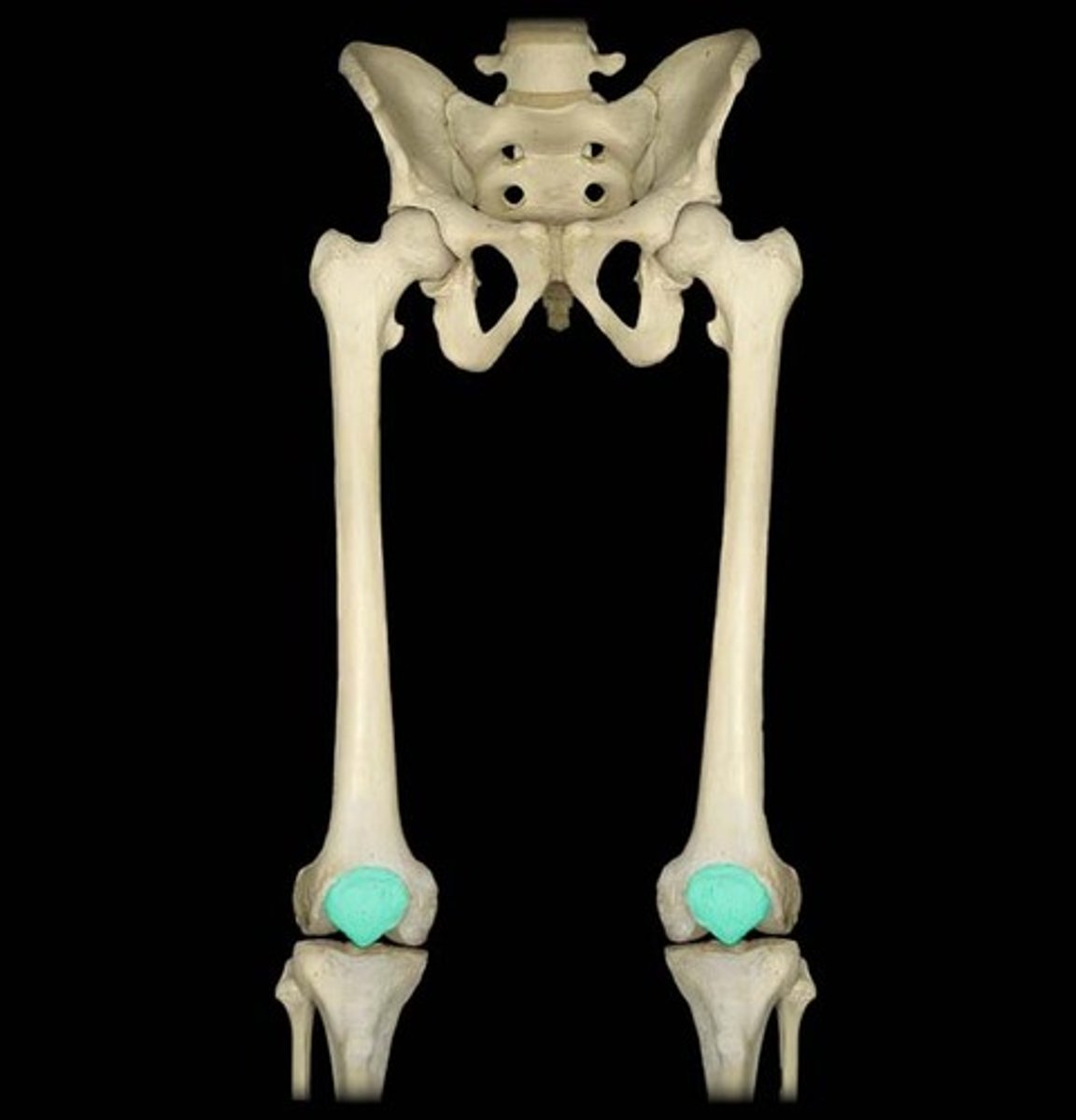
Femur
longest, heaviest, and strongest bone in the body located in thigh region.
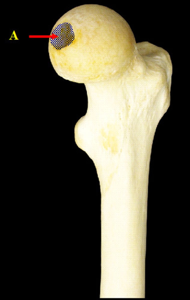
head of femur
articulates with the os coxae at the acetabulum
neck of femur
Joins the shaft of the femur at an angle.

fovea capitis of femur
a small depression in the head of the femur for the attachment of a short ligament that runs to the acetabulum
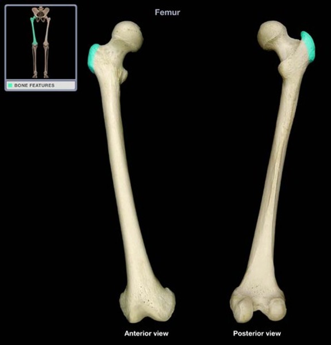
Greater Trochanter
A bony projection on the proximal lateral side of the thigh, just below the hip joint.
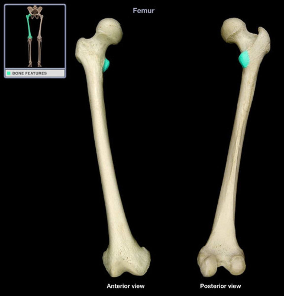
Lesser Trochanter
On the posterior and medial side of femur.
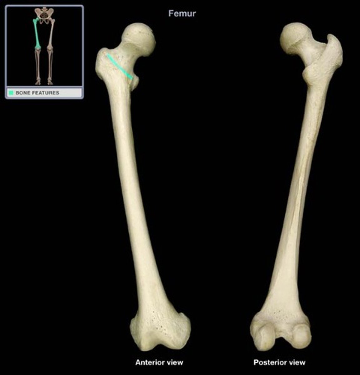
Intertrochanteric line
region formed anteriorly between the trochanters.
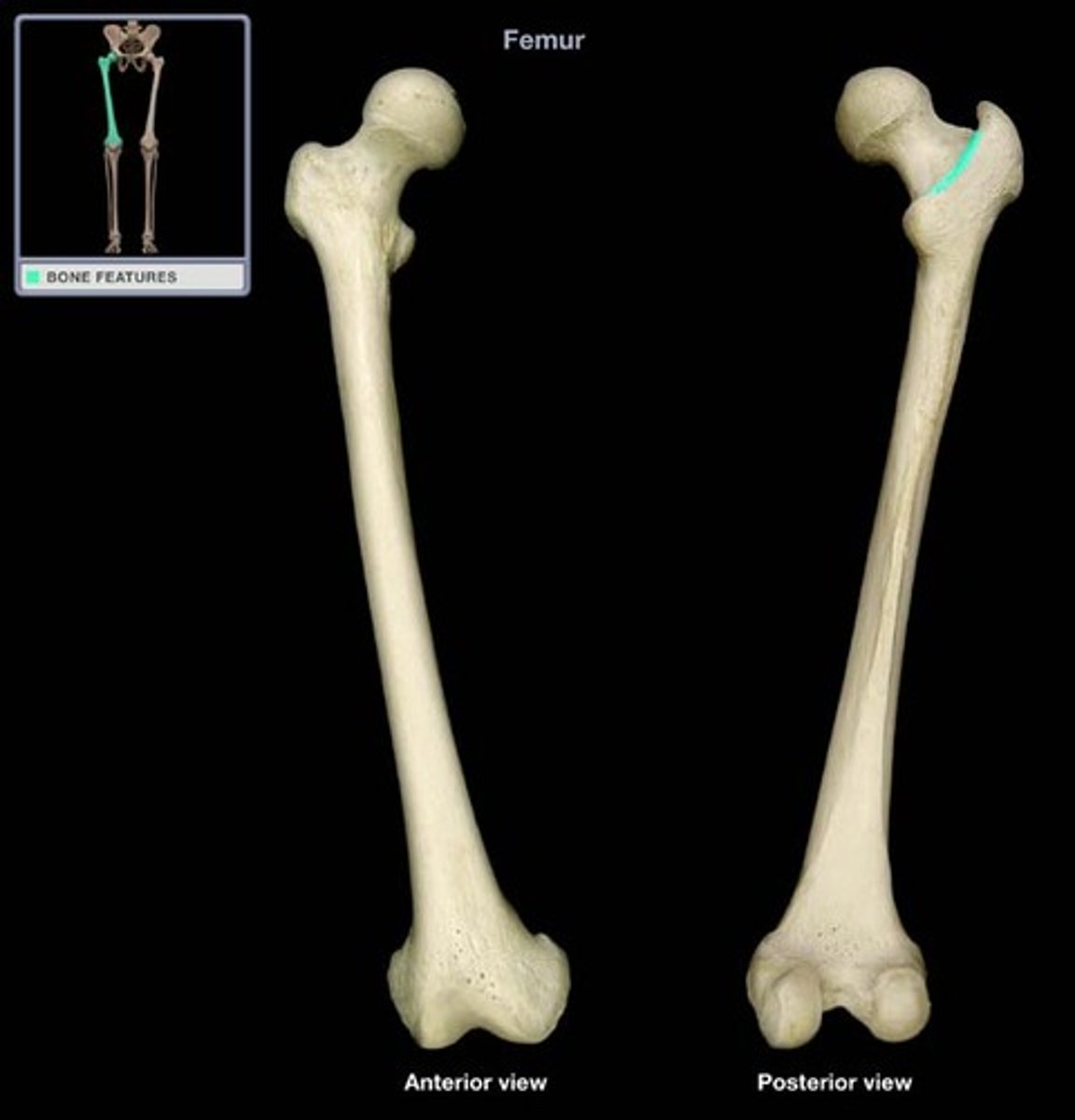
Intertrochanteric crest
oblique region of bone formed posteriorly between the greater and lesser trochanters
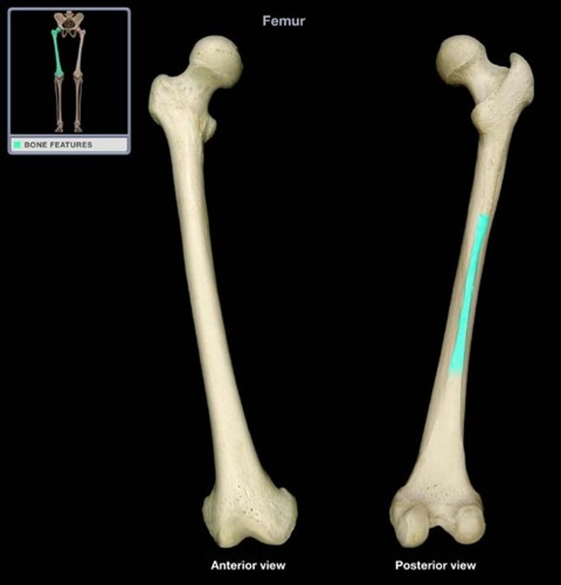
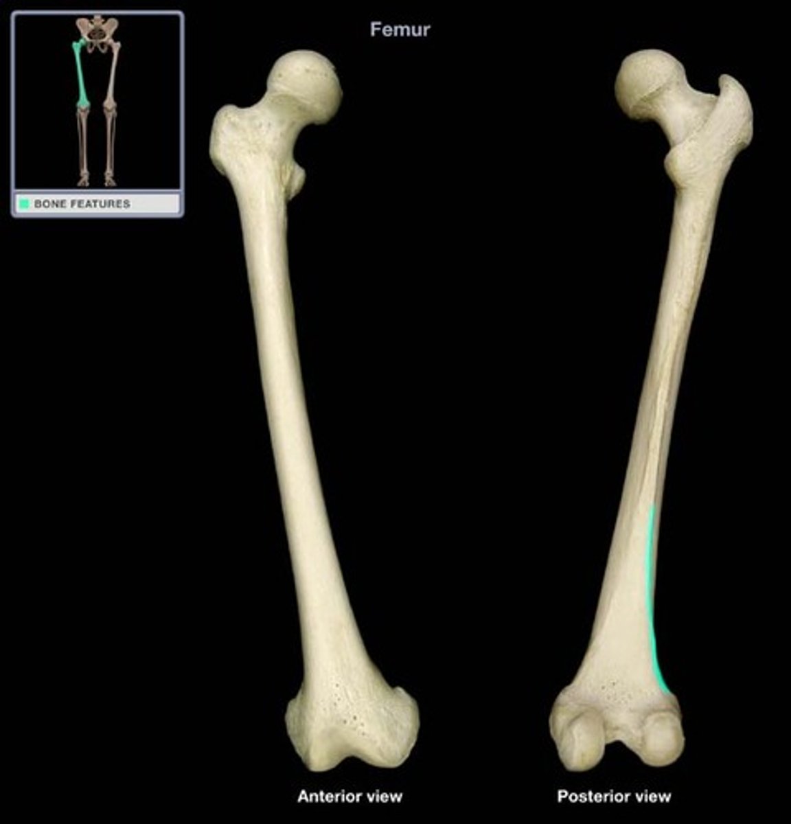
Gluteal tuberosity
back of femur; bump above the linea aspera that is on the upper portion of femur. Attaches gluteus maximus muscle
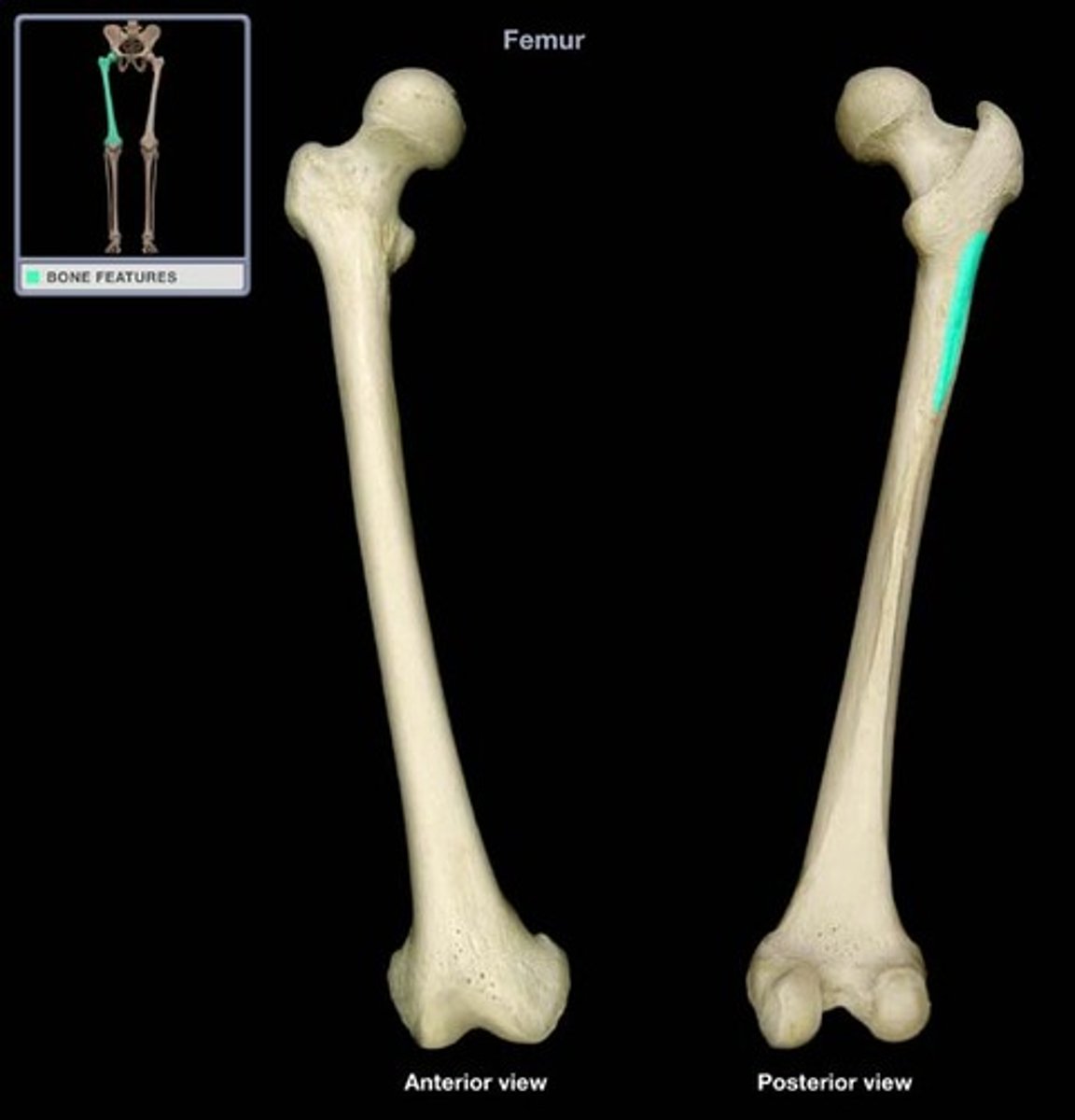
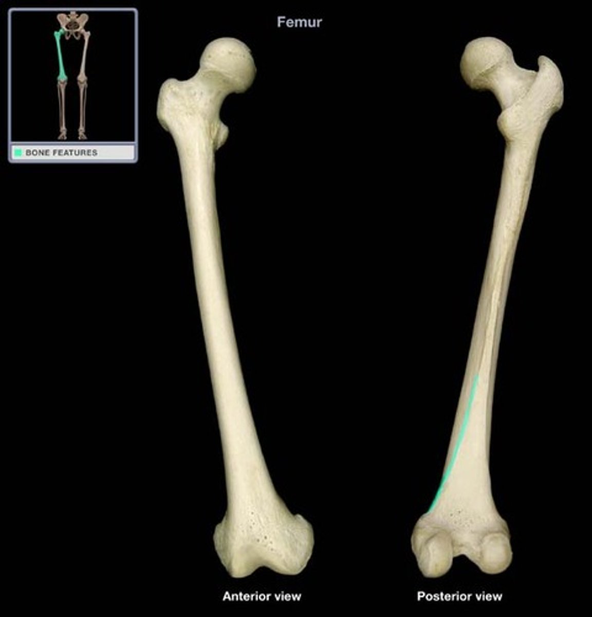
Linea aspera
Elevated midline ridge where thigh muscles attach. branches into medial and lateral supracondylar lines.
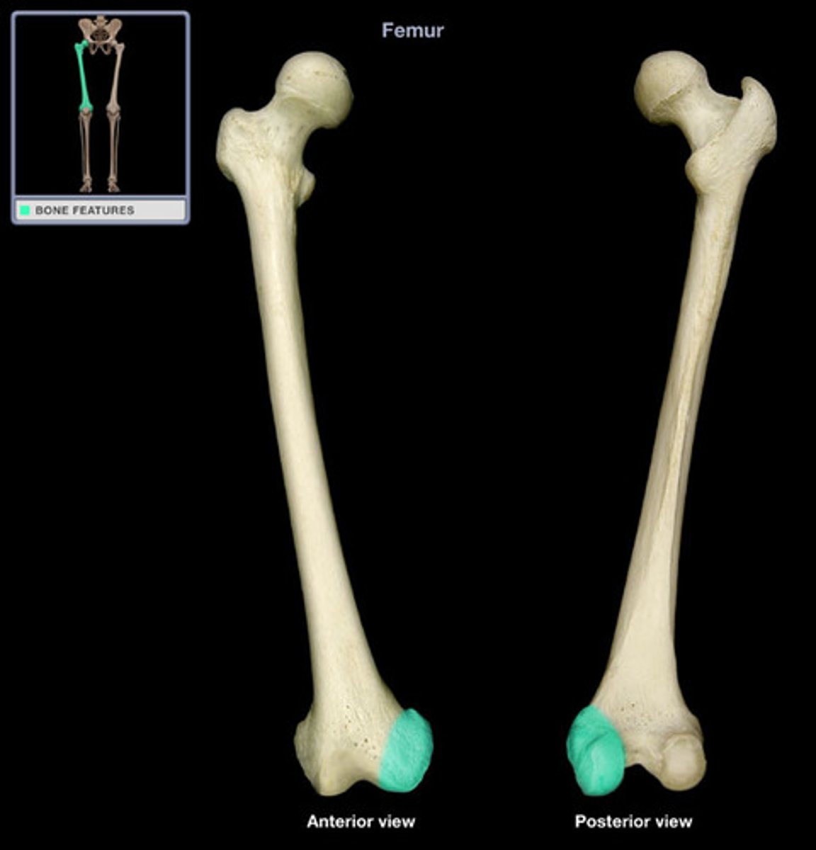
Medial condyle
Separated by deep intercondylar fossa. Inferior to medial epicondyle.
Lateral epicondyle
Separated by deep intercondylar fossa. Inferior to lateral epicondyle.
Medial supracondylar line
a subtle ridge extending from the distal end of the linea aspera and angles medially
Lateral supracondylar line
a subtle ridge extending from the distal end of the linea aspera and angles laterally
Medial epicondyle
Superior to medial condyle
Lateral Epicondyle
Superior to lateral condyle

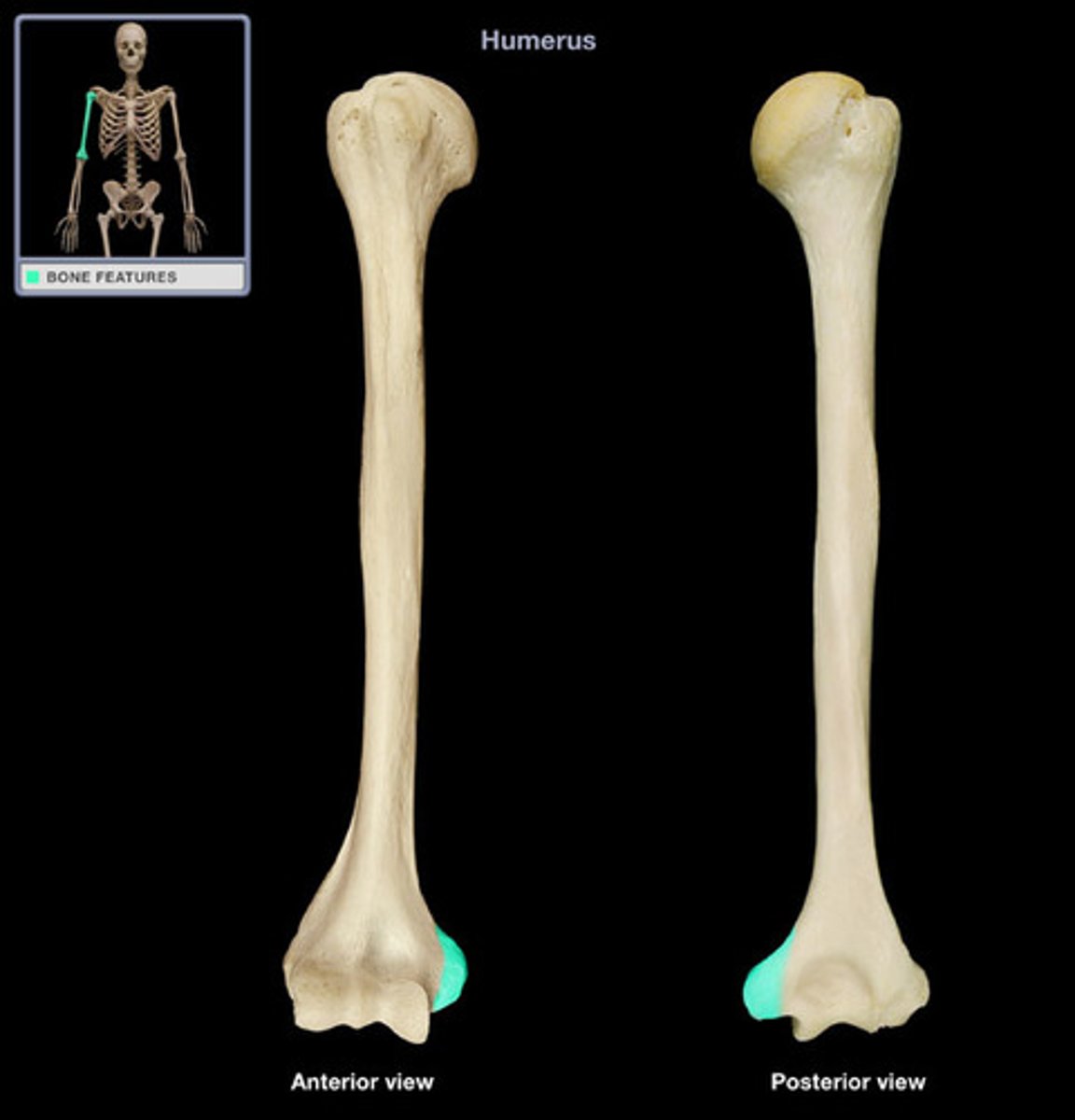
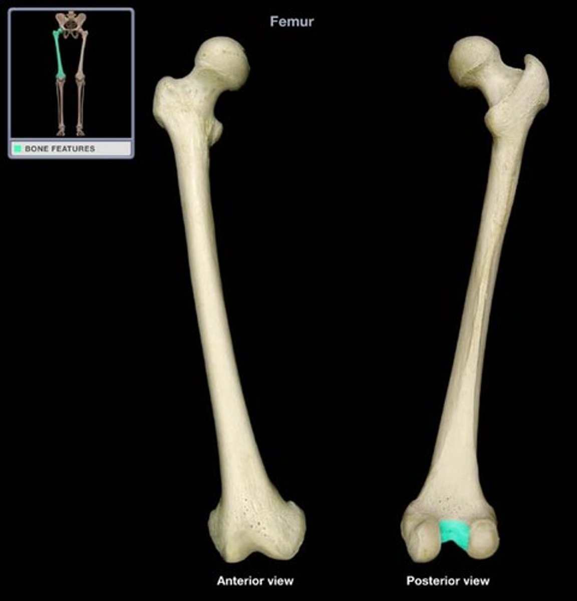
Intercondylar fossa
The depression between the condyles of the femur.
Note for condyles and epicondyles of the femur.
Located distal to the femur.
Patella
kneecap. Base is broader and is superior to apex.
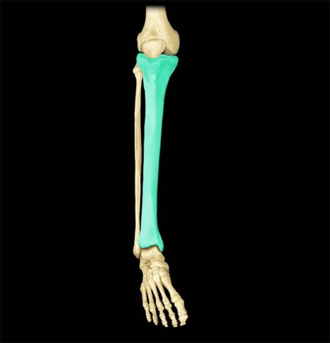
Tibia
Medial bone of the leg. Larger than complement bone. Connects to complementary bone via the interosseous membrane.
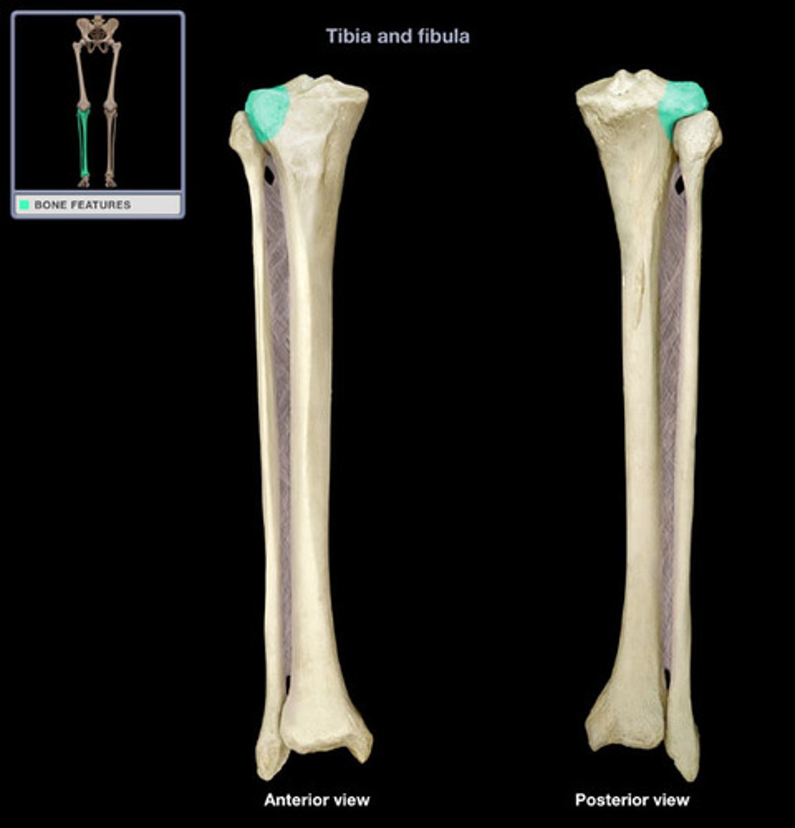
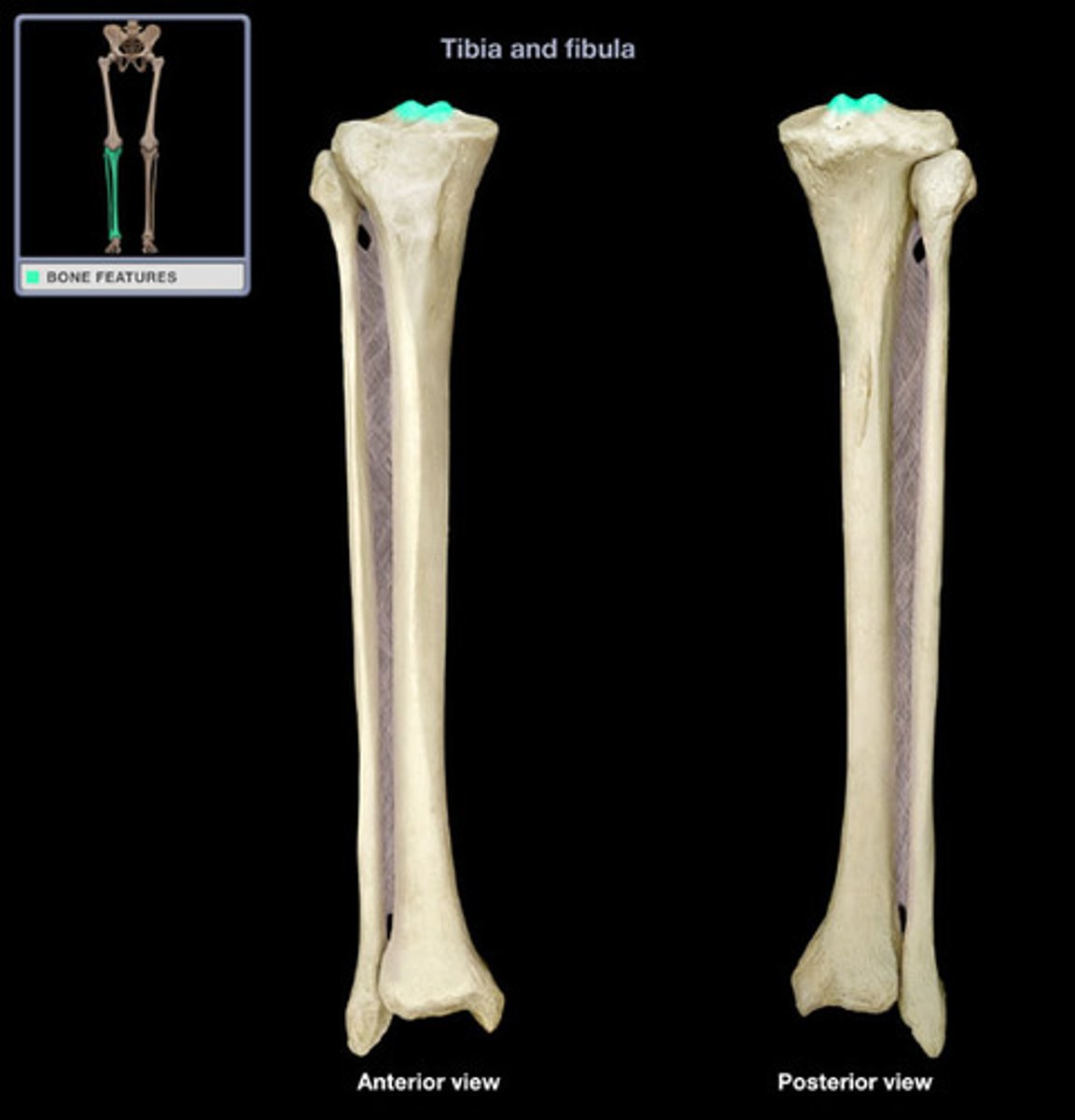
Medial condyle of tibia
Flat surface on superior and media head (proximal of tibia) that articulates with medial condyle of the femur. Separated from lateral condyle by the intercondylar eminence (ridge).
Lateral condyle of tibia
Flat surface on superior and lateral head (proximal of tibia) that articulates with lateral condyle of the femur. Separated from medial condyle by the intercondylar eminence (ridge).
intercondylar eminence of tibia
irregular projection located between the two condyles of the tibia
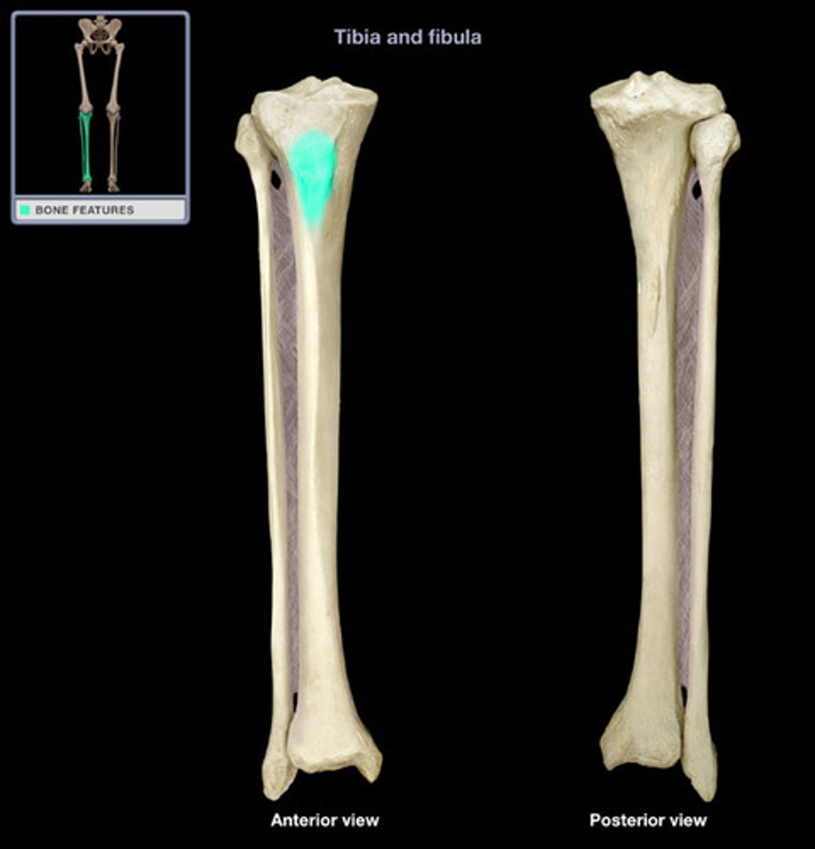
Tibial tuberosity of the tibia
point where the patellar ligament attaches
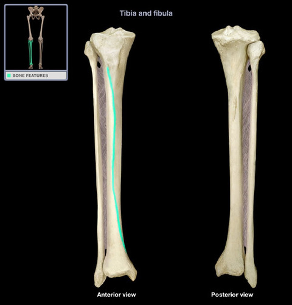
Anterior border of the tibia
The shin. Sharp anterior ridge (anterior crest; tibial crest.)
Medial malleolus of tibia
forms the medial bulge of the ankle
Fibula
Lateral bone of the leg. smaller than complement bone. Connects to complementary bone via the interosseous membrane.

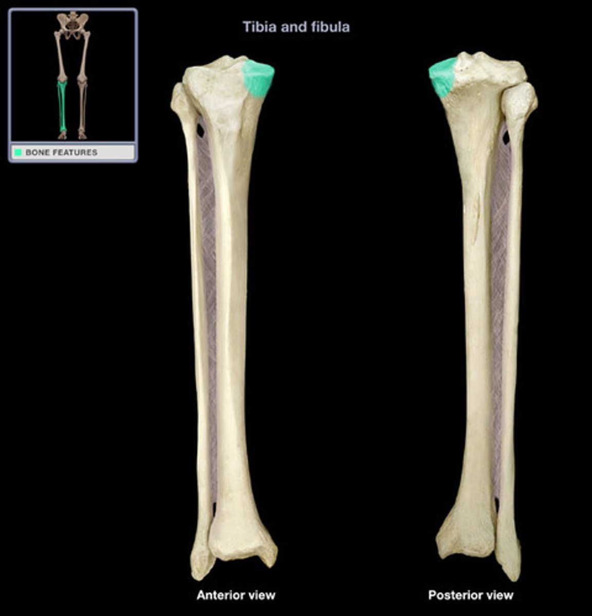
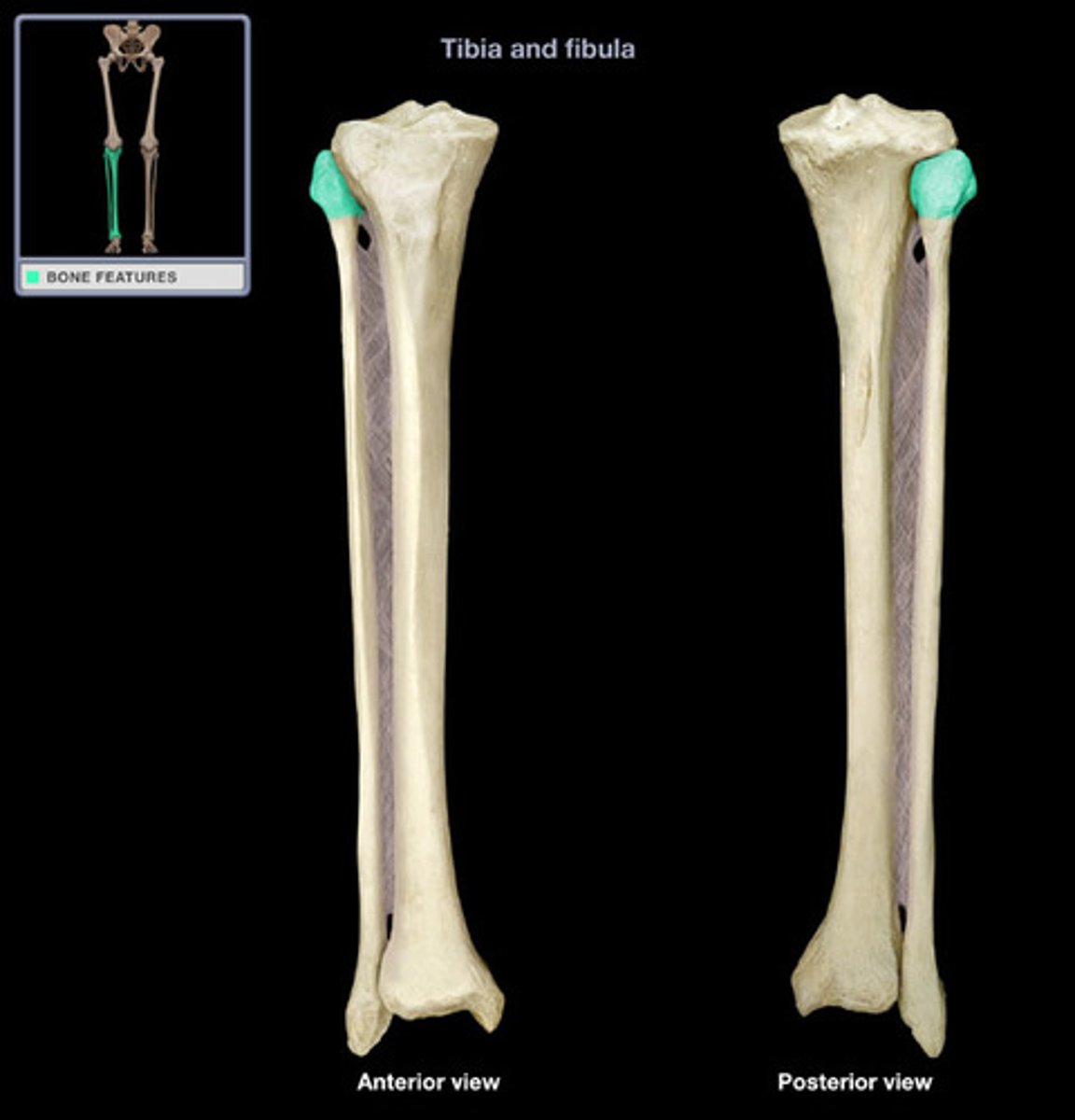
Head of fibula
proximal end of fibula
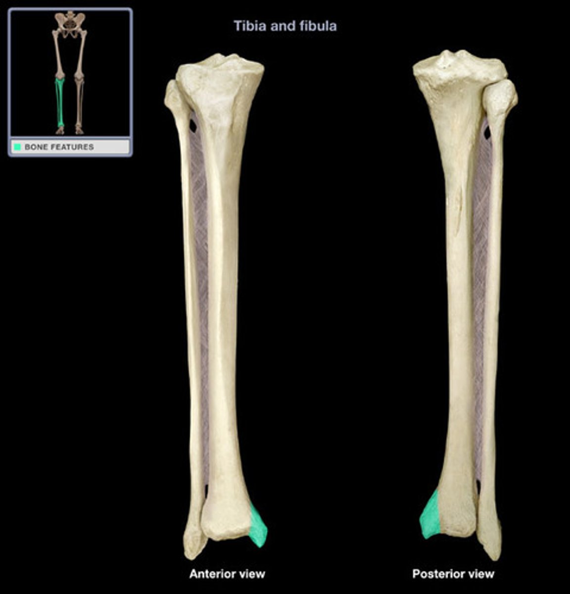
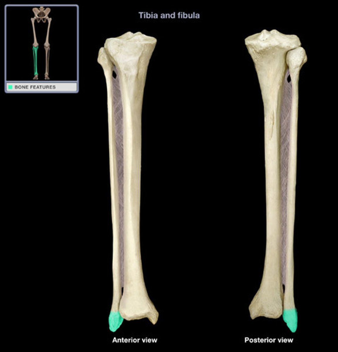
Lateral Malleolus of the fibula
process forming the outer ankle
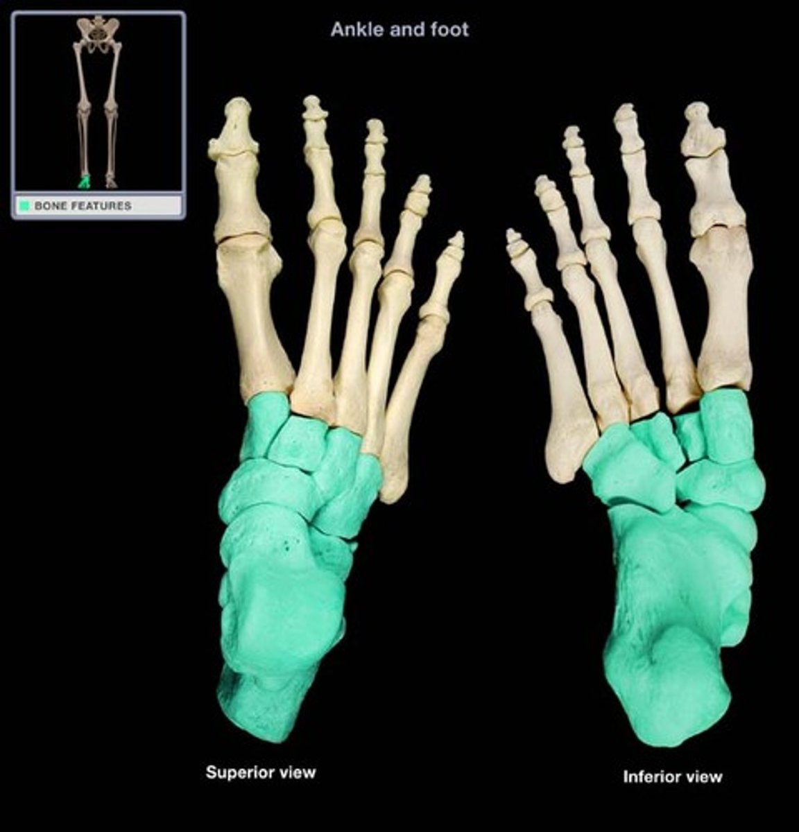
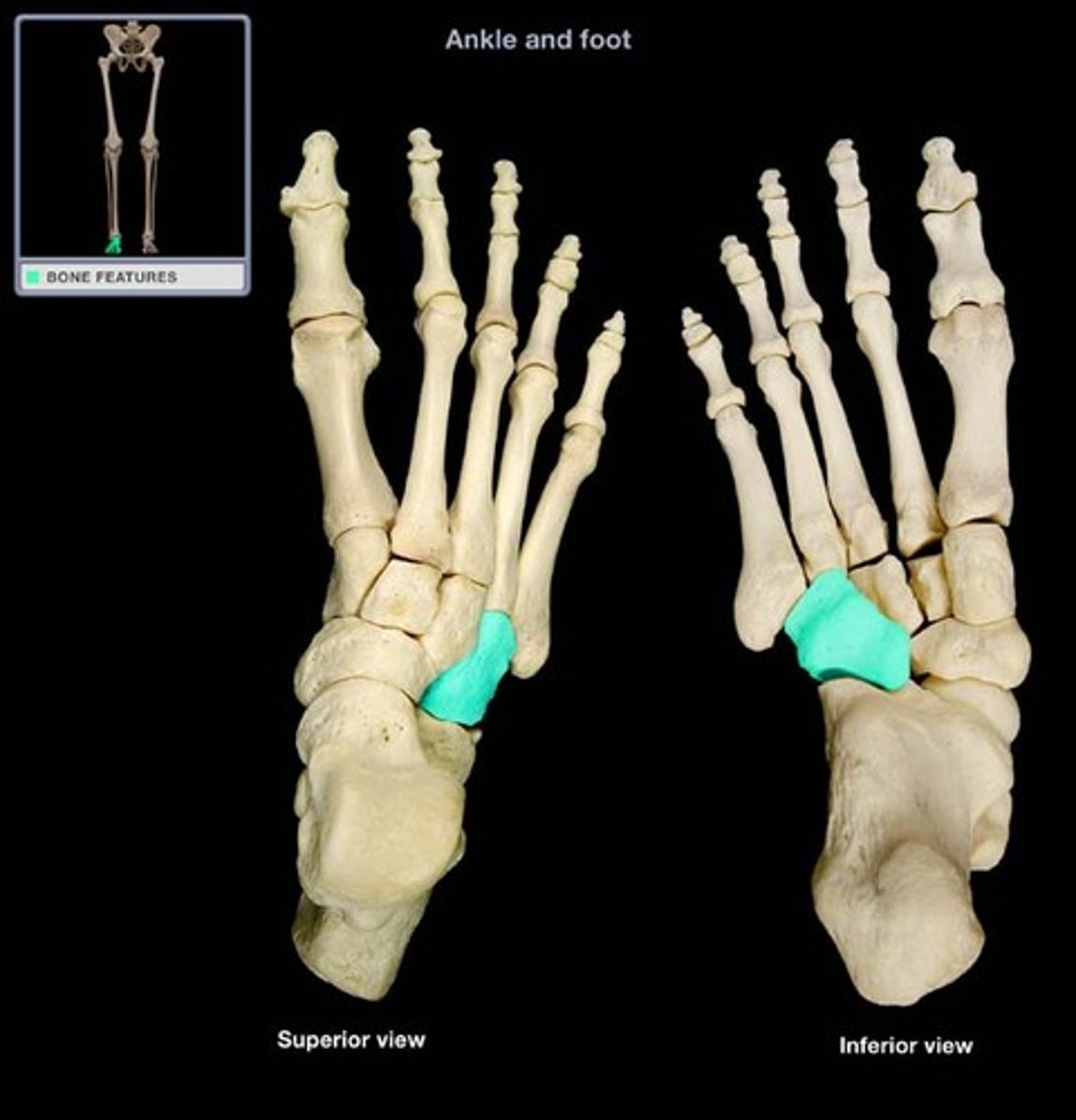
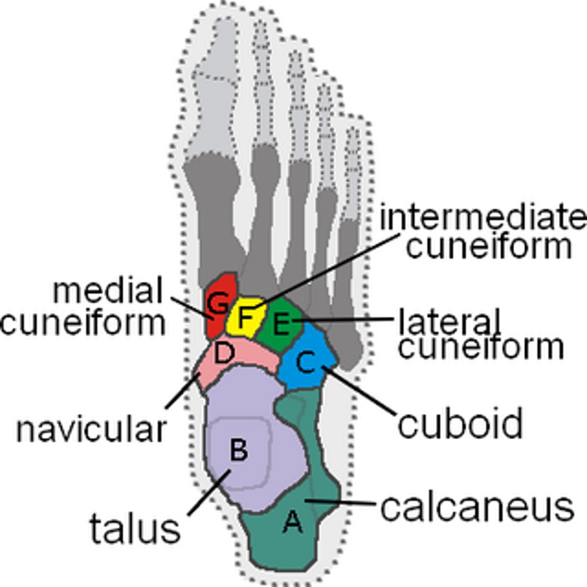
Tarsals
Ankle Bones. 2 sets accounting for both feet. Bones include the Calcaneus, Talus, Cuboid, Medial cuneiform, intermediate cuneiform, and lateral cuneiform.
Mnemonic:
Tiger Cubs Need MILC
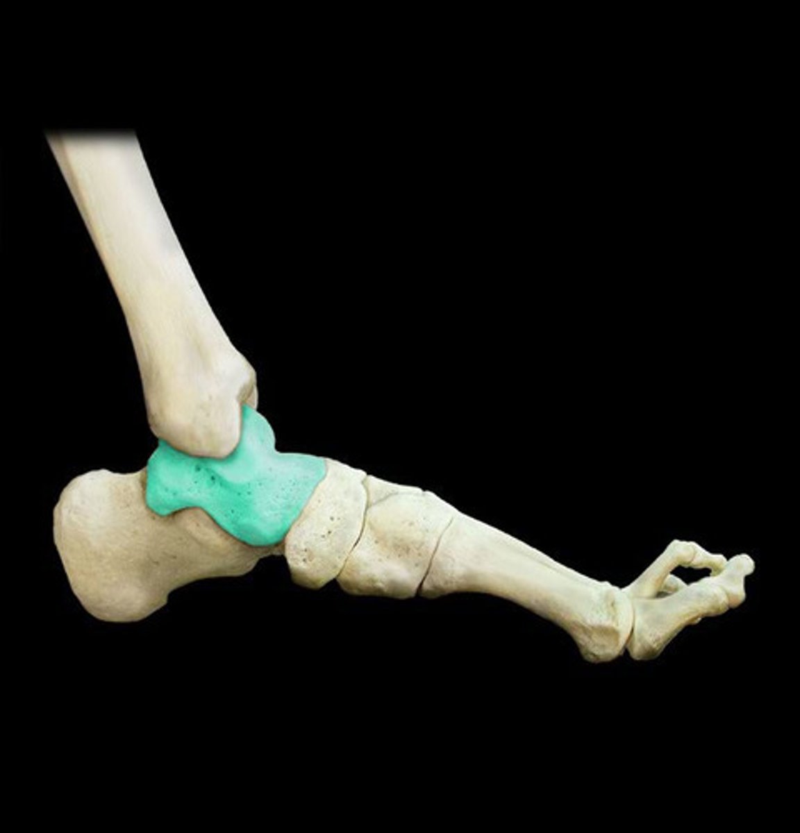
Talus of tibia
Articulates with tibia. Second largest of tarsal bones. "Tiger"
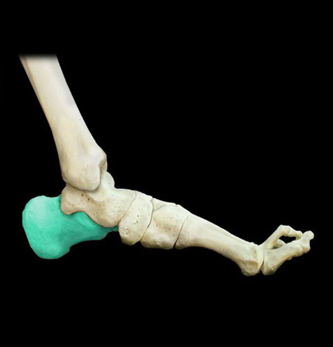
Calcaneus of tarsals
heel bone; largest of the tarsal bones. "Cubs"
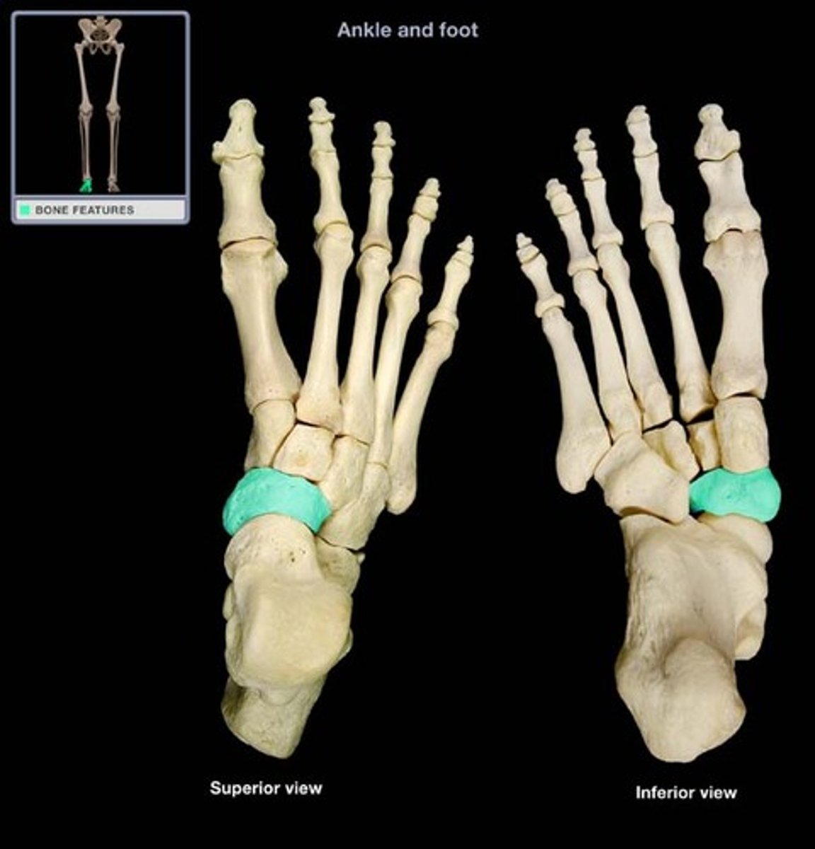
Navicular of tarsals
Medial side of ankle. "Need"
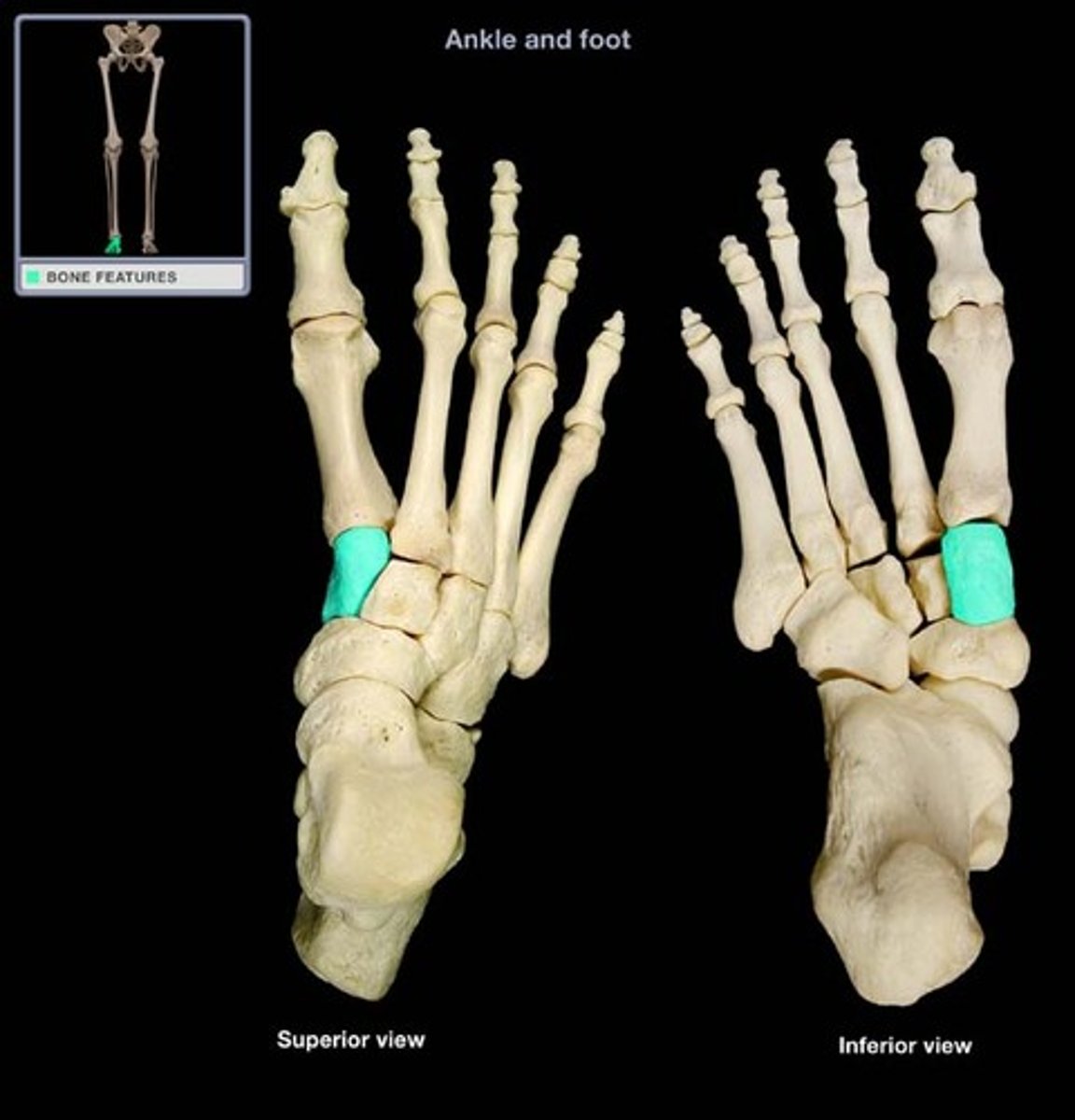
Medial cuneiform of tarsals
Most medial of the cuneiform bones. Articulates with intermediate cuneiform laterally. Positioned anteriorly of navicular bone.
"M".
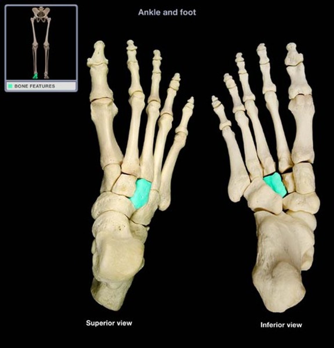
Intermediate cuneiform of tarsals
Between medial and lateral cuneiform of the cuneiform bones. Positioned anteriorly of navicular bone. "I".

Lateral cuneiform of tarsals
Articulates with cuboid bone laterally and intermediate cuneiform medially. "L".
Cuboid of tarsals
Lateral place. Articulates with lateral cuneiform medially and calcaneus posteriorly. "C".
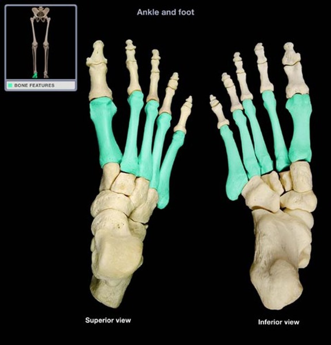
Metatarsals
Foot bone, numbered 1 to 5, from Hallux (big toe) to little toe respectively.
Tiger Cubs need MILC
mnemonic for remembering tarsals.
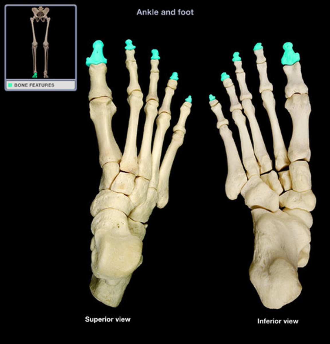
Phalanges
3 types: proximal, middle, and distal.
Proximal Phalanges of foot
Articulates with metatarsals. Digits 1-5 from hallux (big toe) to little toe respectively.
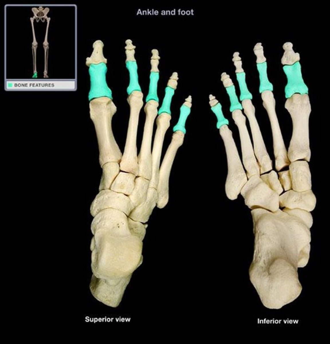
Middle Phalanges of foot
between proximal and distal phalanges of foot. Digits 2-5 from medial to lateral little toe (hallux does not have this) respectively.
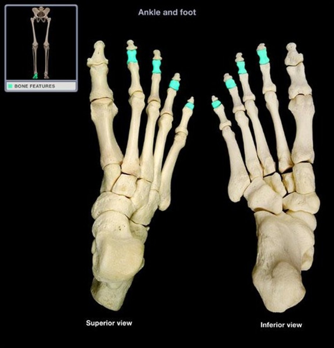
Distal Phalanges of foot.
distal toe bones. Largest is on hallux. Digits 1-5 from hallux to little toe respectively.