Pathophysiology 2, blood cells
1/174
There's no tags or description
Looks like no tags are added yet.
Name | Mastery | Learn | Test | Matching | Spaced | Call with Kai |
|---|
No analytics yet
Send a link to your students to track their progress
175 Terms
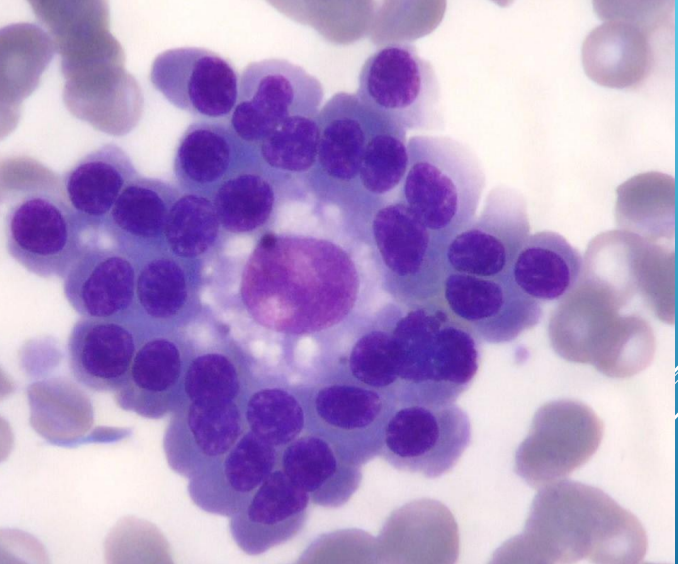
Bone marrow
in the center - macrophage, around - different forms of RBC
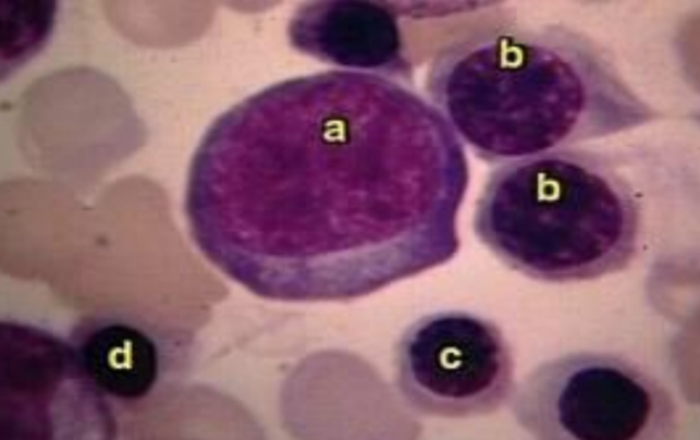
Bone marrow
a - proerythroblast (?), b - basophilic erytroblast, c - polychromatic erytroblast, d - acidophilic erytroblast
Bone marrow
erytroblastic island

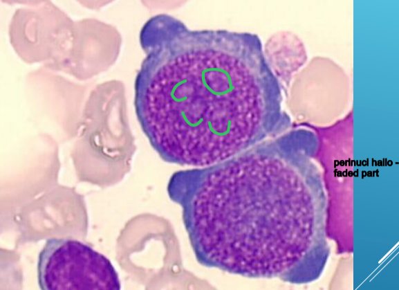
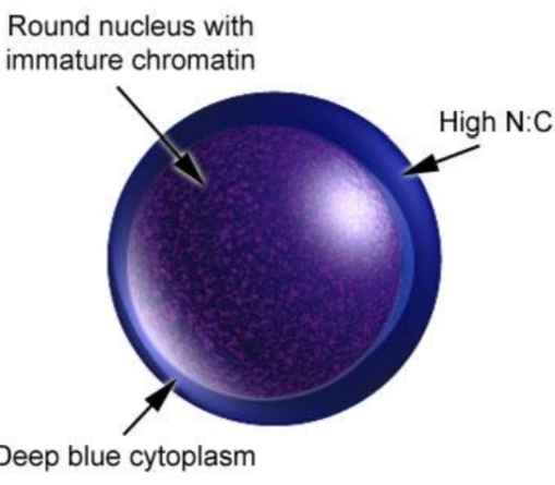
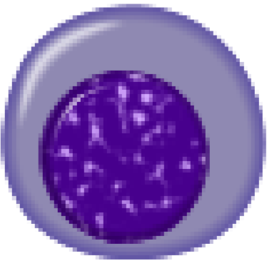
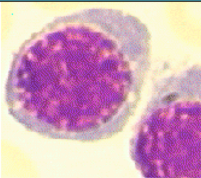
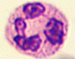
proerythroblast
15-20 μm
round or oval, dark blue cytoplasm, perinuclear
hallo
large nucleus, fine network structure,
chromatin becomes dense with haemoglobin synthesis progression
5 visible nucleoli
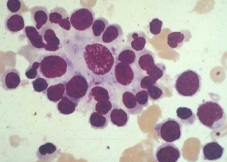
Bone marrow
proerythroblast
Bone marrow
proerytroblast
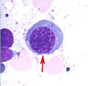
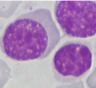
basophilic normoblast (erythroblast)
8-16 μm
round to oval, grey-bluish cytoplasm
hemoglobin synthesis – cytoplasm blue
round nucleus, denser chromatine
no visible nucleoli
Bone marrow
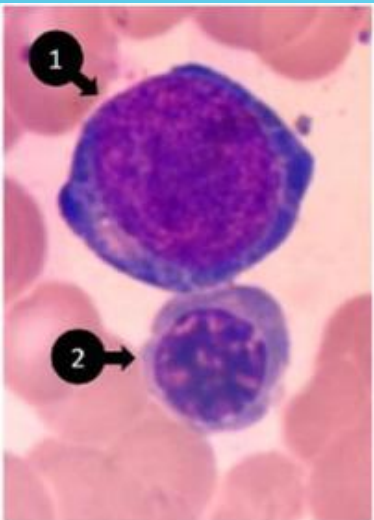
1 - basophilic normoblast, 2 - polychromatic normoblast
Bone marrow
basophilic normoblast (erytroblast)
Polychromatic erythroblast (normoblast)
8-12 μm
round to oval, polychromatic greyish blue cytoplasm
round nucleus, granular, central or eccentric with dark patches of chromatin, without nucleoli
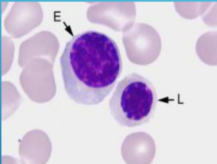
Bone marrow
E - polychromatic normoblast, L - acidophilic normoblast
Acidophilic erythroblast (orthocromatic normoblast)
8-12 μm
round to oval, pink cytoplasm, round or oval nucleus
denser, homogenous, dark blue chromatine
picnotic and excentric nucleus, smaller
changed nucleus is expelled from the cell
cell and description
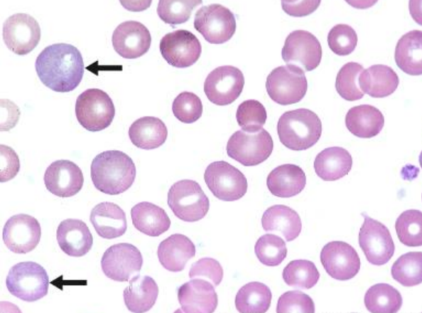
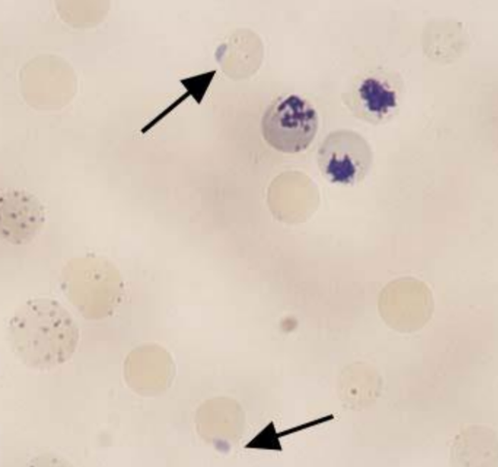
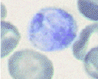
reticulocyte (polychromatophils)
blue pink cytoplasm, no nucleus (remnants)
briliant cresil blue - reticular structure
number increases during bleeding, hemolysis
and succesfull therapies of anemias
reiculocytes
reticulocytes

cell, description
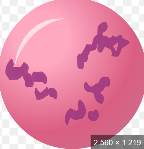
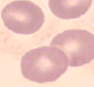
erythrocyte
flexibile, biconcave, discoid cell with
peripherially located hemoglobine and central pale area
red cytoplasm
Bone marrow
proerythroblast
Bone marrow
basophilic normoblast
Bone marrow
polychromatic normoblast
Bone marrow
Orthochromatic normoblast
reticulocyte
erythrocyte
Bone marrow
acidophilic erythroblast
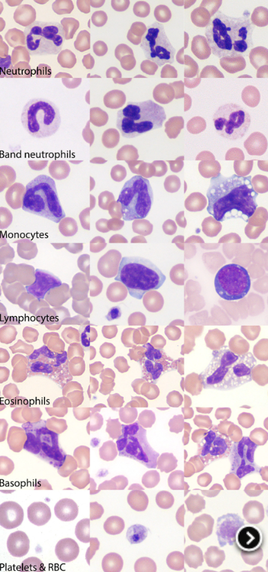
blood;
cell, sp
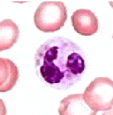
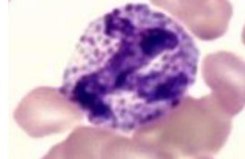
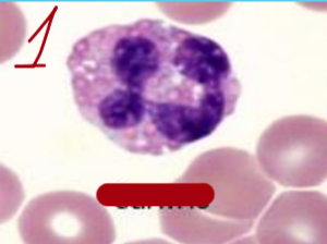
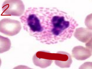
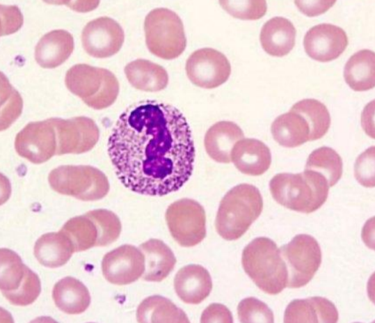
neutrophils (sgmented)
produced in BM, destroyed by macrophages
1st type of Le recruited in inflammation exudate
can’t divide
role: kill microbes. eliminate foreign material, phagocyte, secrete content of granules into exudate
myeloblast
promyelocyte
myelocyte
metamyelocyte
band cell
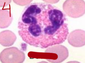
segmented neutrophil

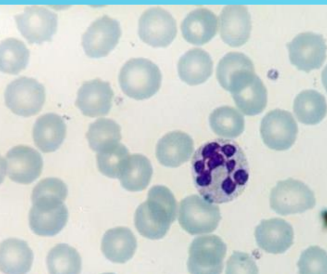
eosinophils (equine)
cell, sp
basophils (feline)
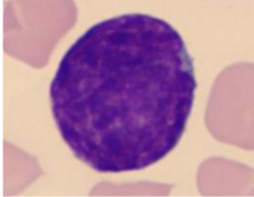
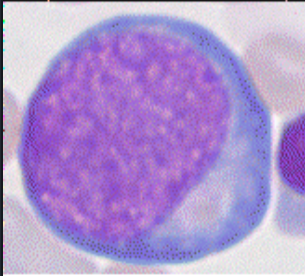
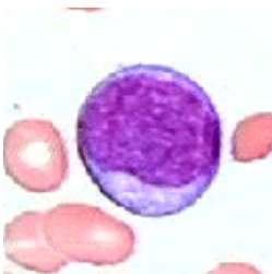
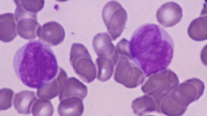
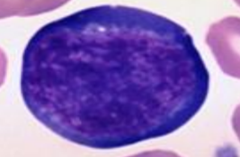
lymphoblast
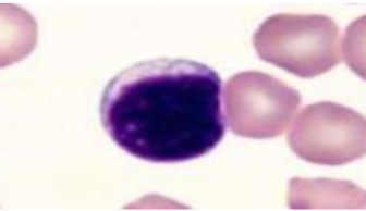
Lymphocyte (feline)
myeloblast
promyelocyte
myelocyte
metamyelocyte
band
segmented
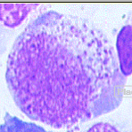
cell and desciption
myeloblast
12-20 μm
round to oval
basophilic cytoplasm, darker at corner
big nucleus with reticular chromatine and visible
nucleoli (2-5)
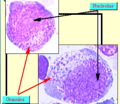
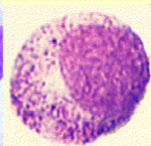
cell, description
promyelocyte
20-25 microm
round to oval
Cytoplasm blue, darker around corners with
primary azurophil (red) and secondary brownish granules
round to oval nucleus sub centrally, indented
promyelocyte
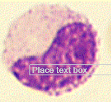
cell, description
myelocyte
11-20 μm
Round to oval, cytoplasm is pale, basophilic in younger,
acidophilic in mature cells
Secondary, specific granules - neutrophilic, eosinophilic, basophilic myelocyte
Round nucleus, excentric
1 - metamyelocyte, 2 - myeloblast
cell, desctiprion
metamyelocyte
12-18 μm
Round to oval, divisions stop
Pink cytoplasm with granules
Kidney shaped nucleus, excentric, no visible
nucleoli
metamyelocyte
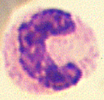
cell, description
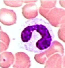
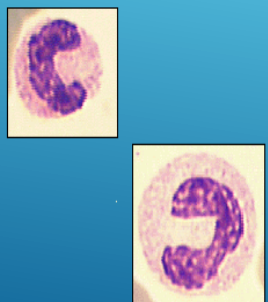
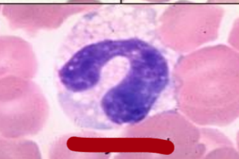
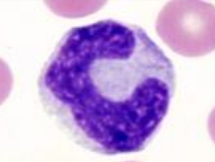
band cells
similar to mature, nucleus in form of C or S letter
chromatin has “leopard-skin” appearance
1 - promyelocyte
2 - neutrophilic myelocyte
3 - metamyelocyte
4 - band cell
cell, sp
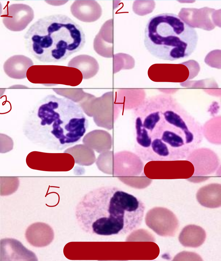
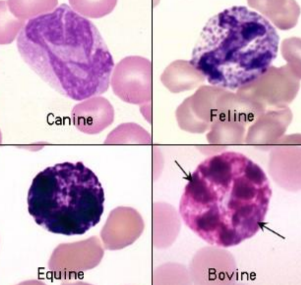
neutrophils
1 - canine, 2 - feline, 3 - equine, 4 - bovine, 5 - band neutrophil
canine eosinophil
cell, sp
feline eosinophils

equine eosinophil
bovine eosinophils
Greyhound eosinophil
basophils (no specifity between species)
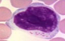
cell, description
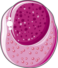
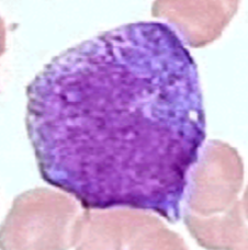
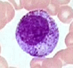
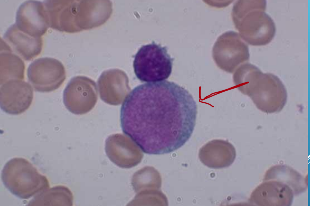
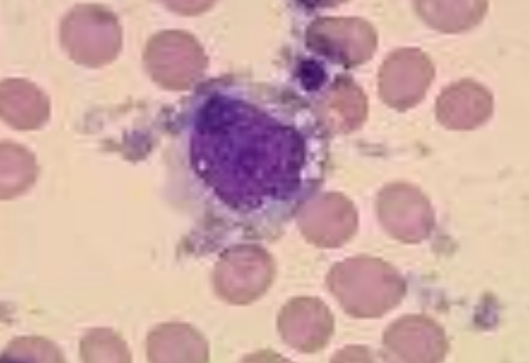
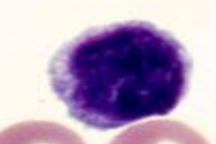
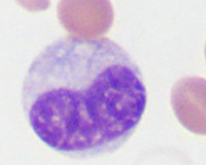
Monoblast
Round to oval, blue or grey cytoplasm,
big indented nucleus with 1-2 nucleoli, heart-shaped
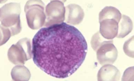
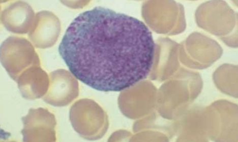
cell, description
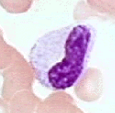
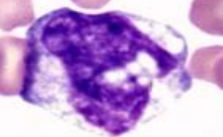
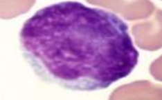
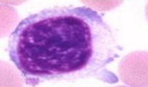
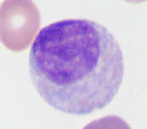
Promonocyte
Round to oval, light blue cytoplasm with big
indented or serrated nucleus and nucleolus
promonocyte, canine
promonocyte, feline
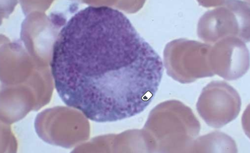
cell, specie
promonocyte, equine
promonocyte, bovine
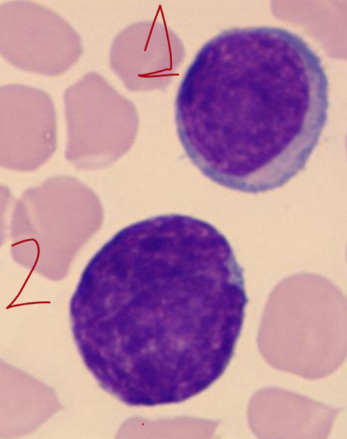
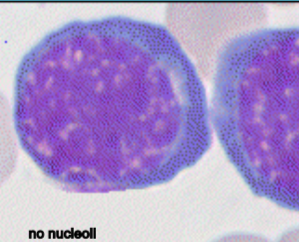
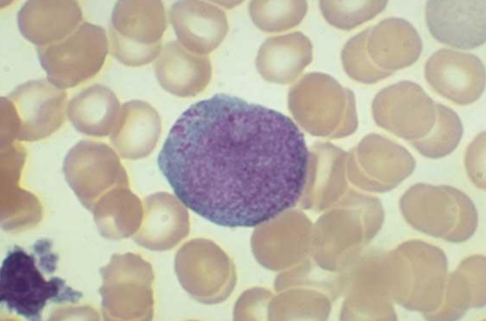
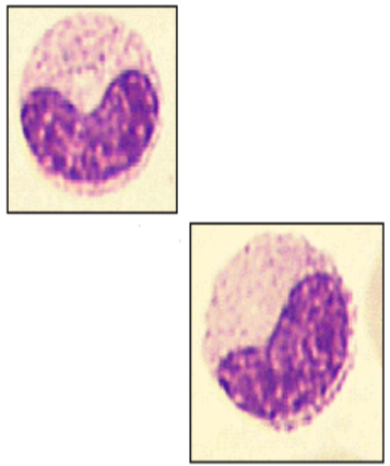
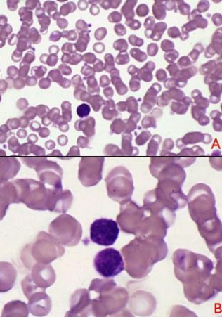
1 - prolymphocyte
2 - lymphoblast
up - could be band cell
down - lymphocyte
cell, sp
canine lymphocyte

feline lymphocyte
equine lymphocyte
bovine lymphocyte
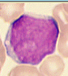
reactive lymphocyte
granular lymphocyte
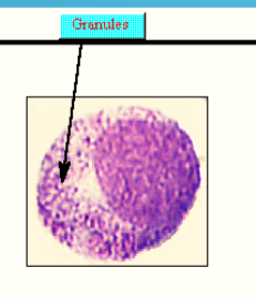
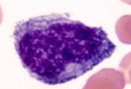
promyelocyte (due to granules)
promyelocyte
myelocyte
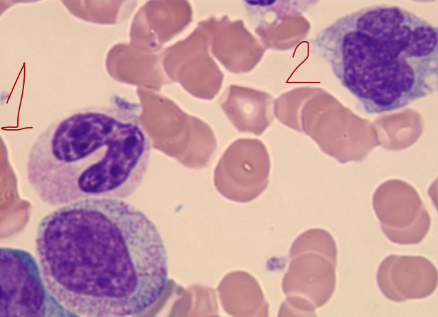
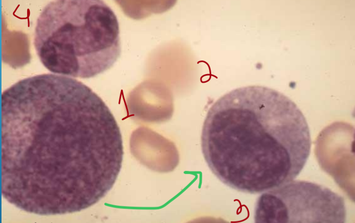
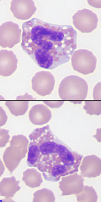
1 - band cell or metamyelocyte (immature)
2 - mature monocyte
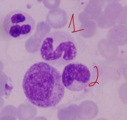
1 - band cell, 2 - metamyelocyte
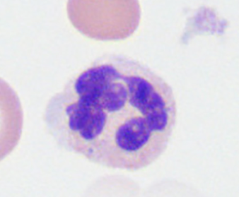
mature neutrophil
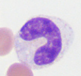
band cell
metamyelocyte
myelocyte
eosinophils (dog)
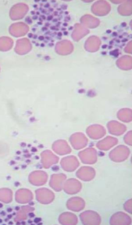
basophils (dog)
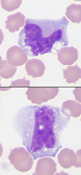
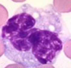
monocyte
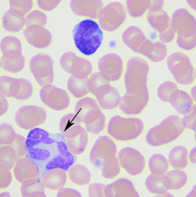
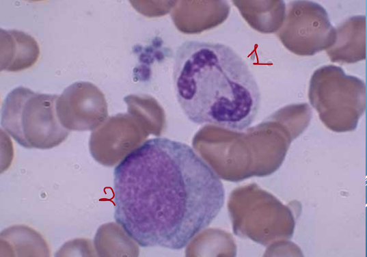
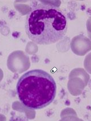
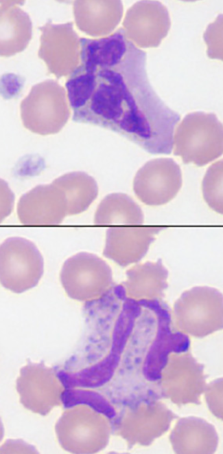
top cell - intermediate reactive lymphocyte, arrow - monocyte (dog)
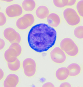
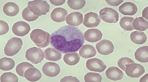
lymphocyte (dog)
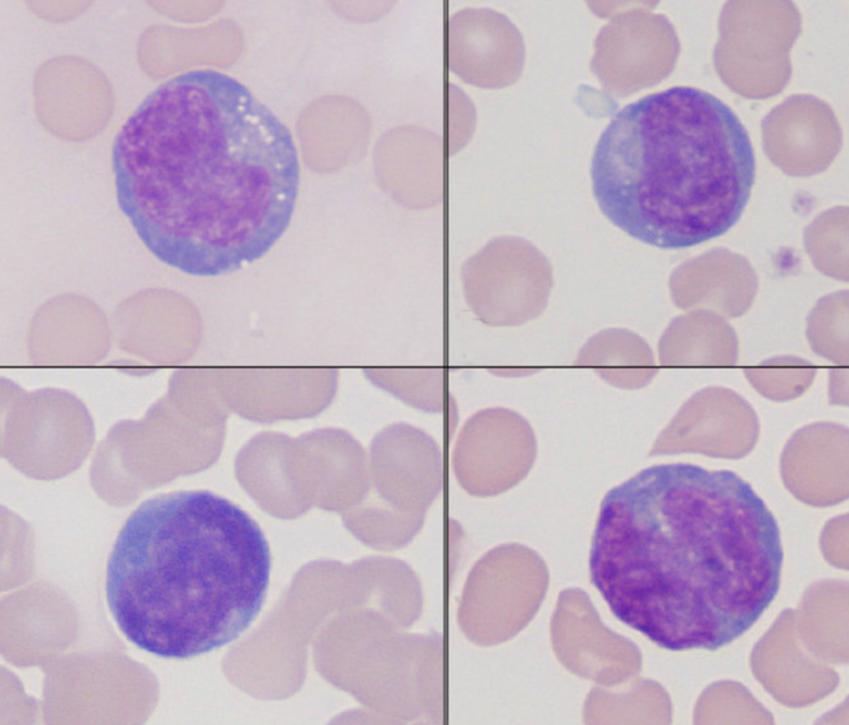
lymphocytes (dog, reactive)
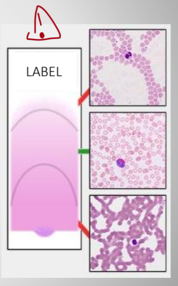
describe
feathered area - cells are flattened and distorted, red cell lose central parlor
monolayer are - cells are in one layer (erythrocytes close to each other but do not overlap),
suitable for differential counting and morphology
body area - Too thick part of blood smear for morphology assessment and differential counting
name the area
body area

name the area
monolayer
name the area
feathered area
name the spiece
dog
name the spiece
dog
name the spiece
bovine
name the spiece
equine
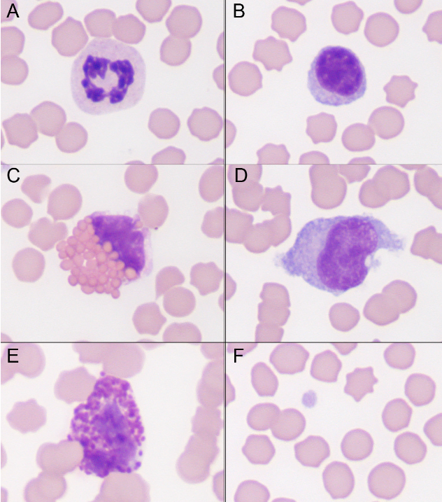
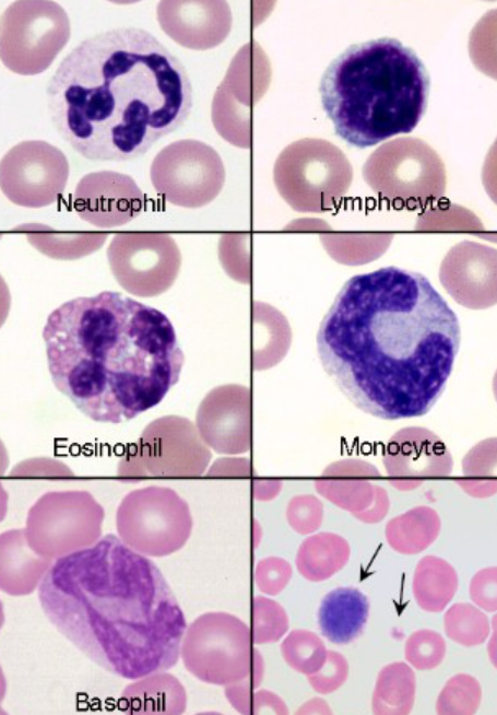
name the spiece and type of cells
equine
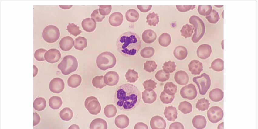
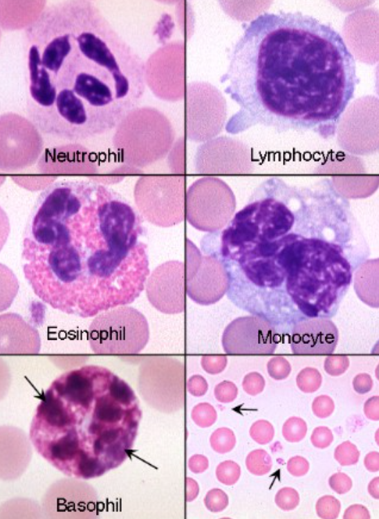
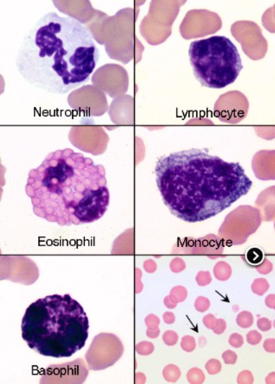
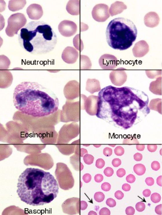
A: Neutrophil; B: Small lymphocyte (mildly reactive - bluer cytoplasm than normal), C: Eosinophil; D: Monocyte; E: Basophil; F: Platelets and red blood cells.
name the spiece
feline
regenerative left shift
Total number of leukocytes is increased, mature cells are
dominant
degenerative left shift
Total number of leukocytes is normal or decreased, Immature cells are dominant
right shift (nuclear hypersegmentation)
5 or more segments
aging, EDTA exposition, glucocorticoids therapy, hyperadrenocorticism, chornic inflam
hypersegmentation
toxic nph
severe inflam, toxemia, c/p basophilia, foamy vacuoals, Dohle’s bodies (1/>more, bs, irregular)