Looks like no one added any tags here yet for you.
Aortic dissection types
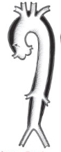
Type 1: All the way to iliac bifurcation
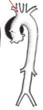
Type 2: Ascending aorta only, ends at innominate artery
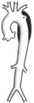
Type 3: Thoracic aorta to subclavian
Name the pathology
Type 1 aortic dissection
Name the pathology
Type 2 aortic dissection
Name the pathology
Type3 aortic dissection
Angina pectoralis
Lack of oxygen to myocardium
Aortic stenosis
Narrowing of aortic valve
What can cause aortic stenosis
Congenital deformity
Rheumatic heart disease
Aging
Arteriosclerosis vs atherosclerosis
Arteriosclerosis - hardening of arteries
Atherosclerosis - plaque in arteries
CABG
Coronary artery bypass graft
Thrombus vs embolism
Thrombus: blood clot formation which obstructs flow
Embolus: Clot breaks free and travels through bloodstream
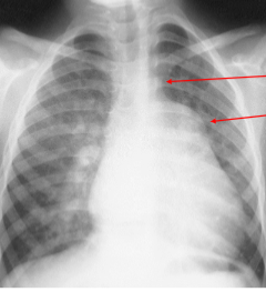
Another name fro AVM
Arteriovenous fistula
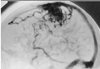
Arteriovenous malformation radiographic appearance
Tangled mess of vessels, seen best with contrast
Name the pathology
AVM
Name the pathology
AVM
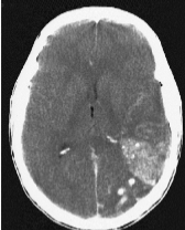
CVD
Cerebrovascular disease - any process which causes abnormality of blood vessels to the brain including hemorrhage, aneurysms and AMV
CHF
Heart is unable to pump sufficient blood to the body
What causes CHF
Abnormality causing defective cardiac filling or emptying
Hypertension
Obstructive processes
Manifestations of CHF (applying to both sides)
Decreased output and stroke volume
Hypoxia
Fatigue, weakness
Dyspnea
Compensation mechanisms for CHF
Tachycardia
vasoconstriction
Oliguria
Right vs left sided CHF
Right - systemic, symptoms in limbs and organs
Left - pulmonary, symptoms related to lungs
Left sided CHF manifestations
Related to pulmonary congestion (fluid buildup):
Dyspnea
Cough
Nocturnal dyspnea
Left sided CHF appearance
Cardiothoracic ratio greater than 50%
Cardiomegaly
Pulmonary edema, effusion
Right sided CHF manifestations
Edema in limbs
hepatomegaly, splenomegaly (digestive disturbance)
Ascites
Acute:
Face flushing
distended neck veins
headache
Visual disruption
Radiographic appearance of right sided CHF
Cardiothoracic ratio greater than 50%
Wide mediastinum
Right hemidiaphragm elevation (hepatomegaly)
Risk factors for coronary artery disease
Hypertension
Obesity
Smoking
High-cholesterol
Sedentary
Causes of coronary artery disease
Arteriosclerosis/atherosclerosis
Angina pectoris
Myocardial infarction
Complications of dexterocardia
Heart defects
Spleen is missing
Strange gallbladder
Issues with lungs and intestines
Congenital heart disease
Cardiac anomalies
Septal defects
valve defects
What types of shunting are there in the heart
Right to left - cyanotic
Left to right - acyanotic
Arterial septal defect
Right and left atria communicating, right to left shunting = increased pulmonary blood flow overloading right ventricle
Radiographic appearance of ASD
Enlarged right ventricle, right atrium and pulmonary trunk
PFO
Patent foramen ovale - communication between between right and left atria due to incomplete closure of foramen ovale
Left to right shunt (pressure higher in left atria)
PFO vs ASD radiographic appearance
Same
Foramen ovale
Opening or shunt in heart tissue allowing blood to flow from right to left atrium
VSD
Ventricular septal defect - left to right shunting between two ventricles
Increased pulmonary blood flow and venous return
VSD radiographic appearance
Pulmonary trunk enlargement, left atrium and ventricle
enlargement but NO right ventricle enlargement
Triangular mediastinum, pulmonary trunk is huge and makes aorta look tiny
Name the pathology
VSD
PDA
Patent ductus arteriosus
Connection between pulmonary arteries and aorta
Left to right shunt
PDA radiographic appearance
Enlarged left atrium, left ventricle and pulmonary arteries
Increased pulmonary vascularity (aortic knob formation)