The Form Sense II: Factors affecting Visual Acuity
1/21
There's no tags or description
Looks like no tags are added yet.
Name | Mastery | Learn | Test | Matching | Spaced |
|---|
No study sessions yet.
22 Terms
name factors that may affect VA (8)
•Refractive error/optical blur
•Retinal location (eccentricity)
•Contrast
•Contour interactions (crowding)
•Luminance
•Orientation/meridional variations
•Movement of the eyes or target
•Developmental aspects
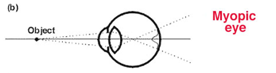
explain the condition of myopia (2)
eye length is too long so a clear image is formed INFRONT of the retina and a blurred one on the retina
eye is too strong and a clear image is formed in front of the retina so the retinal image is blurred
IN FRONT
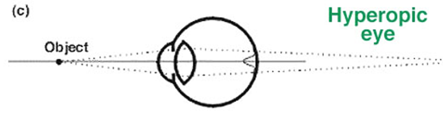
explain the condition of hypermetropia (2)
Eye length is too short
→ So a clear image is formed behind the retina, and a blurred one on the retina.Eye is too weak (not enough optical power)
→ So a clear image is formed behind the retina, making the retinal image blurred.
BEHIND
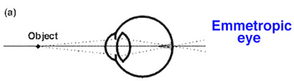
explain what emmetropria is and its effect on VA
•Emmetropia occurs when the optical quality is not diminished and the focus is conjugate with the retina
•Therefore, emmetropia does not impact on VA
explain what refractive error is - 2 types - and its effect on VA
As soon as the optics of the eye become defocussed, the retinal image is also defocused which can result in blurry vision
Myopia (short-sightedness) – one of the main refractive error types causes the retinal image to spread
Hyperopia or hypermetropia (long sightedness) – one of the main refractive error types causes the retinal image to spread
what is the effect of optical blur on VA
as the blurring lens increases in power, VA is degraded/decreases
what is the effect of retinal eccentricity on VA
VA is best at the fovea (00 eccentricity).
The centre of the fovea is where visual acuity is best, which is why our eyes are designed to bring objects into focus at the fovea.
Even small deviations away from the fovea cause a large fall off in VA.
For patients that suffer from ocular diseases that affect the fovea (e.g. macular degeneration), this is an important consideration because central vision is lost
increase in retinal eccentricity = decrease in VA
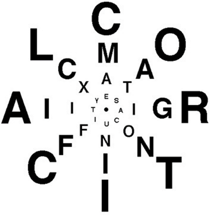
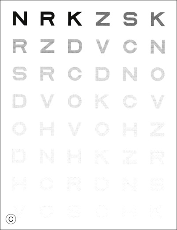
how can we demonstrate the impact of retinal location on VA
•Anstis (1974) devised a vision chart that represents equal letter readability at different retinal locations.
•The chart also shows how VA changes across the field of vision
explain the effect of contrast on VA and how it is specified
Visual acuity also reduces as the letter contrast reduces
Contrast (Weber) is specified as the ratio of ΔL/L.
For ‘black’ on ‘white’ it is sometimes designated. as a minus (-) value e.g. -95%
if luminance of a letter is 1cd / m2 and of the background is 100 cd / m2 - what is the contrast of the letter
work out the difference in luminance between the background and the letter = 99
then that value / 100
as it is black and white it would be -99 / 100 = -0.99 (or -99%)
answer = -99% and 99% is also acceptable
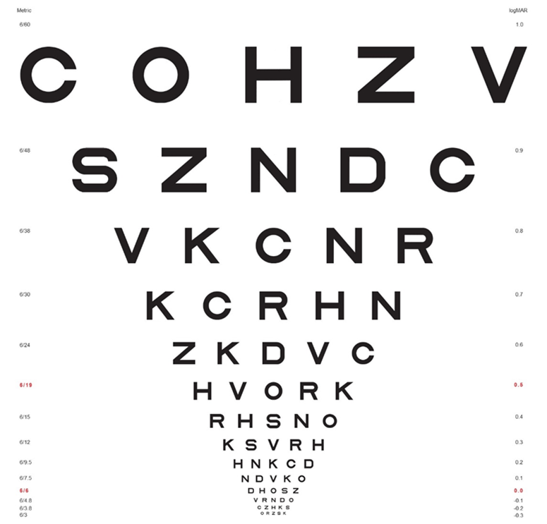
explain how the effects of contrast on VA can be measured using letter charts
Some follow the ETDRS format as shown, where the letters on a particular chart are all a fixed contrast and letter size varies:
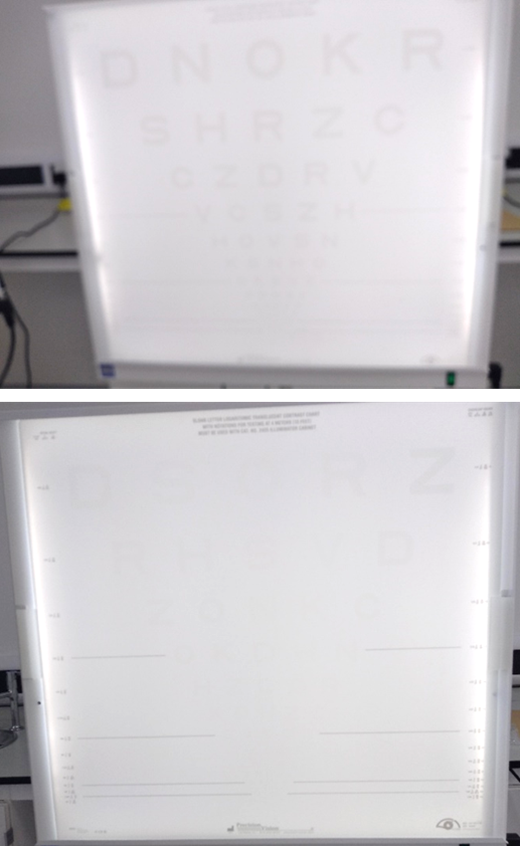
•Other low contrast letter charts have the same size of letter, but contrast varies from high to low, such as the Pelli-Robson chart shown:
usefulness of low contrast VA charts
helpful in assessing patients with low vision (e.g. cataract) and other conditions where normal high contrast charts may show relatively little defect
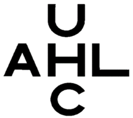
what are flankers and what is their effect on VA
•VA measured with single letters is usually avoided – unless the letters are surrounded by flankers.
•The flankers could be bars or letters as shown in the images.
•The impact of flankers on VA measurements is called crowding.
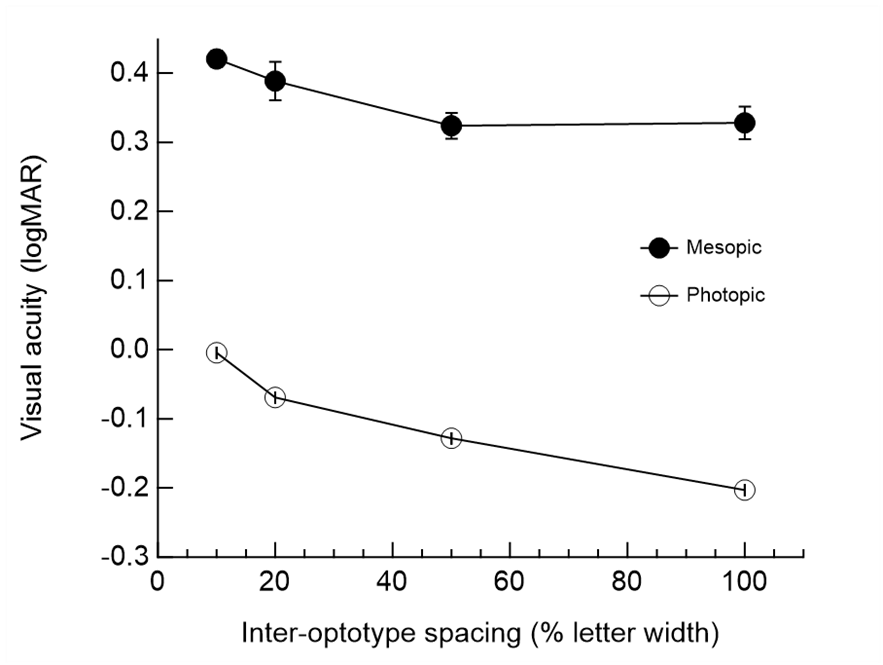
•VA decreases as the distance of the flankers gets closer to the target letter - this is called critical distance
-Critical distance – closer the distance, VA degrades

Presence of neighbouring objects affects vision of target object
The crowding makes it difficult to see the letter H
where is crowding stronger in terms of retinal location and why
Crowding is stronger in the periphery - In peripheral vision (the sides of what you see), your brain can’t separate things as well
At the fovea crowding only occurs over small distances up to about 5 min of arc
what is the best type of chart to use to measure VA
crowded charts
The logMAR form of VA charts are crowded and are preferred particularly when accurate VA measurements are required
The spacing between letters and rows is proportional to the size and is equal.
define the crowding distance
The smallest distance/closest distance between letters where they remain unimpeded is known as the crowding distance, beyond this point the targets struggle to be identified
explain the effect of luminance on VA
need a 100 cd / m2 - ideal luminance - chart luminances should be somewhere between 80-320 cd/m2
It is unlikely that visual acuity will vary much within this range - providing this general luminance is used the VA will not be affected
as luminance drops however, VA will degrade // Visual acuity improves with increasing retinal illuminance
Note that visual acuity remains good for a wide range of photopic luminance’s
explain the clinical measurement of VA - under mesopic conditions
Under mesopic light levels, visual acuity is reduced
due to lower contrast sensitivity and a change in the function of photoreceptors
leads to a decrease in visual acuity on average of 0.48 logMAR (almost 5 lines on a standard ETDRS chart)
VA measurements under mesopic conditions have not been standardised.
explain the effect of orientation on VA - the oblique effect
The resolving power of the eye appears to be less for oblique orientations - degrades VA when oblique orientations are used rather than horizontal
Less sensitive to oblique than horizontal by 15 percent
One of the reasons that use of Landolt C at oblique angles are avoided
explain the effect of the movement of the eyes or the target on VA
•Even under steady fixation, the eyes are in constant motion.
•Under normal conditions, these small (fixational) eye movements do not detract from good visual acuity.
•For larger eye movements or target velocity (greater than about 3 degrees/second, there is a decrease in visual acuity.
what is the effect of development/aging over time on VA
•With age visual acuity gradually declines (over about age 60), probably due to the increase in light scatter within the eye
-Visual acuity varies with age
what age does VA become adult like - from birth
•Following birth visual acuity gradually improves as the visual structures mature and reaches adult like values at around 5-6 years of age